Mineralization-inducing potentials of calcium silicate- based pulp capping materials in human dental pulp cells
Sohee Kang
Department of Dentistry, Yeungnam University Hospital, Daegu, Korea
Background: This study was performed to provide a long-term bacterial seal through the forma- tion of reparative dentin bridge, calcium silicate-based pulp capping materials have been used at sites of pulpal exposure. The aim of this study was to evaluate the mineralization-inducing po- tentials of calcium silicate-based pulp capping materials (ProRoot MTA [PR], Biodentine [BD], and TheraCal LC [TC]) in human dental pulp cells (HDPCs).
Methods: Specimens of test materials were placed in deionized water for various incubation times to measure the pH variation and the concentration of calcium released. The morphology of HDPCs cultured on the specimens was examined using a confocal laser scanning microscope (CLSM). Alizarin red S staining and alkaline phosphatase assays were used to evaluate mineral- ization-inducing potentials of the capping materials.
Results: BD showed the highest calcium release in all test periods, followed by PR and TC.
(p<0.05). All experimental groups showed high alkalinity after 1 day, except at 14 days. BD showed the highest cell viability compared with PR and TC after 1 and 3 days, while TC showed the lowest value (p<0.05). The CLSM analysis showed that cells were well adhered and expressed actin filaments for all pulp capping materials. Mineralization by PR and BD groups was higher than that by TC group based on alizarin red S staining. BD showed significantly higher alkaline phosphatase activity than PR and TC, while TC showed the lowest value (p<0.05).
Conclusion: Within the limitations of the in vitro study, BD had higher mineralization-inducing potential than PR and TC.
Keywords: Biocompatibility testing; Dental material; Dental pulp capping; Dentin
Introduction
At sites of pulpal exposure, dentinal continuity may be restored through the formation of a dentin bridge across the exposure.
There have been reports regarding dentin bridging after pulp cap- ping with agents such as calcium hydroxide. Calcium hydroxide is a preferable material to manage pulp exposure because of its anti- bacterial characteristic and ability to induce reparative dentin for- mation [1]. However, it has been reported that calcium hydroxide
Yeungnam Univ J Med 2020;37(3):217-225 https://doi.org/10.12701/yujm.2020.00248
Received: April 6, 2020 Revised: May 4, 2020 Accepted: May 7, 2020 Corresponding author:
Sohee Kang
Department of Dentistry, Yeungnam University Hospital, 170
Hyeonchung-ro, Namgu, Daegu 42415, Korea
Tel: +82-53-620-3282 Fax: +82-53-629-1772 E-mail: [email protected]
dissolves over time, causing tunnel defects in dentin bridges, and these bridges allow communication between the pulp and cap- ping material [2].
These findings highlight the need to use materials capable of providing a long-term bacterial seal over capped pulps. Among them, mineral trioxide aggregate (MTA) has a better capping ef- fect by stimulating the formation of the perfect reparative dentin bridge without any toxic chemical effects. Previous studies showed that MTA was more effective than calcium hydroxide in
Copyright © 2020 Yeungnam University College of Medicine
This is an Open Access article distributed under the terms of the Creative Commons Attribution Non-Commercial License (http://creativecommons.org/licenses/by-nc/4.0/) which permits unrestricted non-commercial use, distribution, and reproduction in any medium, provided the original work is properly cited.
long-term vital pulp therapy [3,4]. However, high prices, difficul- ties in handling and application, and longer binding duration re- main disadvantages of MTA. Long setting times are an obstacle to using MTA as a pulp capping material since MTA needs to be lay- ered with other materials while it is still fresh [5].
By adding accelerators and modifiers, several researchers at- tempted to decrease the setting reaction time and increase the ef- fectiveness of MTA for direct pulp capping. One calcium sili- cate-based pulp capping material is Biodentine (BD; Septodont, Saint-Maur-des-Fosses, France), which has the advantage of a shorter setting time of 12 minutes. BD is a powder consisting of tricalcium silicate, dicalcium silicate, calcium carbonate, calcium chloride, and zirconium oxide as a radiopacifier. Previous studies showed that BD works similarly to MTA in both in vitro and in vivo. In addition, BD has a positive effect on pulp cells and helps them form reparative dentin [6,7]. However, research data on the mineral-inducing potential of BD is still lacking.
TheraCal LC (TC; Bisco Inc., Schaumburg, IL, USA) is a light- cured, resin-modified calcium silicate-based material designed for use in direct and indirect pulp capping and as a protective liner under various filling materials. The light-cured set permits imme- diate placement and condensation of the restorative material. It contains approximately 45 percentage by weight (wt%) mineral material (type III Portland cement), 10 wt% radiopaque compo- nent, 5 wt% hydrophilic thickening agent (fumed silica), and ap- proximately 45 wt% resin [8]. In previous studies, TC showed good sealing abilities [8] and was well-tolerated by immortalized odontoblast cells [9]. It is necessary to evaluate whether that ma- terial has a mineral-inducing potential that can form a reparative dentin bridge. However, its mineral-inducing potential has not been studied yet.
This study aimed to evaluate the mineralization-inducing po- tentials of calcium silicate-based pulp capping materials on human
dental pulp cells (HDPCs), by the following five outcome mea- sures: (1) the amount of calcium release from the capping materi- als, (2) the pH values of aqueous medium exposed to the extracts of the capping materials which stimulate the pulp cell differentia- tion, (3) the cell viability by an MTT assay, (4) cytoskeletal orga- nization of the HDPCs cultured on the extract of the capping ma- terials and viewed with a microscope, and (5) alizarin red S stain- ing images and alkaline phosphatase (ALP) activity of odonto- blasts cultured on the capping materials.
Materials and methods
1. Specimen preparation
Premixed specimens of ProRoot MTA (PR; Dentsply, Tulsa, OK, USA), BD, and TC were made according to the manufacturers’
instructions (Table 1). Discs (10-mm diameter and 2-mm thick- ness) were made using a stainless-steel frame as a mold. Each disc was allowed to set for 24 hours followed by polishing with #400, 600, and 1,200 grit lapping film (3M lapping film; 3M, Maple- wood, MN, USA). The colors of disc obtained by digital camera show the white color of MTA and TheraCal, and Biodentine has the Ivory color. The specimens were placed in a 48-well tissue cul- ture plate, kept under an ultraviolet clean bench for 24 hours, and further sterilized by ultraviolet irradiation (Figs. 1, 2).
2. Primary culture of human dental pulp cells
HDPCs were provided by the Department of Oral Pathology (School of Dentistry, Kyungpook National University, Daegu, Korea). Fragments of pulp tissue acquired from an extracted hu- man third molar were cultured in minimal essential medium alpha (MEM-α; Invitrogen, Carlsbad, CA, USA) containing 10% fetal bovine serum (FBS; Invitrogen), 100 U/mL penicillin, and 100 U/mL streptomycin (Invitrogen). The cultures were stored in a
Table 1. Materials used in this study
Product Concentration
ProRoot MTA (Dentsply, Tulsa, OK, USA)
Portland cement 75%
Bismuth oxide 20%
Calcium sulfate 5%
Biodentine (Septodont, Saint-Maur-des-Fosses, France)
Powder: tricalcium silicate, dicalcium silicate, calcium carbonate, zirconium oxide, calcium oxide, iron oxide Packaged in capsule (0.7 g)
Liquid: calcium chloride, a hydrosoluble polymer, water Packaged in pipette (0.18 mL)
TheraCal LC (Bisco Inc., Schamburg, IL, USA)
Portland cement type III < 60%
Polyethylene glycol dimethacrylate < 50%
Barium zirconate < 10%
humidified atmosphere of 5% CO
2at 37°C.
3. Measurement of calcium release from pulp capping materials
The calcium release of each specimen was measured to evaluate the mineralization-inducing potentials. The disc specimen was placed in 10 mL of deionized water for 1 and 7 hours and 1, 4, 7, 14, and 21 days. The amount of calcium ion released from the capping materials in the water was measured by inductively cou- pled plasma atomic emission spectroscopy (ICP; Optima 7300DV, PerkinElmer, Shelton, CT, USA). ICP is a technique for detect- ing trace metals. It excites atom and releases ions from electro- magnetic radiation at characteristic wavelengths of a particular element.
4. Measurement of pH value
The pH values of each specimen were measured to assess the amount of eluted hydroxide ions, which stimulate the pulp cell differentiation. The disc specimen was placed in 10 mL of deion-
ized water for 1 and 7 hours and 1, 4, 7, 14, and 21 days. The pH value of the aqueous medium, which was exposed to the extracts of the capping materials, was measured by a pH meter (Orion 3 Star, Thermo Scientific, Singapore). The pH apparatus was cali- brated with solutions of pH 7.0 and 4.0. The electrode was washed with ultrapure water and dried after every measurement.
5. Cell viability test
An MTT assay was conducted to evaluate the effects of the cap- ping materials on cell viability. The amount of HDPCs on each specimen was measured by a colorimetric immune assay that is based on measuring bromodeoxyuridine (BrdU) incorporated during DNA synthesis. BrdU enzyme-linked immunosorbent as- say (ELISA; Roche Molecular Biochemicals, Nutley, NJ, USA) was performed according to the manufacturer’s instructions. Cells were seeded at a density of 2× 10
4cells per well onto a 48-well plate and were cultured for 1 and 3 days with each specimen.
Then, the BrdU-labeling solution was added to each well. The solution was applied to the cells in a CO
2incubator at 37°C for 4
Fig. 1. (A) Stainless-steel frame with five holes, (B) schematic diagram of the specimen, (C) 48-well cell culture plates, having flat bottom which matches the specimen in size.
Fig. 2. Images of specimens. (A) ProRoot MTA, (B) Biodentine, (C) TheraCal LC.
A B C
A B C
10 mm
2 mm
hours. The supernatant of the cell solution was removed by pipet- ting and were washed with phosphate-buffered saline (PBS) twice. After treating the cells with 0.25% trypsin-ethylenedi- aminetetraacetic acid (EDTA; Gibco, Tokyo, Japan), the cells were harvested by centrifugation at 1,000 rpm for 15 minutes. For fixing and denaturing DNA, the harvested cells were mixed with FixDenat (ELISA; Roche Molecular Biochemicals) solution and then incubated for 30 minutes. Diluted anti-BrdU peroxidase (di- lution ratio of 1:100) was added to the cells, which was then incu- bated at 20°C for 2 hours. The unbound antibody conjugate was removed, and 100 μL substrate was added to the 48-well plate.
Then, the cells were incubated for 20 minutes. The reaction was finished by adding 25 μL of H
2SO
4solution (1 M) to the cells.
The solution was moved to a 96-well plate, and the absorbance of the solution was measured using an ELISA plate reader (EL 9800;
Roche Molecular Biochemicals) for 5 minutes at 450 nm with a reference wavelength of 690 nm. The blank reading corresponded to 100 μL of culture medium with or without BrdU.
6. Cytoskeletal organization
To assess the cytoskeletal organization of HDPCs, which were treated with the extracts of the capping materials, double staining was performed. Briefly, a prepared specimen of each cement was stored in 10 mL MEM-α contained 10% FBS for 3 days to pro- duce the extracts used for the treatment of HDPCs. The cells were seeded onto a 48-well plate (2 ×10
4cells/mL) and were cultured for 1 day with the extracts of the specimen. The cells were fixed with 4% paraformaldehyde in PBS and washed with a PBS solution containing 0.05% Tween-20. After permeabiliza- tion with 0.1% Triton X-100 in PBS for 15 minutes at 25°C, the cells were incubated for 30 minutes in PBS containing 1% bovine serum albumin. Subsequently, 5(6)- tetramethyl-rhodamine iso- thiocyanate-conjugated phalloidin (Millipore, Temecula, CA, USA) was added to the 48-well plate, and the cells were incubat- ed for approximately 1 hour. The plates were washed three times (10 minutes each) using the buffer solution and incubated with 4′,6-diamidino-2-phenylindole (Millipore) for 5 minutes. The scaffolds were washed three times (10 minutes each) with the buffer solution, and fluorescence images were visualized by a CLSM (model 700; Carl Zeiss, Oberkochen, Germany).
7. Alizarin red S staining
Mineralized nodules from differentiated cells were visualized through alizarin red S staining to evaluate the mineraliza- tion-inducing potentials of each specimen. Cells were seeded at a density of 2×10
4cells per well onto a 48-well plate and were cultured for 14 days with each specimen. Then, mineralization
of the cells was evaluated through alizarin red S staining (Sig- ma-Aldrich, St. Louis, MO, USA). Briefly, cells were fixed with 4% formalin for 1 hour at 4%, washed three times with deion- ized water, and then stained with 40 mmol/L of alizarin red solution (pH=4.2). The stained cell plate was washed with de- ionized water, and the stained image was obtained with the scanner. To remove the stain, the samples were treated with 10% cetylpyridinium chloride solution (pH=7.0) for 15 min- utes, and absorbance was measured at a wavelength of 540 nm with a standard solution for the quantitative assessment.
8. Alkaline phosphatase activity assay
ALP is the marker of early differentiation and extracellular matrix mineralization. Cells were seeded at a density of 2× 10
4cells per well onto a 48-well plate and were cultured for 14 and 21 days with each specimen. Then, the cells were scraped into cold PBS and sonicated with a cell disruptor (Heat System Ultrasonics, Pla- inview, NJ, USA) in an ice-cold bath. ALP activity in the superna- tant was determined using p-nitrophenyl phosphate as a substrate.
Absorbance was measured at 410 nm using an ELISA plate reader.
9. Statistical analysis
Statistical analysis of the data from calcium ion release, pH, MTT assay, and ALP activity test was performed using IBM SPSS version 20.0 (IBM Corp., Armonk, NY, USA). One-way ANOVA, followed by the Tukey test, was performed. The level of significance was established at 0.05.
Results
1. Measurement of calcium release
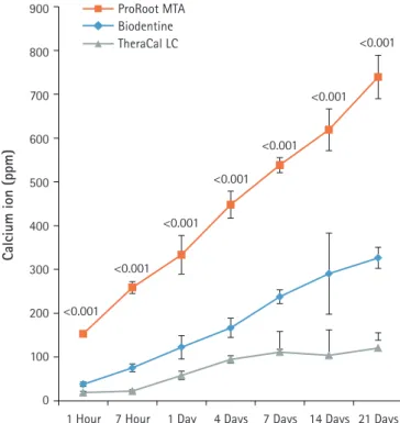
BD showed significantly higher calcium release compared to PR and TC in all test periods (p<0.05) (Fig. 3). Calcium release from PR was significantly higher than TC (p <0.05). The amount of calcium released from both BD and PR constantly increased with increased immersing time. However, the calcium release from TC slowly increased up to 4 days and thereafter al- most stopped.
2. Measurement of pH value
After 1-day immersion, PR and BD had similar pH values, but TC showed lower pH value compared with others (Fig. 4). In the ear- ly stage, PR showed the highest pH value compared with others.
All experimental groups showed high alkalinity near pH 11.0 after
1 day, except at 14 days. There was no significant difference in pH
values between PR, BD, and TC after 1 day, except at 14 days
(p>0.05).
3. Cell viability test
As shown in Fig. 5, BD showed the highest cell viability compared with PR and TC on both 1 day and 3 days. TC showed signifi- cantly lower cell viability than BD, but there was no significant difference between PR and TC (p<0.05).
4. Cell morphologic analysis
Cells were observed by confocal laser scanning microscopy to evaluate cell growth and morphology. As shown in Fig. 6, cells are well adhered in the shape of the fusiform and expressed actin fila- ments for all pulp capping materials.
5. Alizarin red S staining and alkaline phosphatase activity test
There was a high increase in mineralization in the PR and BD groups compared with the TC group based on alizarin red S stain- ing for calcium (Fig. 7). However, there was no difference in min- eralization between PR and BD. In terms of ALP activity, there was a significant difference between the three groups. BD showed Fig. 3. The amount of calcium released from ProRoot MTA,
Biodentine, and TheraCal LC in deionized water as a function of immersion time. The amount of calcium released from ProRoot MTA and Biodentine increased constantly with the immersion time, whereas the released calcium from TheraCal LC had a tendency to increase until day 4, and almost stopped increasing.
For each of the three types of samples, five samples were used at each time of the experiment. Error bars indicate standard errors of the means.
Fig. 5. Effects of ProRoot MTA, Biodentine, and TheraCal LC on cell viability measured by MTT assay. On both 1 and 3 days, the cell viability for Biodentine was highest. However, ProRoot MTA and TheraCal LC showed no significant difference (p<0.05). For each of the three types of samples, six samples were used at each time of the experiment. Error bars indicate standard errors of the means. MTT OD value: cell viability absorbance (490 nm).
Fig. 4. pH values of aqueous medium exposed to the extracts of the capping materials as a function of immersion time. pH values for ProRoot MTA, Biodentine, and TheraCal LC were stable in near 11.0 after 1 day, except at 14 days. Values not sharing a common letter (a, b) are significantly different (p<0.05). For each of the three types of samples, five samples were used at each time of the experiment. Error bars indicate standard errors of the means.
900
800
700
600
500
400
300
200
100
0 1 Hour
<0.001
<0.001
<0.001
<0.001
<0.001
<0.001
<0.001
4 Days
7 Hour 1 Day 7 Days 14 Days 21 Days
Calcium ion (ppm)
ProRoot MTA Biodentine TheraCal LC
■ ProRoot MTA ■ Biodentine ■ TheraCal LC
MTT OD value
Incubation time 0.25
0.20
0.15
0.10
0.05
0.00
1 Day
<0.001
0.005
<0.001
<0.001
3 Days
14
13
12
11
10
9
8
7
0
1 Hour 7 Hour 1 Day 4 Days 7 Days 14 Days 21 Days
pH value
0.01
0.005
0.002
a
a
a a
b b
b
b b
ProRoot MTA Biodentine TheraCal LC
a significantly higher value than PR and TC, while TC showed the lowest activity compared to the others (p<0.05) (Fig. 8).
Discussion
This study was designed to investigate the mineralization-induc- ing potentials of pulp capping materials on cultured HDPCs.
In regards to the calcium ion release, BD showed a markedly higher release of free calcium ions compared with PR and TC.
The increased calcium release from BD has been considered to be correlated with the presence of a calcium silicate component, calcium chloride, and low solubility (11.93%). This is likely linked to a superplasticizer that is commonly used to reduce the
Fig. 6. Confocal laser scanning microscopic images (actin [red], nucleus [blue]) of HDPCs cultured on (A) ProRoot MTA, (B) Biodentine, and (C) TheraCal LC which were incubated in the extract of the materials for 3 days. Cells were well adhered and expressed actin filaments for all pulp capping materials (×200).
Fig. 7. Alizarin red S staining images of odontoblasts cultured on (A) ProRoot MTA, (B) Biodentine, and (C) TheraCal LC for 14 days. There was a high increase in mineralization in the ProRoot MTA and Biodentine groups compared with the TheraCal LC group based on alizarin red S staining for calcium.
20 μg 20 μg 20 μg
A
A
B
B
C
C
amount of water required (L/P 0.257) to disperse the particles and to enhance the fluidity, making the cement self-consolidat- ing. In addition, the fast hydration reaction of tricalcium silicate can be correlated with the low solubility and high calcium re- lease at early endpoints [8]. In another previous study, BD ex- hibited advanced hydration both in vitro and after pulp capping.
This was evident from the densely hydrated product in the ce- ment matrix and the absence of unhydrated cement particles af- ter 14 days of hydration [10].
In contrast, TC exhibited the lowest calcium ion leaching. This
result can be attributed to the fact that the release of calcium ions
is limited due to the presence of a resin-modified matrix in the
structure of TC after setting. The restriction of fluid absorption
pate in the mineralization process [16,17].
The release of calcium hydroxide is mainly the result of the stimulation of odontoblast activity and subsequent mineraliza- tion. The pulp capping materials that are based on tricalcium sili- cate all allegedly release calcium hydroxide as a by-product of hy- dration. This has been demonstrated for MTA [18] and BD [19].
Calcium, as well as hydroxyl ions released from the capping mate- rials, regulate the event leading to tertiary dentinogenesis. For the biological effects of calcium hydroxide, the release of bioactive molecules, either through direct stimulation of cells or by solubili- zation of dentin extracellular matrix, is vital. Calcium silicate ce- ment, together with microcrystals deposited on its surface, pro- vides a biologically active substrate for the adsorption of biomole- cules and adhesion of odontoblasts [2].
In this study, the number of calcium ions released from the cap- ping materials and pH value of the medium were measured in de- ionized water rather than simulated body fluid in order to stan- dardize the test conditions and hence allow a comparison of the data with other future studies.
Prior to investigating the mineralization-inducing potentials of the materials, the biocompatibility was compared by evaluating the effects of the capping materials on cell morphology and cell viability in this study. BD showed better cell viability than PR and TC on the MTT assay. The result of the current study corre- sponded with an investigation by Poggio et al. [20], which showed BD induced a more favorable cell response due to high mitochondrial activity compared with MTA and TC during the 72 hours of incubation. Meanwhile, other researchers reported that BD showed similar cell viability to MTA [5,21].
Confocal laser scanning microscopic images were used to compare cell morphology and cytoskeletal organization. When HDPCs were cultured on the capping materials for 1 day, the cultured cells appeared flat and showed well-defined cytoplas- mic extensions, indicating all materials allowed cell attachment and growth. The actin microfilament cytoskeleton is involved in cell processes, cell shape, and cell attachment. As the cell ad- heres to a substrate material, filopodia are formed. They are moved into place by actin acting upon the plasma membrane.
The actin is observed in the filopodia as directed tight parallel bundles. Contractile stress fibers are seen once the filopodia are attached [22]. Our results showed that the cytoskeletal organi- zation of cells was observed, as shown in Fig. 6. The phenotype of differentiation-induced HDPCs is known to resemble several crucial characteristics of odontoblasts, such as increased ALP activity, mRNA expression of differentiation markers genes, and the formation of mineralized matrix in vitro [23].
ALP is the most frequently used marker of odontoblastic cell Fig. 8. Alkaline phosphatase (ALP) activity of odontoblasts
cultured on pulp capping materials for 14 and 21 days. The ALP activity of odontoblasts in Biodentine group was highest on both observation days. However, the odontblasts in TheraCal LC group showed the lowest ALP activity (p<0.05). For each of the three types of samples, five samples were used at each time of the experiment. Values not sharing a common letter (a, b, c) are significantly different (p<0.05). Error bars indicate standard errors of the means.
■ ProRoot MTA ■ Biodentine ■ TheraCal LC
ALP (nmol/30 min/μg)
Incubation time 0.50
0.40
0.30
0.20
0.10
0
15 Days b
a b
a c
c
21 Days