Evaluation of the effect of two different
occlusal splints on maximum occlusal force in patients with sleep bruxism: a pilot study
Duygu Karakis*, Arife Dogan, Bulent Bek
Department of Prosthodontics, Faculty of Dentistry, Gazi University, Ankara, Turkey
PURPOSE. The occlusal splint has been used for many years as an effective treatment of sleep bruxism. Several methods have been used to evaluate efficiency of the occlusal splints. However, the effect of the occlusal splints on occlusal force has not been clarified sufficiently. The purpose of this study was to evaluate the effect of occlusal splints on maximum occlusal force in patients with sleep bruxism and compare two type of splints that are Bruxogard-soft splint and canine protected hard stabilization splint. MATERIALS AND METHODS. Twelve students with sleep bruxism were participated in the present study. All participants used two different occlusal splints during sleep for 6 weeks. Maximum occlusal force was measured with two miniature strain-gage transducers before, 3 and 6 weeks after insertion of occlusal splints. Clinical examination of temporomandibular disorders was performed for all individuals according to the Craniomandibular Index (CMI) before and 6 weeks after the insertion of splints. The changes in mean occlusal force before, 3 and 6 weeks after insertion of both splints were analysed with paired sample t-test. The Wilcoxon test was used for the comparison of the CMI values before and 6 weeks after the insertion of splints. RESULTS. Participants using stabilization splints showed no statistically significant changes in occlusal force before, 3, and 6 weeks after insertion of splint (P>.05) and participants using Bruxogard-soft splint had statistically significant decreased occlusal force 6 weeks after insertion of splint (P<.05). There was statistically significant improvement in the CMI value of the participants in both of the splint groups (P<.05). CONCLUSION. Participants who used Bruxogard-soft splint showed decreases in occlusal force 6 weeks after insertion of splint. The use of both splints led to a significant reduction in the clinical symptoms. [J Adv Prosthodont 2014;6:103-8]
KEY WORDS: Occlusal splint; Bruxogard-Soft splint; Occlusal force; Sleep bruxism; Strain-gage
INTRODUCTION
Sleep bruxism is an involuntary activity of the masticatory muscles that is characterized by clenching and/or grinding
of the teeth during sleep.1 The occlusal splint has been fre- quently used as an effective treatment of sleep bruxism to protect teeth from damage caused by forceful jaw muscle contractions or to reduce orafacial pain, if present.2 However, little is known regarding the mechanism of the action of occlusal splints.3-5 Most of the studies have exam- ined changes in the masticatory muscles activity before and after application of an occlusal splint4,6-11 and they showed that occlusal splint treatment resulted in a decrease in noc- turnal masticatory muscle activities in patients with brux- ism.2,6,12,13 Furthermore, Kurita et al.3 suggested that the use of the stabilization splint has an effect of reducing hyper- activity and asymmetry in the activity of jaw elevator mus- cles and consequently brings a stable and physiologically optimal occlusal force from the muscles. In addition, Holmgren et al.14 showed that splints redistribute the load borne by the teeth and masticatory system. It has been sug-
Corresponding author:
Duygu Karakis
Department of Prosthodontics, Faculty of Dentistry, University of Gazi, Ankara, 06510, Turkey
Tel. 9 0312 203 41 96: e-mail, [email protected]
Received May 22, 2013 / Last Revision February 20, 2014 / Accepted March 4, 2014
© 2014 The Korean Academy of Prosthodontics
This is an Open Access article distributed under the terms of the Creative Commons Attribution Non-Commercial License (http://creativecommons.
org/licenses/by-nc/3.0) which permits unrestricted non-commercial use, distribution, and reproduction in any medium, provided the original work is properly cited.
Present study was supported by Cumhuriyet University, Foundation of Scientific Research Projection.
gested that the relief of bruxism symptoms with splint treatment may be a result of redistribution of overload- ing.15,16 According to these suggestions, redistribution of forces and reduction of bruxism symptoms with use of occlusal splints may change maximum occlusal force that has often been studied as an indicator of functional state of the masticatory system.17,18 Nevertheless, the effect of use of the occlusal splints on occlusal force has not been clarified sufficiently. There is only one study available that evaluated the effect of occlusal splints on occlusal force in patients with masticatory muscles disorders.3
Various splints have been used for treatment of brux- ism that are made up of basically two different materials.
Although, these occlusal splints have slightly different appearances and properties, in fact, scientific evidence sup- ports the use of hard acrylic resin occlusal splints12,19-21 that are preferentially of full coverage, located in the maxillary arch, with simultaneous, even, and bilateral contacts, and have anterior guidance and canine protected articulation.22,23 On the other hand, soft appliances have been less docu- mented in the scientific literature, however some research- ers suggest the use of soft-resin appliances24-27 that are easi- ly fabricated, and may be inserted at an initial appointment, so practitioners may desire the use of soft appliances. One of these soft splints is easily tolerable prefabricated Bruxogard Soft splint that fit tightly to upper arch and pro- vides flat special plane to protect dentition of sleep brux- ism patients.
The present study designed to evaluate the effect of the use of occlusal splints on maximum occlusal force of patients with sleep bruxism. For this purpose, the effect of two splints basically fabricated from two different materials that are Bruxogard-soft splint and canine protected hard stabilization splints were evaluated and compared. The null hypothesis was that the use of both occlusal splints would reorganize occlusal force in patients with bruxism in accor- dance with the improvements of symptoms; however these improvements would stand out more in hard occlusal splint group.
MATERIALS AND METHODS
Twelve students of Dental School of the University of Gazi (ranging age from 18 to 27) with sleep bruxism partic- ipated in the present study. This research was reviewed and approved by the Ethics Committee of University of Gazi (Process, 6/7/2007-3). Informed consent was obtained from each participant according to the ethical guidelines recommended by The Helsinki Declaration. The initial inclusion criteria were: Patients who had no reported sys- temic disease or apparent facial asymmetry, no craniofacial trauma or surgery, no use of any medication, and had full permanent dentition (not including third molars), vital first molars without mesio-occluso-distal restorations. The selected sleep bruxism patients had the following criteria suggested by Rompre et al.28: (a) a history of frequent tooth grinding occurring at least 3 nights per week for the preced-
ing 6 months, as confirmed by a sleep partner; (b) clinical presence of tooth wear; (c) masseter muscle hypertrophy;
and (d) report of jaw muscle fatigue or tenderness in the morning.
After these initial inclusion criteria, clinical examination of temporomandibular joint (TMJ) was performed for all the individuals according to the Craniomandibular Index (CMI) of Fricton and Schiffman29 before and 6 weeks after the insertion of splints. The CMI measures tenderness and dysfunction in the stomatognathic system and divided to the subscales: Dysfunction Index (DI) and the Palpation Index (PI). Dysfunction Index (DI) includes items related to limits in range of motion, deviation and/or pain in movement, TMJ noise during movement, TMJ tenderness, and Palpation Index (PI) reflects the muscle tenderness at intraoral jaw muscles and extraoral jaw and neck muscles.
In this way, these indexes separate joint problems from muscle problems. CMI has a 0-1 scale and is calculated according to the formula (DI + PI) / 2.
Two types of maxillary occlusal splints were constructed:
canine protected hard stabilization splint and Bruxogard-soft splint (Myofunctional Research Co, Waalwijk, Netherlands).
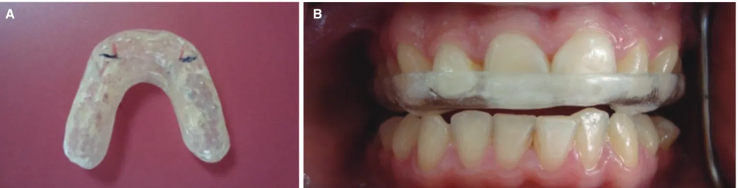
Participants were randomly assigned to either of two splint groups. Six of bruxism patients received hard stabilization splints fabricated from autopolymerizing acrylic resin (Akribel, Belmar, Izmir, Turkey). The splints were fabricat- ed covering the maxillary teeth completely, with 2 mm thickness of acrylic resin between maxillary and mandibular first molar teeth. The hard stabilization splint had even, simultaneous occlusal contacts with mandibular buccal cusp tips and incisal edges in the centric relation and had canine protection to separate the opposing posterior teeth during eccentric movement (Fig. 1).
Six bruxism patients were used Bruxogard-soft splints that were prepared easily without impression. It was placed into the boiled water for 30 seconds and then soft inner surface of splint inserted to mouth that V-cut of splint was in the centre line of anterior teeth. Then, it was pushed with fingers to seat. Patients were asked close upper and lower teeth slightly just to hold splint in position. The hard- er occluded surface of splint compared to the inner surface was flat without indents of the lower teeth. Then, Bruxogard soft splint was removed from the mouth and placed in cold water for 20 seconds (Fig. 2).
All splints were made and adjusted by the same clini- cians. All participants used occlusal splints for 6 weeks, dur- ing sleep, at least 8 hours. Participants were instructed not to take any medications, such as muscle relaxants, sleeping pills, transquilizers and antidepressants during the treatment.
Maximum occlusal force was measured from each side of the dental arch using two miniature strain-gage transduc- ers (Model VLPB, Load Cell Central, Monroeton, PA, USA) with stainless steel cases as described previously (Fig. 3).30 Two transducers were placed bilaterally on a flat metal arch.
The metal surfaces of the arch were covered with plaster (Betasan, Kocaeli, Turkey). The metal arch and transducers were further covered by a disposable latex finger cot to
avoid contamination during measurements. Each transducer has a height of 4 mm and a diameter of 12 mm; with these applications transducers reached a height of 6 mm.
Occlusal force was detected as a two-channel signal from each side with a bio-signal acquisition device designed by Kardiosis (Tepa Inc., Kardiosis Ltd., Ankara, Turkey). The force signals were monitored online and then measured on a computer screen as kg (kilogramme), using a specific soft- ware program developed by the same company. The cali- bration of the transducers was performed by loading the transducer with known force values, the deviation from lin- earity with a load of 5 kg was + 2.66% in right transducer and + 4% in left transducer, 15 kg was + 5.2% in right transducer and + 2.6% in left transducer.
Occlusal force of all participants were performed using 2 mm hard occlusal splints fabricated from hard acrylic res- in to avoid damage of teeth, to prevent metal contact of teeth that suppressing maximum occlusal force and to stan- dardize transducers on during measurement of maximum occlusal force.
During the test, participants were seated in an upright position with the head in a natural posture, keeping the Frankfort horizontal plane approximately parallel to the floor. Initially, bilateral transducers that positioned on the
metal plate were placed between the first molar teeth on both sides. The transducers were also maintained parallel to the Frankfort horizontal plane during recordings.
Participants were instructed to bite as forcefully as possible three times. Before the recordings, the participants were trained to perform their highest possible occlusal force.
The highest value of each clenching was recorded and the mean value of the three highest clenching measurements was considered to be the maximum occlusal force. The sum of the right and left occlusal force values was considered to be the maximum occlusal force.
The experimental protocol including clinical examina- tion and recordings of occlusal force was performed by the same investigator. Occlusal force values of participants were recorded before, 3 and 6 weeks after insertion of the splints.
The evaluation of the changes in mean occlusal force values before, 3 weeks and 6 weeks after insertion of both splints were analysed with paired sample t-test. The analysis of differences of occlusal force values between Stabilization and Bruxogard soft splint before treatment, after 3 weeks and 6 weeks was performed with independent samples of t-test. The changes in the amount and percentage of occlu- sal force, before and 6 weeks after treatment, were analyzed Fig. 1. Canine protected hard stabilization splint (A) extraoral view and (B) inraoral view.
A B
Fig. 3. Strain gages.
Fig. 2. Broxogard Soft splint.
with paired sample t-test for each splint groups. The Wilcoxon test was used for the comparison of the Craniomandibular index values before and 6 weeks after the insertion of splints. Comparison CMI values of both splints before and after treatments were performed with Mann-Whithney U test. Differences at the 5% level of probability were considered statistically significant. All sta- tistical analyses were performed by the SPSS 11.5 (Statistical Package for Social Sciences Software Inc, Chicago, IL, USA).
RESULTS
The mean occlusal force of both splint groups before, 3 and 6 weeeks after insertion of splints are listed in Table 1.
There was no statistically significant differences in any intervals in canine protected hard stabilization splint group (Paired t-test; P>.05). There was only a significant differ- ence in occlusal force value between before and 6 weeks after insertion of Bruxogard-soft splints. (Paired t-test;
P<.05, P=.032). Independent samples t-test showed no dif- ference between both splint groups before treatment, 3 and 6 weeks after treatment (P>.05: P=.27, P=.73, P=.28, respectively).
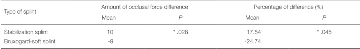
The amount and percentage of changes in mean occlu- sal force between before and 6 weeks after insertion of both splint groups, were presented in Table 2. The stabiliza- tion splint group showed 10 kg and 17% of mean occlusal
force increase and the participants used Bruxogard-soft splint group showed 9 kg and 24% of mean occlusal force decrease, and it was statistically significant (P<.05).
Participants included Bruxogard-soft splint and canine protected hard stabilization splint had statistically signifi- cant improvement in Cranimandibular index value after treatment (Table 3) (P<.05: P=.043, P=.046, respectively).
The comparison of CMI values in both splint groups before and 6 weeks after insertion of splints showed no difference (P>.05: P=.69, P=.81, respectively).
DISCUSSION
The null hypothesis was partially accepted that the use of both occlusal splint showed an improvement of the symp- toms, however, contrary to our hypothesis soft splints showed statistical significant alterations in occlusal force.
In the clinical researches, several methods have been used to evaluate efficiency of the occlusal splints. The most of these studies have evaluated the masticatory muscle activities.2,4-6,13 It has been stated that the use of occlusal splint reduces the activities of the masseter2 and anterior temporalis muscle;8-10,14 and improves the degree of asym- metry in the muscle activity;11 lessens the forces placed on the TMJs and other structures within the masticatory sys- tem and in this way lessens the symptoms of bruxism.2 Although it has not been used widely, another method that is measuring the maximum occlusal force may be used to
Table 3. Mean values of Craniomandibular Index (CMI) in participants and P values
Before treatment
After
treatment P
Stabilization splint 0.21 0.09 .046*
Bruxogard-soft splint 0.22 0.12 .043*
*: Significant at P<.05.
Table 1. Mean occlusal force (kg) and P value of participants before, 3 and 6 weeks after insertion of Bruxogard-soft splints and Stabilization splints
Groups
Bruxogard- soft splint
mean
Stabilization splint mean
P
Before treatment 35.66a 28 .27
3 weeks after 27.75 31.3 .73
6 weeks after 23.16a 32.66 .28
(n=6, paired t-test)
a: Significant at P<.05.
Table 2. Comparison of the changes in the amount and percentage of the mean occlusal force (kg) for both splint groups after the splint treatment
Type of splint Amount of occlusal force difference Percentage of difference (%)
Mean P Mean P
Stabilization splint 10 * .028 17.54 * .045
Bruxogard-soft splint -9 -24.74
(Mann Whitney U-Test)
*: Significant at P<.05.
evaluate efficiency of occlusal splints. On the other hand, Holmgren et al.14 stated that the therapeutic mechanism of the occlusal splint should be related to factors that modify and reduce parafunctional activity and/or redistribute its overloading in the masticatory system. In addition, Dylina16 stated that occlusal splints balanced the force distribution in the entire masticatory system. In fact, maximum occlusal force has an influence on muscle efficiency and functional state of masticatory system.17,18 However, there is only one study performed by Kurita et al.3 They have recorded the occlusal force of the patients in masticatory muscle disor- ders before, 2 and 4 weeks after the use of the stabilization splints. As different from the present study, Kurita et al.3 used Dental Prescale system for measurement of occlusal force. They stated that the occlusal loads in the higher level decreased and, in contrast, those in the lower level increased with the use of the stabilization splint and con- cluded that the use of splints has the effect of normalizing the occlusal force in patients with masticatory muscle disor- ders. In the present study, occlusal force of participants who used stabilization splint showed no statistically signifi- cant differences after the splint treatment. According to the results of our previous study, the mean maximum occlusal force of healthy participants was 30.16 kg30 and in the pres- ent study, the occlusal force value of participants recorded before and after the use of stabilization splint was close to the occlusal force of the healthy participants. It was consid- ered that the lack of difference might be related to the severity of the bruxism and small number of the participat- ed subjects.
On the other hand, CMI values in both splint groups were similar before insertion of splints, and after 6 weeks, participants who used Bruxogard-soft splint and hard stabi- lization splint showed statistically significant improvements in clinical signs and symptoms. Bruxogard-soft splint has not been studied in the literature. Therefore, the studies related to the soft splints were included to this study to give an idea. In accordance with the result of current study, Wright et al.24 stated that the soft splint was an effective short-term treatment for reducing the signs and symptoms of masticatory muscle pain in the patients. Accordingly, Pettengill et al.27 suggested that soft and hard appliances may be equally useful in reducing the temporomandibular symptoms in short-term appliance therapy.
It has been stated that splints redistributed the load induced by the teeth and masticatory system.14,15 In the cur- rent study, while the participants used stabilization splints exerted no significant changes in occlusal force, the use of Bruxogard soft splints led to decreasing of occlusal force.
Narita et al.31 stated that jaw clenching with a soft occlusal splint caused a significant decreases in occlusal force as well as significant increase in awareness of tiredness of the mus- cles and in contrast, the usage of the hard occlusal splint did not cause significant change in the occlusal force and an awareness of tiredness. The occlusal force results of that study is in agreement with the findings of the present study and the current study indicates that both of the splints
might have different mechanism on occlusal force distribu- tion for improvement of the clinical symptoms. These splints might redistribute occlusal force in a different way to the stomatognatic system because of their different mechanism that could depend on the splint material or occlusion of splints. However, considering the main limita- tion of present study which was the small number of sub- jects, further studies with longer evaluation period should be performed in a larger patient population in order to yield more consistent results regarding the effect of occlu- sal splints on occlusal force.
CONCLUSION
The use of canine protected hard stabilization splint has not showed significant differences in maximum occlusal force and use of a Bruxogard-soft splint was accompanied by decrease in occlusal force in patients with sleep bruxism.
Treatments with both of splints were acceptably successful and led to a significant reduction in the clinical symptoms.
REFERENCES
1. Cosme DC, Baldisserotto SM, Canabarro Sde A, Shinkai RS.
Bruxism and voluntary maximal bite force in young dentate adults. Int J Prosthodont 2005;18:328-32.
2. Harada T, Ichiki R, Tsukiyama Y, Koyano K. The effect of oral splint devices on sleep bruxism: a 6-week observation with an ambulatory electromyographic recording device. J Oral Rehabil 2006;33:482-8.
3. Kurita H, Ikeda K, Kurashina K. Evaluation of the effect of a stabilization splint on occlusal force in patients with masti- catory muscle disorders. J Oral Rehabil 2000;27:79-82.
4. Roark AL, Glaros AG, O’Mahony AM. Effects of interocclu- sal appliances on EMG activity during parafunctional tooth contact. J Oral Rehabil 2003;30:573-7.
5. Al-Saad M, Akeel RF. EMG and pain severity evaluation in patients with TMD using two different occlusal devices. Int J Prosthodont 2001;14:15-21.
6. Hiyama S, Ono T, Ishiwata Y, Kato Y, Kuroda T. First night effect of an interocclusal appliance on nocturnal masticatory muscle activity. J Oral Rehabil 2003;30:139-45.
7. Yap AU. Effects of stabilization appliances on nocturnal parafunctional activities in patients with and without signs of temporomandibular disorders. J Oral Rehabil 1998;25:64-8.
8. Williamson EH, Lundquist DO. Anterior guidance: its effect on electromyographic activity of the temporal and masseter muscles. J Prosthet Dent 1983;49:816-23.
9. Fitins D, Sheikholeslam A. Effect of canine guidance of maxillary occlusal splint on level of activation of masticatory muscles. Swed Dent J 1993;17:235-41.
10. Sheikholeslam A, Holmgren K, Riise C. Therapeutic effects of the plane occlusal splint on signs and symptoms of crani- omandibular disorders in patients with nocturnal bruxism. J Oral Rehabil 1993;20:473-82.
11. McCarroll RS, Naeije M, Kim YK, Hansson TL. Short-term effect of a stabilization splint on the asymmetry of submaxi-
mal masticatory muscle activity. J Oral Rehabil 1989;16:171-6.
12. Okeson JP. The effects of hard and soft occlusal splints on nocturnal bruxism. J Am Dent Assoc 1987;114:788-91.
13. Roark AL, Glaros AG, O’Mahony AM. Effects of interocclu- sal appliances on EMG activity during parafunctional tooth contact. J Oral Rehabil 2003;30:573-7.
14. Holmgren K, Sheikholeslam A, Riise C. Effect of a full-arch maxillary occlusal splint on parafunctional activity during sleep in patients with nocturnal bruxism and signs and symp- toms of craniomandibular disorders. J Prosthet Dent 1993;
69:293-7.
15. Zarb GA, Speck JE. The treatment of temporomandibular joint dysfunction: a retrospective study. J Prosthet Dent 1977;38:420-32.
16. Dylina TJ. A common-sense approach to splint therapy. J Prosthet Dent 2001;86:539-45.
17. Braun S, Bantleon HP, Hnat WP, Freudenthaler JW, Marcotte MR, Johnson BE. A study of bite force, part 1: Relationship to various physical characteristics. Angle Orthod 1995;65:
367-72.
18. Kogawa EM, Calderon PS, Lauris JR, Araujo CR, Conti PC.
Evaluation of maximal bite force in temporomandibular dis- orders patients. J Oral Rehabil 2006;33:559-65.
19. Lobbezoo F, van der Zaag J, van Selms MK, Hamburger HL, Naeije M. Principles for the management of bruxism. J Oral Rehabil 2008;35:509-23.
20. Sheikholeslam A, Holmgren K, Riise C. A clinical and elec- tromyographic study of the long-term effects of an occlusal splint on the temporal and masseter muscles in patients with functional disorders and nocturnal bruxism. J Oral Rehabil 1986;13:137-45.
21. al-Quran FA, Lyons MF. The immediate effect of hard and soft splints on the EMG activity of the masseter and tempo- ralis muscles. J Oral Rehabil 1999;26:559-63.
22. Bonfante G, Ramos Júnior L, Bonfante EA. Restoration of canine guidance on an occlusal splint using amalgam: a clini- cal report. J Prosthet Dent 2003;90:420-3.
23. Klasser GD, Greene CS. Oral appliances in the management of temporomandibular disorders. Oral Surg Oral Med Oral Pathol Oral Radiol Endod 2009;107:212-23.
24. Wright E, Anderson G, Schulte J. A randomized clinical trial of intraoral soft splints and palliative treatment for mastica- tory muscle pain. J Orofac Pain 1995;9:192-9.
25. Wright EF. Using soft splints in your dental practice. Gen Dent 1999;47:506-10.
26. Giedrys-Leeper E. Night guards and occlusal splints. Dent Update 1990;17:325-9.
27. Pettengill CA, Growney MR Jr, Schoff R, Kenworthy CR. A pilot study comparing the efficacy of hard and soft stabiliz- ing appliances in treating patients with temporomandibular disorders. J Prosthet Dent 1998;79:165-8.
28. Rompré PH, Daigle-Landry D, Guitard F, Montplaisir JY, Lavigne GJ. Identification of a sleep bruxism subgroup with a higher risk of pain. J Dent Res 2007;86:837-42.
29. Fricton JR, Schiffman EL. Reliability of a craniomandibular index. J Dent Res 1986;65:1359-64.
30. Koc D, Dogan A, Bek B, Yucel M. Effects of increasing the
jaw opening on the maximum bite force and electromyo- graphic activities of jaw muscles. J Dent Sci 2012;7:14-9.
31. Narita N, Funato M, Ishii T, Kamiya K, Matsumoto T.
Effects of jaw clenching while wearing an occlusal splint on awareness of tiredness, bite force, and EEG power spec- trum. J Prosthodont Res 2009;53:120-5.