http://dx.doi.org/10.3988/jcn.2011.7.4.203 J Clin Neurol 2011;7:203-209
Introduction
Dyslipidemia contributes to the development of atherosclerosis,1-3 and there is accumulating evidence that atherosclerosis is promot-
ed by remnant lipoproteins (RLPs).4-13 These are products of par- tially catabolized chylomicrons and very-low-density lipopro- teins, from which some triglycerides (TGs) have been removed by the action of lipoprotein lipase and, to a lesser extent, by he- patic lipase. These particles have reduced TGs, but are enriched with cholesterol and apolipoprotein (apo)E; they are smaller and denser than the parent particles, and are believed to be more strong- ly atherogenic than larger TG-rich lipoproteins.4-9
Elevated RLP cholesterol (RLP-C) levels were reported to be
High Levels of Remnant Lipoprotein Cholesterol Is a Risk Factor for Large Artery Atherosclerotic Stroke
Jeong-Yeon Kim,a* Jong-Ho Park,b* Sang-Wuk Jeong,a Dawid Schellingerhout,c Jin-Eok Park,a Dong Kun Lee,a Won Jun Choi,a Seok-Lae Chae,d Dong-Eog Kima
aDepartment of Neurology and MINER (Molecular Imaging & Neurovascular Research) Laboratory, Dongguk University Ilsan Hospital, Goyang, Korea
bStroke Center, Kwandong University Myongji Hospital, Goyang, Korea
cDepartment of Radiology, University of Texas M.D. Anderson Cancer Center, Houston, Texas, USA
dDepartment of Laboratory Medicine, Dongguk University Ilsan Hospital, Goyang, Korea
Received December 31, 2010 Revised March 16, 2011 Accepted March 16, 2011 Correspondence Dong-Eog Kim, MD, PhD Department of Neurology, Dongguk University College of Medicine,
Dongguk University Ilsan Hospital, 814 Siksa-dong, Ilsandong-gu, Goyang 410-773, Korea Tel +82-31-961-7211 Fax +82-31-961-7212 E-mail [email protected]
*J.-Y.K. and J.-H.P. contributed equally to this study.
Background and PurposezzRemnant lipoproteins (RLPs) are products of partially catabolized chylomicrons and very-low-density lipoprotein, from which some triglycerides have been removed.
These particles are smaller and denser than the parent particles and are believed to be strongly ath- erogenic. We explored the association between RLP cholesterol (RLP-C) and ischemic stroke, in- cluding stroke subtypes.
MethodszzA cohort of 142 ischemic stroke patients (90 men and 52 women; age, 65.2±12.8 years, mean±SD) was enrolled; all had acute infarcts confirmed by diffusion-weighted MRI, and had fast- ing lipograms. A full stroke-related evaluation was conducted on each patient. An outpatient popu- lation of 88 subjects without a history of cerebrovascular or cardiovascular disease served as a control group. Serum RLP fractions were isolated using an immunoaffinity gel containing specific antiapoli- poprotein (anti-apo)B-100 and anti-apoA-I antibodies. RLP-C values were considered to be high when they were in the highest quartile of all values in the study.
ResultszzHigh RLP-C values were more common in stroke patients than in control patients (31.0%
vs. 14.8%, p=0.01), when 5.6 mg/dL (>75th percentile) was used as the cutoff value. Multivariable analyses indicated that RLP-C was a risk factor for stroke, with an odds ratio of 2.54 (p=0.045). The RLP-C level was higher in the large artery atherosclerosis subgroup (5.7±3.9 mg/dL) than in any other stroke subgroup (small vessel occlusion, 4.9±5.9 mg/dL; cardioembolism, 1.8±2.3 mg/dL;
stroke of undetermined etiology, 3.1±2.9 mg/dL).
ConclusionszzWe have found an association between high RLP-C levels and ischemic stroke, and in particular large artery atherosclerotic stroke.
J Clin Neurol 2011;7:203-209 Key Wordszz remnant lipoprotein cholesterol, ischemic stroke, large artery atherosclerosis.
Open Access
cc This is an Open Access article distributed under the terms of the Cre- ative Commons Attribution Non-Commercial License (http://creative- commons.org/licenses/by-nc/3.0) which permits unrestricted non-com- mercial use, distribution, and reproduction in any medium, provided the ori- ginal work is properly cited.
associated with endothelial dysfunction, an early marker for ath- erosclerotic disease.10 The Framingham Heart Study found that an increase in RLP-C levels was a significant risk factor for cor- onary artery disease in women.11 In another study with a 3-year follow-up period, the incidence of cardiovascular events in the high RLP-C group (RLP-C >5.1 mg/dL) was higher than that in the low RLP-C group (RLP-C ≤3.3 mg/dL).12 As a result, in 2000 the United States Food and Drug Administration recognized that an increase of RLP-C was a risk factor for coronary heart disease.13
Nakamura et al.14 recently reported that high levels of RLPs predict ischemic stroke in patients with metabolic syndrome and mild carotid atherosclerosis. However, the patient population in that study comprised a selected subgroup of patients who were admitted for cardiac catheterization for chest pain or electrocardio- graphic ischemic changes. In the present study of consecutive pa- tients with ischemic stroke, we aimed to add new information re- garding the association between RLP-C and ischemic stroke and, given that stroke is a heterogeneous disease with more than 150 known causes,15 to identify the correlations of RLP-C levels with various stroke subtypes.
Methods
Study patients
This study was approved by the institutional review board of the Dongguk University Ilsan Hospital (DUIH), Korea. From Febru- ary to September 2007, 198 consecutive patients who were ad- mitted to the DUIH with acute ischemic stroke (≤1 week) were prospectively screened for enrollment. Acute ischemic lesions were assessed by diffusion-weighted imaging with the apparent diffusion coefficient. We excluded three patients with incom- plete evaluations due to severely impaired consciousness or ear- ly discharge, four patients with transient ischemic attack with- out visible lesions on diffusion-weighted imaging, and six pa- tients with cerebral infarction due to venous thrombosis, arterial dissection, or moyamoya disease. Sixteen patients were exclud- ed because their lipid profiles included only total cholesterol.
We excluded a further 27 patients who did not fast for at least 9-12 hours before the measurement of lipid or RLP-C levels. Thus, a total of 142 patients (90 men and 52 women; age, 65.2±12.8 years, mean±SD) were entered into the study. All patients un- derwent systemic investigations including assessment of medi- cation history, magnetic resonance imaging (MRI) with MR an- giography, carotid duplex ultrasonography, transthoracic echocardiography, 24-h Holter monitoring, and other routine ad- mission laboratory tests. We prospectively collected demographic data, prior medication history, and the presence of vascular risk factors including hypertension, diabetes mellitus, heart disease, previous stroke, and smoking.
Assessment of stroke risk factors
Hypertension was considered to be present if a subject had one of the following conditions: repeated blood pressure readings above 140/90 mm Hg at 1-2 weeks after stroke onset, a history of hypertension, or use of antihypertensives. Diabetes mellitus was defined as a glycated hemoglobin (HbA1C) level of ≥6.5%, a history of diabetes mellitus, or use of diabetic medication. Smok- ers included current smokers, who smoked daily or only occasion- ally. Heart disease as a potential source of cerebral embolism16 was considered to be present if a subject had a left atrial or ventricular thrombus, atrial fibrillation or sick sinus syndrome, recent myo- cardial infarction within 1 month, rheumatoid mitral or aortic valve disease, bioprosthetic or mechanical heart valves, chronic myo- cardial infarction with a low ejection fraction of <28%, symptom- atic congestive heart failure with an ejection fraction of <30%, dilated cardiomyopathy, aneurysms or akinetic segments of the left ventricular wall, or nonbacterial thrombotic endocarditis.
Electrocardiography, transthoracic echocardiography, and 24-h Holter monitoring were performed to identify these cardioem- bolic sources. Transesophageal echocardiography or a transcra- nial Doppler bubble study was also performed in selected pa- tients.
Blood samples were taken in the morning after an overnight fast of 9-12 h (12.0±3.2 h in the stroke group and 12.0±0.1 h in the control group). In the stroke subjects, the median sampling time from stroke onset was 39.8 h. Total cholesterol, TG, high- density lipoprotein cholesterol, and low-density lipoprotein cho- lesterol levels were assayed by enzymatic techniques. RLP-C was measured in triplicate after isolation, as reported previous- ly,17 by using an immunoaffinity gel containing monoclonal anti- bodies to human apoB-100 and apoA-I (Jimro-II RLP-C kit, Otsuka, Japan). Mean values were used for the analysis. Serum RLP-C levels were considered to be high when they were within the highest quartile (>75th percentile)18 of the values of all of the study participants.
Stroke subtype analysis
Subtypes of index stroke were determined by the consensus of three neurologists (M.G.J., S.W.J., and D.E.K.) as follows: large ar- tery atherosclerosis (LAA), small vessel occlusion, cardioembolism (CE), and ischemic stroke of undetermined etiology, as describ- ed in the Trial of Org 10172 in Acute Stroke Treatment study.16 Control subjects
As a control group, we chose 88 consecutive subjects (34 men and 54 women; age, 54.4±9.3 years) 1) who visited the DUIH Healthcare Center for health screening during the same study pe- riod, 2) who did not have a history of stroke, transient ischemic attack, myocardial infarction, or angina, and 3) whose lipid pro- files and RLP-C were measured after 9-12 h of fasting.
Statistical analyses
Data are presented as mean±SD values. Comparisons of con- tinuous variables between groups were performed using Stu- dent’s t-test, one-way analysis of variance (ANOVA) with Dun- nett’s post hoc test, or Kruskal-Wallis ANOVA with post hoc Mann-Whitney test. The chi-square test or Fisher’s exact test was used to compare proportions between groups. Bivariate correlations were calculated to quantify the relationships between pairs of variables. Multivariable regression analyses were con- ducted to determine if high RLP-C level was an independent risk factor for ischemic stroke or an independent predictor for LAA stroke after adjustment for known vascular risk factors. All statistical analyses were conducted using the SPSS software package (SPSS 18.0, Chicago, IL, USA). A value of p<0.05 was considered statistically significant.
Results
Univariable analysis showed that RLP-C is a risk factor for ischemic stroke
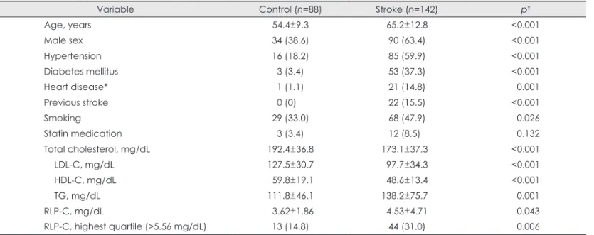
Table 1 summarizes the demographic and clinical features of the study subjects. Compared with the control group, the stroke group had higher mean values or prevalences of the vascular risk fac- tors including age, male sex, hypertension, diabetes, heart dis- ease, previous stroke, smoking, and TG level (all p<0.05). How- ever, the serum levels of total cholesterol, low-density lipoprotein cholesterol, and high-density lipoprotein cholesterol were high- er in the control group than in the stroke group (all p<0.05). In line with our hypothesis, the univariable analysis showed that RLP-C levels were higher in the stroke group than in the con-
trol group (p=0.043). Similarly, when RLP-C levels were dichot- omized at 5.56 mg/dL (which was the 75th percentile boundary), a significantly higher proportion of the stroke group had high RLP-C levels (31.0% vs. 14.8%, p=0.006).
Multivariable analyses showed that a high RLP-C may be an independent risk factor for ischemic stroke
The association between RLP-C and stroke could be due to var- ious confounding factors; therefore, logistic regression analy- ses were conducted, where those variables with p<0.15 in the univariable analyses were included. The continuous variable
“RLP-C” did not satisfy the linearity assumption of logistic re- gression; therefore, the dichotomized variable “high RLP-C” was entered into the model. After adjusting for age, sex, hyperten- sion, diabetes mellitus, smoking, heart disease, previous stroke, total cholesterol, and statin medication, the odds ratio for the as- sociation of high RLP-C with stroke was 2.54 (p=0.045). When TG was also entered into the model, a high RLP-C was not sig- nificantly associated with stroke (p=0.24). TG was also not sig- nificantly associated with stroke (p=0.21). The scatterplot and Pearson correlation analysis (Fig. 1) revealed that there was a linear relationship between RLP-C and TG (p<0.01, r=0.67).
RLP-C is an independent predictor
for LAA stroke versus the other stroke subtypes As indicated in Table 3, which presents the risk factors in the stroke subgroups based on the Trial of Org 10172 in Acute Stroke Treat- ment classification, the RLP-C level was highest in the LAA sub- group (5.73±3.94 mg/dL) and lowest in the CE subgroup (1.76
Table 1. Univariable analyses: factors associated with stroke vs. control
Variable Control (n=88) Stroke (n=142) p†
Age, years 54.4±9.3 65.2±12.8 <0.001
Male sex 34 (38.6) 90 (63.4) <0.001
Hypertension 16 (18.2) 85 (59.9) <0.001
Diabetes mellitus 3 (3.4) 53 (37.3) <0.001
Heart disease* 1 (1.1) 21 (14.8) 00.001
Previous stroke 0 (0)0. 22 (15.5) <0.001
Smoking 29 (33.0) 68 (47.9) 00.026
Statin medication 3 (3.4) 12 (8.5)0 00.132
Total cholesterol, mg/dL 192.4±36.8 173.1±37.30 <0.001
LDL-C, mg/dL 127.5±30.7 97.7±34.3 <0.001
HDL-C, mg/dL 059.8±19.1 48.6±13.4 <0.001
TG, mg/dL 111.8±46.1 138.2±75.70 00.001
RLP-C, mg/dL 03.62±1.86 4.53±4.71 00.043
RLP-C, highest quartile (>5.56 mg/dL) 13 (14.8) 44 (31.0) 00.006
Data are number (percentage) or mean±SD of patients.
*Atrial fibrillation (n=18) or left-ventricular akinesia (n=3), †p for Student’s t-test or chi-square test.
LDL-C: low-density lipoprotein cholesterol, TG: triglyceride, HDL-C: high-density lipoprotein cholesterol, RLP-C: remnant lipoprotein cholesterol.
±2.23 mg/dL). As illustrated in the box plots and demonstrated by the Kruskal-Wallis ANOVA with Mann-Whitney post hoc tests (Fig. 2), the RLP-C level was significantly higher in the LAA subgroup than in the subgroups (all p<0.05). Similarly, the RLP- C level was more frequently in the highest quartile in the LAA subgroup than in the other groups (43.8% vs. 10.5-31.5%). Other stroke risk factors and traditional lipid profiles, with the excep- tion of TG levels, did not differ significantly among the subtypes.
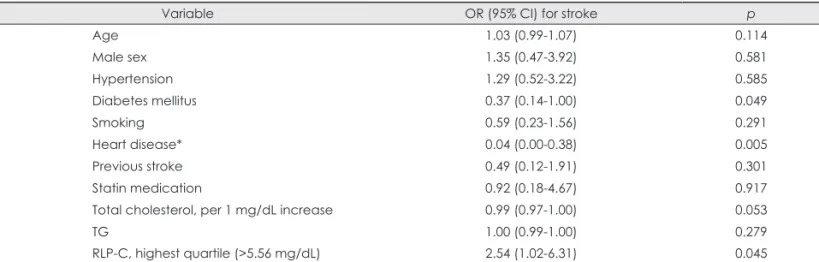
Unlike for the RLP-C levels, the serum TG levels of the LAA sub- group were not higher than those of the other subgroups except the CE subgroup. Table 4 presents the independent factors associated with LAA vs. other stroke subtypes. After adjusting for TG as well as age, sex, hypertension, diabetes mellitus, smoking, heart dis- ease, previous stroke, total cholesterol, and statin medication, a high RLP-C was the only significant predictor that was positively associated with the LAA stroke vs. the other stroke subtypes (p=
0.045). Heart disease and diabetes were negatively associated with LAA stroke (p=0.005 and 0.049, respectively).
Discussion
In this study we observed a significant association between high- er RLC-C levels and ischemic stroke in general, with a particu- lar association between RLP-C and LAA stroke. A high RLP- C was four times more prevalent in patients with LAA stroke (44%) than in the CE subgroup (11%). In multivariable analy- sis, a high RLP-C was linked with ischemic stroke after adjust- ing for prior statin medication and known vascular risk factors.
TG levels were correlated with RLP-C levels, and after adjust- ing for TG as well as other risk factors, a high RLP-C was the only significant predictor that was positively associated with LAA stroke. This suggests a separate significance for RLP-C levels independent of TG levels. Together these findings indi- cate the potential utility of examining RLP-C levels for stroke risk assessment.
RLP-C has not been widely studied due to a lack of routine laboratory assays; until the recent development of a simple and reliable technique for measurements based on an immunosep- aration method,17 it has been difficult to assay levels of RLP-C.
In the field of stroke, only one recent study14 has demonstrated that high RLP-C levels (≥5.8 mg/dL) were related to echolu- cent carotid plaques and shown to be an independent risk factor for future ischemic strokes in a subgroup of patients admitted for cardiac catheterization, who had metabolic syndrome and evidence of carotid plaques producing stenosis of less than 50%.
The present cross-sectional study found that an ischemic stroke was about 2.5 times more likely in subjects with RLP-C levels higher than 5.56 mg/dL than in the controls. This sup- ports the notion that RLP-C may be related to the genesis of ce- rebral vascular events,14 which could be mediated by the pro- inflammatory and proatherothrombogenic effects of RLP-C:18-21 inhibition of nitric-oxide-mediated arterial dilation, and up-reg- Table 2. Multivariable analysis: factors associated with stroke vs. control
Variable OR (95% CI) for stroke p
Age 1.06 (1.03-1.10)0 0.001
Male sex 4.73 (1.70-13.13) 0.003
Hypertension 3.59 (1.52-8.49)0 0.004
Diabetes mellitus 5.57 (1.36-22.79) 0.017
Smoking 1.09 (0.40-2.93)0 0.868
Heart disease* 11.12 (1.04-119.35) 0.047
Previous stroke 0.00 (0.00-0.00)0 0.998
Statin medication 4.42 (0.77-25.22) 0.095
Total cholesterol, per 1 mg/dL increase 00.99 (0.977-0.997) 0.014
RLP-C, highest quartile (>5.56 mg/dL) 2.54 (1.02-6.31)0 0.045
Variables were selected for inclusion in the model based on the results of univariate analyses (p<0.15). The Hosmer-Lemeshow good- ness-of-fit test showed χ2=7.97 and p=0.44, demonstrating a good model fit.
*Atrial fibrillation (n=18) or left-ventricular akinesia (n=3).
OR: odds ratio, CI: confidence interval, RLP-C: remnant lipoprotein cholesterol.
Fig. 1. Correlation between remnant lipoprotein cholesterol (RLP- C) and triglyceride levels.
0 100 200 300 400 500 600
30
25 r=0.67
20 15 10
0 5
RLPC (mg/dL)
Triglyceride (mg/dL)
ulation of endothelial expression of intracellular adhesion mol- ecule-1 and vascular cell adhesion molecule-1.
The finding that high RLP-C levels were more frequently encountered in patients with LAA stroke supports a role for RLP-C in proatherosclerotic endothelial dysfunction,18-20 which would be expected to play a larger role in this group than in pa- tients with embolic vascular occlusions. It is possible that future studies will allow RLP-C levels to be used for determining the contribution of endothelial dysfunction to stroke, particularly when it is not clear if in situ atherosclerosis or embolism was re- sponsible for a lesion. This may have important clinical implica-
tions because the management and prognosis of stroke patients are influenced by the ischemic stroke subtype.
The mean RLP-C level of our control group was 3.62 mg/dL, which is similar to the values reported for Japanese control sub- jects (3.3 or 3.7 mg/dL).19-23 The mean RLP-C level of the stroke group was 4.5 mg/dL, while the LAA subgroup had the highest mean value, at 5.73 mg/dL. These findings are in line with previ- ous reports that patients with RLP-C levels higher than 4.7-5.1 mg/
dL are at increased risk of cardiovascular events.12 The stroke pa- tients in our CE subgroup had the lowest mean RLP-C level, 1.84 mg/dL, which was lower than the mean value for the control group.
The patients in the CE subgroup had atrial fibrillation or ventricu- lar akinesia as a cause of the index stroke; they did not have sig- nificant atherosclerotic narrowing of the large artery supplying the infarct territory. These data indicate that high RLP-C levels are associated with atherosclerotic stroke, not cardioembolic stroke.
There are several mechanisms by which RLPs can lead to en- dothelial dysfunction and stroke. The findings of a previous in vitro experiment suggested that macrophage foam cells in ath- erosclerotic plaques, which could secrete inflammatory prote- ases in or around the complicated areas of the human atheroma- ta,24 are derived from the cellular uptake of chylomicron rem- nants.25 In addition, high levels of RLPs were reported to cause endothelial vasomotor dysfunction in human coronary arteries.18 RLP-C was also shown to up-regulate the expressions of both intracellular adhesion molecule-1 and vascular cell adhesion molecule-1 in cultured human endothelial cells,19 which could Table 3. Risk-factor analysis according to the stroke subtype
LAA (n=48) SVO (n=54) CE (n=19) SUE (n=21) p†
Age, years 66.0±12.5 61.4±11.9 71.5±13.2 67.8±12.8 0.013
Male sex 33 (68.8) 37 (68.5) 9 (47.4) 11 (52.4) 0.221
Hypertension 31 (64.6) 30 (55.6) 10 (52.6)0 14 (66.7) 0.641
Diabetes mellitus 14 (29.2) 23 (42.6) 6 (31.6) 10 (47.6) 0.358
Heart disease* 1 (2.1) 0 (0) 18 (94.7)0 2 (9.5) <0.0010
Previous stroke 06 (12.5) 08 (14.8) 4 (21.1) 04 (19.0) 0.790
Smoking 23 (47.9) 31 (57.4) 7 (36.8) 07 (33.3) 0.197
Statin medication 4 (8.3) 4 (7.4) 2 (10.5) 2 (9.5) 1.000
Total cholesterol, mg/dL 168.8±31.90 183.2±40.20 160.2±39.60 168.3±34.70 0.064
LDL-C, mg/dL 93.6±28.6 104.5±37.10 88.8±40.4 97.2±31.1 0.263
TG, mg/dL 143.2±66.7§ 154.8±92.30 98.3±43.7 121.0±52.50 .0.021‡
HDL-C, mg/dL 48.1±13.2 48.7±13.9 51.7±14.2 46.9±12.1 0.710
RLP-C, mg/dL .5.73±3.94ǁ 4.97±5.96 1.76±2.23 3.15±2.88 <0.001‡.
RLP-C (>5.56 mg/dL) 21 (43.8) 17 (31.5) 2 (10.5) 04 (19.0) 0.032
Data are number (percentage) or mean±SD of patients. The Trial of Org 10172 in Acute Stroke Treatment classification was used for the determination of stroke etiology: LAA, SVO, CE, SUE.
*Atrial fibrillation (n=18) or left-ventricular akinesia (n=3), †p for ANOVA, ‡Kruskal-Wallis ANOVA, chi-square, or Fisher’s exact test, §p=0.005 vs. CE (post hoc Mann-Whitney test), ǁp=0.023, 0.000, and 0.005 vs. SVO, CE, and SUE, respectively (post hoc Mann-Whitney test).
LAA: large-artery atherosclerosis, SVO: small-vessel occlusion, CE: cardioembolism, SUE: stroke of undetermined etiology, LDL-C: low- density lipoprotein-cholesterol, TG: triglyceride, HDL-C: high-density lipoprotein-cholesterol, RLP-C: remnant lipoprotein cholesterol, ANOVA: analysis of variance.
Control LAA SVO CE SUE
0.00 5.00 10.00 15.00 20.00
RLPC (mg/dL)
TOAST classification
*
Fig. 2. Box plots of remnant lipoprotein cholesterol (RLP-C) levels of the control group and stroke subgroups. *p<0.05 for large-artery atherosclerotic (LAA) stroke vs. control by Mann-Whitney test, and LAA stroke vs. small-vessel occlusion (SVO), cardioembolism (CE), or stroke of undetermined etiology (SUE) by Kruskal-Wallis ANO- VA with Mann-Whitney post hoc tests. TOAST: Trial of Org 10172 in Acute Stroke Treatment.
promote atherosclerosis.20,21 Moreover, high levels of RLP-C may contribute to the development and thrombotic complica- tions of atherosclerosis via the combined effects of up-regula- tion of endothelium-derived proatherothrombogenic molecules and enhanced platelet reactivity.22 Our finding that high RLP-C levels were associated with LAA stroke is consistent with and provides a potential explanation for the findings of the aforemen- tioned studies.
Our study was subject to some limitations. First, the study had a cross-sectional design and therefore did not allow us to make causal inferences. Second, the control group was not fully rep- resentative of the source population from which the stroke pa- tients were selected, although all the subjects were consecutive- ly enrolled from the same hospital during the same study period, and multivariable analyses were used to adjust for potential con- founders.
In conclusion, we have demonstrated an association between elevated levels of RLP-C and LAA stroke.
Conflicts of Interest
The authors have no financial conflicts of interest.
Acknowledgements
This study was supported by a grant from the Korea Healthcare Technolo- gy R&D Project (A101904, to Dr. Dong-Eog kim), Ministry of Health &
Welfare, Republic of Korea. Jimro-II RLP-C kit was kindly provided by Korea Otsuka Pharmaceutical Co., LTD. Authors thank Dr. Myung-Goo Ji, Dr. Sang-Mi Noh, and Stroke Research Nurses (Eun-Kyoung Hwang, Dong- Hee Kang, and Jeong-Eun Park) for helping prospective collection of the clinical data for the study subjects.
REFERENCES
1. Gotto AM Jr. Evolving concepts of dyslipidemia, atherosclerosis, and cardiovascular disease: the Louis F. Bishop Lecture. J Am Coll Cardiol 2005;46:1219-1224.
2. Illingworth DR, Durrington PN. Dyslipidemia and atherosclerosis: how
much more evidence do we need? Curr Opin Lipidol 1999;10:383-386.
3. Amarenco P, Labreuche J. Lipid management in the prevention of stroke: review and updated meta-analysis of statins for stroke preven- tion. Lancet Neurol 2009;8:453-463.
4. Devaraj S, Vega G, Lange R, Grundy SM, Jialal I. Remnant-like parti- cle cholesterol levels in patients with dysbetalipoproteinemia or coro- nary artery disease. Am J Med 1998;104:445-450.
5. Karpe F, Taskinen MR, Nieminen MS, Frick MH, Kesäniemi YA, Pasternack A, et al. Remnant-like lipoprotein particle cholesterol con- centration and progression of coronary and vein-graft atherosclerosis in response to gemfibrozil treatment. Atherosclerosis 2001;157:181-187.
6. Jialal I, Devaraj S. Remnant lipoproteins: measurement and clinical significance. Clin Chem 2002;48:217-219.
7. Krauss RM. Atherogenicity of triglyceride-rich lipoproteins. Am J Car- diol 1998;81:13B-17B.
8. Hodis HN. Triglyceride-rich lipoprotein remnant particles and risk of atherosclerosis. Circulation 1999;99:2852-2854.
9. Cohn JS, Marcoux C, Davignon J. Detection, quantification, and char- acterization of potentially atherogenic triglyceride-rich remnant lipo- proteins. Arterioscler Thromb Vasc Biol 1999;19:2474-2486.
10. Karpe F, Boquist S, Tang R, Bond GM, de Faire U, Hamsten A. Rem- nant lipoproteins are related to intima-media thickness of the carotid ar- tery independently of LDL cholesterol and plasma triglycerides. J Lip- id Res 2001;42:17-21.
11. McNamara JR, Shah PK, Nakajima K, Cupples LA, Wilson PW, Or- dovas JM, et al. Remnant-like particle (RLP) cholesterol is an indepen- dent cardiovascular disease risk factor in women: results from the Fram- ingham Heart Study. Atherosclerosis 2001;154:229-236.
12. Kugiyama K, Doi H, Takazoe K, Kawano H, Soejima H, Mizuno Y, et al. Remnant lipoprotein levels in fasting serum predict coronary events in patients with coronary artery disease. Circulation 1999;99:2858-2860.
13. Tanaka A. Postprandial hyperlipidemia and atherosclerosis. J Athero- scler Thromb 2004;11:322-329.
14. Nakamura T, Obata JE, Takano H, Kawabata K, Sano K, Kobayashi T, et al. High serum levels of remnant lipoproteins predict ischemic stroke in patients with metabolic syndrome and mild carotid atherosclerosis.
Atherosclerosis 2009;202:234-240.
15. Amarenco P, Bogousslavsky J, Caplan LR, Donnan GA, Hennerici MG.
Classification of stroke subtypes. Cerebrovasc Dis 2009;27:493-501.
16. Adams HP Jr, Bendixen BH, Kappelle LJ, Biller J, Love BB, Gordon DL, et al. Classification of subtype of acute ischemic stroke. Definitions for use in a multicenter clinical trial. TOAST. Trial of Org 10172 in Acute Stroke Treatment. Stroke 1993;24:35-41.
Table 4. Multivariable analysis: factors associated with large artery atherosclerotic stroke vs. other stroke subtypes
Variable OR (95% CI) for stroke p
Age 1.03 (0.99-1.07) 0.114
Male sex 1.35 (0.47-3.92) 0.581
Hypertension 1.29 (0.52-3.22) 0.585
Diabetes mellitus 0.37 (0.14-1.00) 0.049
Smoking 0.59 (0.23-1.56) 0.291
Heart disease* 0.04 (0.00-0.38) 0.005
Previous stroke 0.49 (0.12-1.91) 0.301
Statin medication 0.92 (0.18-4.67) 0.917
Total cholesterol, per 1 mg/dL increase 0.99 (0.97-1.00) 0.053
TG 1.00 (0.99-1.00) 0.279
RLP-C, highest quartile (>5.56 mg/dL) 2.54 (1.02-6.31) 0.045
TG as well as the stroke risk factors used in Table 2 were included in the model. The Hosmer-Lemeshow goodness-of-fit test showed χ2=6.22 and p=0.62, demonstrating a good model fit.
*Atrial fibrillation (n=18) or left-ventricular akinesia (n=3).
CI: confidence interval, OR: odds ratio, RLP-C: remnant lipoprotein cholesterol, TG: triglyceride.
17. Nakajima K, Okazaki M, Tanaka A, Pullinger CR, Wang T, Nakano T, et al. Separation and determination of remnant-like particles in human serum using monoclonal antibodies to apo B-100 and apo A-I. J Clin Ligand Assay 1997;19:177-183.
18. Kugiyama K, Doi H, Motoyama T, Soejima H, Misumi K, Kawano H, et al. Association of remnant lipoprotein levels with impairment of en- dothelium-dependent vasomotor function in human coronary arteries.
Circulation 1998;97:2519-2526.
19. Doi H, Kugiyama K, Oka H, Sugiyama S, Ogata N, Koide SI, et al.
Remnant lipoproteins induce proatherothrombogenic molecules in en- dothelial cells through a redox-sensitive mechanism. Circulation 2000;
102:670-676.
20. Griendling KK, Alexander RW. Endothelial control of the cardiovas- cular system: recent advances. FASEB J 1996;10:283-292.
21. Fukushima H, Sugiyama S, Honda O, Koide S, Nakamura S, Sakamo- to T, et al. Prognostic value of remnant-like lipoprotein particle levels
in patients with coronary artery disease and type II diabetes mellitus. J Am Coll Cardiol 2004;43:2219-2224.
22. Satoh A, Adachi H, Tsuruta M, Hirai Y, Hiratsuka A, Enomoto M, et al.
High plasma level of remnant-like particle cholesterol in the metabolic syndrome. Diabetes Care 2005;28:2514-2518.
23. Cybulsky MI, Gimbrone MA Jr. Endothelial expression of a mononu- clear leukocyte adhesion molecule during atherogenesis. Science 1991;
251:788-791.
24. Kim DE, Kim JY, Schellingerhout D, Kim EJ, Kim HK, Lee S, et al.
Protease imaging of human atheromata captures molecular information of atherosclerosis, complementing anatomic imaging. Arterioscler Thromb Vasc Biol 2010;30:449-456.
25. Ellsworth JL, Kraemer FB, Cooper AD. Transport of beta-very low den- sity lipoproteins and chylomicron remnants by macrophages is mediat- ed by the low density lipoprotein receptor pathway. J Biol Chem 1987;
262:2316-2325.