ORIGINAL ARTICLE J Cardiovasc Ultrasound 2017;25(3):91-97
• Received: May 20, 2017 • Revised: August 10, 2017 • Accepted: September 1, 2017
• Address for Correspondence: Jae-Hyeong Park, Division of Cardiology, Department of Internal Medicine, Chungnam National University Hospital, Chungnam National University School of Medicine, 282 Munhwa-ro, Jung-gu, Daejeon 35015, Korea Tel: +82-42-280-7187, Fax: +82-42-280-8238, E-mail: [email protected]
• This is an Open Access article distributed under the terms of the Creative Commons Attribution Non-Commercial License (http://creativecommons.org/licenses/by-nc/4.0) which permits unrestricted non-commercial use, distribution, and reproduction in any medium, provided the original work is properly cited.
Current Awareness and Use of the Strain Echocardiography in Routine Clinical
Practices: Result of a Nationwide Survey in Korea
Ju-Hee Lee, MD1, Jae-Hyeong Park, MD2, Seung Woo Park, MD3, Woo-Shik Kim, MD4, Il Suk Sohn, MD5, Jung Yeon Chin, MD6, Jung Sun Cho, MD7, Ho-Joong Youn, MD8, Hae Ok Jung, MD8, Sun Hwa Lee, MD9, Seong-Hwan Kim, MD10, Wook-Jin Chung, MD11, Chi Young Shim, MD12, Jin-Won Jeong, MD13, Eui-Young Choi, MD14, Se-Joong Rim, MD14, Jang-Young Kim, MD15, Kye Hun Kim, MD16, Joon-Han Shin, MD17, Dae-Hee Kim, MD18, Ung Jeon, MD19, Jung Hyun Choi, MD20, Yong-Jin Kim, MD21, Seung Jae Joo, MD22, Ki-Hong Kim, MD23, Kyoung Im Cho, MD24, and Goo-Yeong Cho, MD25
1Division of Cardiology, Department of Internal Medicine, Chungbuk National University School of Medicine, Cheongju, Korea
2Division of Cardiology, Department of Internal Medicine, Chungnam National University Hospital, Chungnam National University School of Medicine, Daejeon, Korea
3Division of Cardiology, Department of Medicine, Samsung Medical Center, Sungkyunkwan University School of Medicine, Seoul, Korea
4Cardiovascular Center, Department of Internal Medicine, Kyung Hee University Medical Center, Kyung Hee University School of Medicine, Seoul, Korea
5Department of Cardiology, Kyung Hee University School of Medicine, Kyung Hee University Hospital at Gangdong, Seoul, Korea
6Division of Cardiology, Eulji University School of Medicine, Daejeon, Korea
7Division of Cardiology, Department of Internal Medicine, Daejeon St. Mary’s Hospital, College of Medicine, The Catholic University of Korea, Daejeon, Korea
8Department of Internal Medicine, Seoul St. Mary’s Hospital, College of Medicine, The Catholic University of Korea, Seoul, Korea
9Department of Cardiology, Chonbuk National University Hospital, Chonbuk National University, Jeonju, Korea
10Division of Cardiology, Department of Medicine, Korea University Ansan Hospital, Korea University College of Medicine, Ansan, Korea
11Division of Cardiology, Department of Internal Medicine, Gil Hospital, Gachon University of Medicine and Science, Incheon, Korea
12Division of Cardiology, Severance Cardiovascular Hospital, Yonsei University College of Medicine, Seoul, Korea
13Department of Internal Medicine, Wonkwang University Hospital, Institute of Wonkwang Medical Science, Iksan, Korea
14Heart Center, Gangnam Severance Hospital, Yonsei University College of Medicine, Seoul, Korea
15Department of Internal Medicine, Wonju College of Medicine, Yonsei University, Wonju, Korea
16Department of Cardiology, Chonnam National University Hospital, Gwangju, Korea
17Department of Cardiology, Ajou University Medical Centre, Suwon, Korea
18Department of Cardiology, Asan Medical Center, University of Ulsan College of Medicine, Seoul, Korea
19Department of Internal Medicine, Soonchunhyang University Cheonan Hospital, Soonchunhyang University College of Medicine, Cheonan, Korea
20Division of Cardiology, Department of Internal Medicine, Pusan National University School of Medicine, Busan, Korea
21Division of Cardiology, Department of Internal Medicine, Cardiovascular Center, Seoul National University College of Medicine, Seoul, Korea
22Division of Cardiology, Department of Internal Medicine, Jeju National University School of Medicine, Jeju, Korea
23Division of Cardiology, Heart Center, College of Medicine, Konyang University, Daejeon, Korea
24Division of Cardiology, Department of Internal Medicine, Kosin University College of Medicine, Busan, Korea
25Division of Cardiology, Department of Internal Medicine, Seoul National University and Cardiovascular Center, Seoul National University Bundang Hospital, Seongnam, Korea
Introduction
Because presence of left ventricular (LV) systolic dysfunction and right ventricular (RV) systolic dysfunction are well-known to be poor prognostic markers in many cardiovascular diseases, the assessment of ventricular systolic function can give useful information in the clinical setting.1-5) Although left ventricular ejection fraction (LVEF) is the most commonly used echocar- diographic marker, LVEF has some limitations.6)7) Because it is a volumetric indicator and its assessment is subjective and op- erator dependent, it can be affected by cardiac loading condition and heart rate, and demands good visualization of the endocar- dial borders.8) Unlike the LV, RV has complex morphology and systolic motion, so the echocardiographic measurement of RV systolic function is challenging in routine clinical practice.9) There are several indices of RV systolic function including RV fractional area change, tricuspid annular plane systolic excursion and RV myocardial performance index.10) However, these conventional echocardiographic parameters do not represent intrinsic myo- cardial function.
Thus, strain echocardiography has been introduced in the clin- ical fields to afford a noninvasive and objective marker of myo- cardial contractility. LV and RV strains can measure myocardial mechanical deformation and represent regional and global myo- cardial systolic function.11) The strain echocardiography can de- tect subclinical myocardial dysfunction in their early stages,12) and can give prognostic information in many cardiovascular diseases.13-15)
However, there are also several limitations of using echocar- diographic strain in the clinical practice. We wanted to find out more information on the current status of awareness and use of
strain echocardiography in routine clinical practice through a na- tionwide survey.
Methods
Study population
We conducted a nationwide survey to evaluate current use and awareness of the practice of strain echocardiography. The ques- tionnaire surveyed awareness, current status of clinical prac- tice, and the perceived future of strain echocardiography (Sup- plement Materials) and obtained results from members of the Korean Society of Echocardiography.
Because this study was a questionnaire based survey, it was not approved by the Institutional Review Board, and the in- formed content was waived from the study population.
Statistical analysis
We used commercial software including SPSS Statistics ver- sion 22 (IBM Corp., Armonk, NY, USA) in the statistical anal- ysis. Data was expressed as mean ± standard deviation for con- tinuous data and as frequencies (percentages) for categorical variables. Differences between groups were assessed using a chi- square test for categorical variables, and paired t-test for con- tinuous variables. p values < 0.05 were considered statistically significant.
Results
We gathered a total of 321 questionnaires from 25 cardiolo- gy centers in Korea. All participants were able to perform or interpret echocardiographic examinations. All participating Background: Because conventional echocardiographic parameters have several limitations, strain echocardiography has often been introduced in clinical practice. However, there are also obstacles in using it in clinical practice. Therefore, we wanted to find the current status of awareness on using strain echocardiography in Korea.
Methods: We conducted a nationwide survey to evaluate current use and awareness of strain echocardiography from the mem- bers of the Korean Society of Echocardiography.
Results: We gathered total 321 questionnaires from 25 cardiology centers in Korea. All participants were able to perform or interpret echocardiographic examinations. All participating institutions performed strain echocardiography. Most of our study participants (97%) were aware of speckle tracking echocardiography and 185 (58%) performed it for clinical and research purpos- es. Two-dimensional strain echocardiography was the most commonly used modality and left ventricle (LV) was the most com- monly used cardiac chamber (99%) for clinical purposes. Most of the participants (89%) did not think LV strain can replace LV ejection fraction (LVEF) in their clinical practice. The common reasons for not performing routine use of strain echocardiography was diversity of strain measurements and lack of normal reference value. Many participants had a favorable view of the future of strain echocardiography.
Conclusion: Most of our study participants were aware of strain echocardiography, and all institutions performed strain echo- cardiography for clinical and research purposes. However, they did not think the LV strain values could replace LVEF. The diver- sity of strain measurements and lack of normal reference values were common reasons for not using strain echocardiography in clinical practice.
KEY WORDS: Awareness · Strain echocardiography · Survey.
institutions performed strain echocardiography. Their baseline characteristics are listed in Table 1. In this present study, we in- cluded a total of 153 cardiologists, 12 general internists, and 156 echocardiographers. The total participants’ career in echocar- diography was 7.1 ± 5.8 years in length, and the participants who performed strain echocardiography had longer echocar- diographic careers (8.8 ± 5.6 years vs. 4.8 ± 5.3 years, p < 0.001).
Table 1. Baseline characteristics
Characteristics N = 321
Male gender, n (%) 131 (41)
Age 20’s 30’s 40’s
More than 50-years-old
16 (5) 201 (63)
85 (26) 19 (6) Occupation, n (%)
Physician
General cardiologist Imaging specialist Interventional cardiologist Electrophysiologist Heart failure specialist General internist
165 41 (25) 41 (25) 50 (31) 16 (10) 5 (3) 12 (7)
Echocardiographer 156
Working hospital, n (%) Tertiary teaching hospital Secondary general hospital
293 (91) 28 (9) Career in echocardiography (year) 7.1 ± 5.8 (range: 0.3–30)
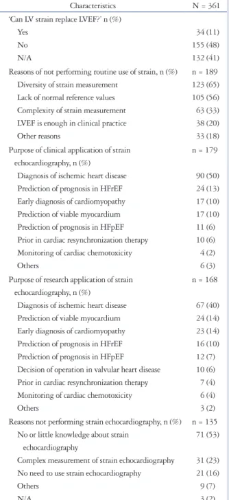
Table 2. Current status of clinical application of strain echocardiog- raphy (continued)
Characteristics N = 361
‘Can LV strain replace LVEF?’ n (%) Yes
No N/A
34 (11) 155 (48) 132 (41) Reasons of not performing routine use of strain, n (%)
Diversity of strain measurement Lack of normal reference values Complexity of strain measurement LVEF is enough in clinical practice Other reasons
n = 189 123 (65) 105 (56) 63 (33) 38 (20) 33 (18) Purpose of clinical application of strain
echocardiography, n (%)
Diagnosis of ischemic heart disease Prediction of prognosis in HFrEF Early diagnosis of cardiomyopathy Prediction of viable myocardium Prediction of prognosis in HFpEF Prior in cardiac resynchronization therapy Monitoring of cardiac chemotoxicity Others
n = 179
90 (50) 24 (13) 17 (10) 17 (10) 11 (6) 10 (6) 4 (2) 6 (3) Purpose of research application of strain
echocardiography, n (%)
Diagnosis of ischemic heart disease Prediction of viable myocardium Early diagnosis of cardiomyopathy Prediction of prognosis in HFrEF Prediction of prognosis in HFpEF
Decision of operation in valvular heart disease Prior in cardiac resynchronization therapy Monitoring of cardiac chemotoxicity Others
n = 168
67 (40) 24 (14) 23 (14) 16 (10) 12 (7) 10 (6) 7 (4) 6 (4) 3 (2) Reasons not performing strain echocardiography, n (%)
No or little knowledge about strain echocardiography
Complex measurement of strain echocardiography No need to use strain echocardiography Others
N/A
n = 135 71 (53)
31 (23) 21 (16) 9 (7) 3 (2) LV: left ventricle, RV: right ventricle, LA: left atrium, RA: right atrium, LVEF: left ventricular ejection fraction, HFrEF: heart failure with reduced ejection fraction, HFpEF: heart failure with preserved ejection fraction Table 2. Current status of clinical application of strain echocardiog-
raphy
Characteristics N = 361
Awareness of strain echocardiography, n (%) No idea
Moderate knowledge High knowledge
11 (3) 81 (25) 229 (71) Performance of strain echocardiography, n (%)
Physician Echocardiographer
186 (58) 53 (32) 133 (85) Purpose of strain echocardiography, n (%)
For research For clinical use
n = 186 138 (74) 126 (68) Modalities of strain echocardiography, n (%)
Tissue Doppler imaging
Two-dimensional strain echocardiography Three-dimensional strain echocardiography
n = 186 93 (50) 175 (94) 25 (13) Vendors of strain echocardiography, n (%)
GE Philips Siemens Toshiba Tomtec
n = 186 181 (97) 87 (27) 35 (19) 2 (1) 22 (12) Measurement of cardiac chambers, n (%)
LV RV LA RA
n = 186 184 (99) 71 (38) 25 (13) 3 (2)
Their awareness of and clinical use of the strain echocardiog- raphy are summarized in Table 2. Most of our study partici- pants (97%) were aware of the concept of the strain echocar- diography. Most of imaging and heart failure specialists (96%) had high-level knowledge about it.
Of the participants, 186 (58%) performed strain echocar- diography. Interestingly, echocardiographers measured strain values more frequently than physicians (85% vs. 32%, p <
0.001). Among cardiologists (n = 164), imaging specialists and heart failure specialists performed strain echocardiography more frequently than other cardiologists or general internists
(85% vs. 12%, p < 0.001). The participants used strain echo- cardiography for clinical and research purposes. However, re- search purposes seemed to be more frequent. Two-dimensional strain echocardiography was the most commonly used modal- ity in strain echocardiography, and GE was the most frequent- ly used algorithm.
LV was the most commonly used cardiac chamber (99%) for clinical purposes. When RV, left atrium and right atrium were measured, it was usually for research purposes.
Most of the participants did not think LV strain can replace LVEF in their clinical practice, and only 11% of them thought LV
Fig. 1. Result of questionnaire about clinical usefulness of strain echocardiography in the future.
The responders were asked on a scale from 1 to 10, and the higher score means the more favorable response to the question. IHD: ischemic heart disease, HFrEF: heart failure with reduced ejection fraction, CRT: cardiac resynchronization therapy, HFpEF: heart failure with preserved ejection fraction, CMP: cardiomyopathy, OP: operation, VHD: valvular heart disease.
35 30 25 20 15 10 5 0
1 2 3 4 5 6 7 8 9 10 11 In systernic diseases
Relative frequency (%)
Mean = 5.33 SD = 1.99
25 20 15 10 5 0
1 2 3 4 5 6 7 8 9 10 11 Mean = 6.44
SD = 1.93
Prognosis in IHD
Relative frequency (%)
25 20 15 10 5 0
1 2 3 4 5 6 7 8 9 10 11 Mean = 6.53
SD = 1.95
Predicting viable myocardium
Relative frequency (%)
25 20 15 10 5 0
1 2 3 4 5 6 7 8 9 10 11 Mean = 6.74
SD = 1.95
Diagnosis of IHD
Relative frequency (%)
25 20 15 10 5 0
1 2 3 4 5 6 7 8 9 10 11 Mean = 6.46
SD = 1.96
In CRT
Relative frequency (%)
25 20 15 10 5 0
1 2 3 4 5 6 7 8 9 10 11 Mean = 6.61
SD = 1.83
Prognosis in HFrEF
Relative frequency (%)
25 20 15 10 5 0
1 2 3 4 5 6 7 8 9 10 11 Prognosis in HFpEF
Relative frequency (%)
Mean = 6.56 SD = 1.90
25 20 15 10 5 0
1 2 3 4 5 6 7 8 9 10 11 Early diagnosis of CMP
Relative frequency (%)
40 35 30 25 20 15 10 5 0
1 2 3 4 5 6 7 8 9 10 11 Prognosis in VHD
Relative frequency (%)
Mean = 5.39 SD = 1.88 35
30 25 20 15 10 5 0
1 2 3 4 5 6 7 8 9 10 11 Decision of OP in VHD
Relative frequency (%)
Mean = 5.33 SD = 1.88 Mean = 6.7
SD = 1.92
strain can be used as LVEF. The most common reason for not performing routine use of strain echocardiography was diversi- ty of strain measurement (65%). Lack of normal reference val- ue and complexity of strain measurement were other reasons.
Imaging specialists and heart failure specialists thought diver- sity is the most common reason for not doing strain echocardiog- raphy than other physician (84% vs. 16%, p = 0.008). There was no difference in other reasons.
The most common purpose for clinical and research applica- tion of strain echocardiography was the diagnosis of ischemic heart disease (50%). Other purposes of use included prediction of the prognosis of heart failure with reduced ejection fraction (HFrEF), early diagnosis of cardiomyopathy, and prediction of viable myocardium. Imaging specialists and heart failure spe- cialists use strain echocardiography in the diagnosis of isch- emic heart disease most commonly. Moreover, they use strain echocardiography in the prediction of viable myocardium, early diagnosis of cardiomyopathy and prediction of prognosis in pa- tients with heart failure frequently. However, there was no sta- tistical difference.
The most common reason of not using strain measurement was no knowledge about the strain echocardiography in general physician (61% vs. 20%, p = 0.002). However, imaging and heart failure specialists did not perform strain measurement because of its complexity and lack of their time (60% vs. 19%, p = 0.002).
Many participants had a favorable view of the future of strain echocardiography (Fig. 1). They thought strain echocardiogra- phy will be most useful in the diagnosis of ischemic heart dis- eases. Other fields in which strain echocardiography was expect- ed to be useful in included the early diagnosis of cardiomyopathy, the prediction of prognosis in HFrEF and heart failure with preserved ejection fraction, and the prediction of viable myo- cardium. However, they did not think strain echocardiography will be more useful in the decision of operation time in valvular heart diseases, and application in systemic diseases and predic- tion of prognosis in valvular heart diseases. Compared to other physicians, imaging specialists and heart failure specialists be- lieved that strain echocardiography will be more useful in al- most all fields besides cardiac resynchronization therapy.
Discussion
Most of the participants of this survey had knowledge about strain echocardiography, and all participating institutions per- formed strain echocardiography in their clinical practice and re- search fields. Most of them measured LV in their clinical purpose.
However, they did not believe that LV strains could replace LVEF for many reasons.
Strain echocardiography is a relatively new echocardiograph- ic modality for measuring myocardial deformations. In Korea, initial introduction of strain echocardiography took place in the research field in the late 1990’s. The first Korean article using strain echocardiography was published by Cho et al.16)
in 2003. At the time, tissue Doppler imaging was the first mo- dality used to measure myocardial strains. Two-dimensional and three-dimensional strain echocardiography became readily available in current clinical practice with technical improve- ment. In this survey, all participating institutions performed the strain echocardiography for clinical and research purposes.
Interestingly, echocardiographers who measured strains were about 85% of the total number of echocardiographers (132/156).
However, only about 33% of the physicians performed strain measurements (54/165). We think that this result was due to the current practice in which echocardiographic examinations are done mostly by echocardiographers, while cardiologists usu- ally confirm the results. However, imaging specialists and heart failure specialists performed strain echocardiography in about 85% in their clinical practice (39/46), so these results should be interpreted with caution.
Interestingly, most of our participants had knowledge of the concept of myocardial strain and strain echocardiography.
Moreover, almost all imaging specialists and heart failure spe- cialists thought they have a high level of knowledge. This may be a result of several lectures on strain echocardiography to im- aging and heart failure specialists and increased the number of articles using strain echocardiography. However, the percent- age of participants who responded that they have no knowledge or moderate level of knowledge was higher in general internists and even in cardiologists other than in imaging and heart failure specialists (53% vs. 4%, p < 0.001). This may be a result of sev- eral lectures on strain echocardiography to imaging and heart failure specialists and increased number of articles using strain echocardiography. Education programs showing the strengths and the weaknesses of strain echocardiography will be needed to increase their interest and knowledge, especially for non- imaging and heart failure specialists and general internists. The difference in personal areas of interest can be another reason for this result.
Most of our participants measured LV strain usually for clini- cal use. This is probably because there are many articles show- ing LV strain as a good prognostic marker in many cardiovascu- lar diseases.1)2)17) Although there are many study results showing that LV strain has many advantages over LVEF, and many car- diologists are aware of strain echocardiography, the cardiolo- gists generally did not believe that LV strains will replace LVEF in their clinical practice. Because LVEF is the echocardiograph- ic parameter most commonly used to represent LV systolic func- tion, it has been used as a good prognostic parameter in rou- tine clinical practice.1)2) Despite this firm belief, LVEF does have several limitations, and strain echocardiography has been shown to solve these problems in clinical and research fields.6)7) However, only 11% of our participants thought LV strain can be used like LVEF. To change this thinking, more research showing the prognostic significance of LV strains will be nec- essary. Other studies are also needed to show the strength of strain echocardiography in the detection of subclinical myocar-
dial dysfunction and viable myocardium.
Most participants measured strains of other chambers like RV for research purposes. The main reason for not using RV strains in clinical use was that the estimation of RV systolic function usually depends on visual assessment and strain measurement lacked standardization and normal reference values.18)
Although many still do not think that strain values can be used as an auxiliary indicator of ventricular systolic function, many respondents believed that in the future, strain echocar- diography will be more useful in their clinical practice. Imag- ing specialists had a more favorable outlook on the future of strain echocardiography. To overcome this difference between the high awareness of advantages of strain echocardiography and low application of strain values in actual clinical practice, there are several problems to overcome. The respondents men- tioned diversity of strain measurements as the most common reason for not using LV strains instead of LVEF. The second reason was the lack of reference values. To increase the use of the strain echocardiography in clinical practice, it is necessary to use the integrated strain algorithms to solve vendor diversity, as well as to define normal reference values of the strain.
Limitation
This study had several limitations. First of all, this study was based on a questionnaire. Because a survey study allows for gen- eralizable statements about the strain, this study can provide a general sense on the current use of strain echocardiography in the clinical field. However, it may merely represent the data at the present time and give little information on the precise meaning of the data. Moreover, there was room for researcher bias, especially in the preparing of the questionnaire. Secondly, the use of strain echocardiography was dependent on the char- acteristics of each participating institution. The fact that the institutions that participated in this study were mainly tertia- ry teaching hospitals may also have influenced the results.
Conclusion
Most of the participants of this study were aware of strain echocardiography, and all institutions performed strain echo- cardiography for clinical and research purposes. However, the participants did not believe LV strain values could replace LVEF.
Because the diversity of strain measurements and lack of nor- mal reference values were common reasons for not using strain echocardiography in the clinical practice, researchers should pay attentions to solve these problems.
Supplementary Materials
The online-only Data Supplement is available with this arti- cle at https://doi.org/10.4250/jcu.2017.25.3.91.
References
1. Joyce E, Hoogslag GE, Leong DP, Debonnaire P, Katsanos S, Boden H, Schalij MJ, Marsan NA, Bax JJ, Delgado V. Association between left
ventricular global longitudinal strain and adverse left ventricular dilatation after ST-segment-elevation myocardial infarction. Circ Cardiovasc Imaging 2014;7:74-81.
2. Choi SW, Park JH, Sun BJ, Park Y, Kim YJ, Lee IS, Kim MS, Kim JH, Lee JH, Jeong JO, Kwon IS, Seong IW. Impaired two-dimensional global longitudinal strain of left ventricle predicts adverse long-term clinical outcomes in patients with acute myocardial infarction. Int J Cardiol 2015;
196:165-7.
3. Park SJ, Park JH, Lee HS, Kim MS, Park YK, Park Y, Kim YJ, Lee JH, Choi SW, Jeong JO, Kwon IS, Seong IW. Impaired RV global lon- gitudinal strain is associated with poor long-term clinical outcomes in patients with acute inferior STEMI. JACC Cardiovasc Imaging 2015;8:161-9.
4. Zornoff LA, Skali H, Pfeffer MA, St John Sutton M, Rouleau JL, La- mas GA, Plappert T, Rouleau JR, Moyé LA, Lewis SJ, Braunwald E, Solomon SD; SAVE Investigators. Right ventricular dysfunction and risk of heart failure and mortality after myocardial infarction. J Am Coll Cardiol 2002;39:1450-5.
5. Park JH, Park MM, Farha S, Sharp J, Lundgrin E, Comhair S, Tang WH, Erzurum SC, Thomas JD. Impaired global right ventricular longi- tudinal strain predicts long-term adverse outcomes in patients with pulmonary arterial hypertension. J Cardiovasc Ultrasound 2015;23:91-9.
6. White HD, Norris RM, Brown MA, Brandt PW, Whitlock RM, Wild CJ. Left ventricular end-systolic volume as the major determinant of survival after recovery from myocardial infarction. Circulation 1987;76:44- 51.
7. White HD, Norris RM, Brown MA, Takayama M, Maslowski A, Bass NM, Ormiston JA, Whitlock T. Effect of intravenous streptokinase on left ventricular function and early survival after acute myocardial infarc- tion. N Engl J Med 1987;317:850-5.
8. Hoit BD. Strain and strain rate echocardiography and coronary artery dis- ease. Circ Cardiovasc Imaging 2011;4:179-90.
9. Haddad F, Hunt SA, Rosenthal DN, Murphy DJ. Right ventricular function in cardiovascular disease, part I: anatomy, physiology, aging, and functional assessment of the right ventricle. Circulation 2008;117:1436-48.
10. Lang RM, Badano LP, Mor-Avi V, Afilalo J, Armstrong A, Ernande L, Flachskampf FA, Foster E, Goldstein SA, Kuznetsova T, Lancel- lotti P, Muraru D, Picard MH, Rietzschel ER, Rudski L, Spencer KT, Tsang W, Voigt JU. Recommendations for cardiac chamber quantification by echocardiography in adults: an update from the American Society of Echo- cardiography and the European Association of Cardiovascular Imaging. J Am Soc Echocardiogr 2015;28:1-39.e14.
11. Pirat B, McCulloch ML, Zoghbi WA. Evaluation of global and regional right ventricular systolic function in patients with pulmonary hypertension using a novel speckle tracking method. Am J Cardiol 2006;98:699-704.
12. Sarvari SI, Haugaa KH, Anfinsen OG, Leren TP, Smiseth OA, Kongsgaard E, Amlie JP, Edvardsen T. Right ventricular mechanical dispersion is related to malignant arrhythmias: a study of patients with ar- rhythmogenic right ventricular cardiomyopathy and subclinical right ven- tricular dysfunction. Eur Heart J 2011;32:1089-96.
13. Thomas JD, Popović ZB. Assessment of left ventricular function by cardiac ultrasound. J Am Coll Cardiol 2006;48:2012-25.
14. Geyer H, Caracciolo G, Abe H, Wilansky S, Carerj S, Gentile F, Ness- er HJ, Khandheria B, Narula J, Sengupta PP. Assessment of myocardial mechanics using speckle tracking echocardiography: fundamentals and clinical applications. J Am Soc Echocardiogr 2010;23:351-69.
15. Nahum J, Bensaid A, Dussault C, Macron L, Clémence D, Bouhe- mad B, Monin JL, Rande JL, Gueret P, Lim P. Impact of longitudinal myocardial deformation on the prognosis of chronic heart failure patients.
Circ Cardiovasc Imaging 2010;3:249-56.
16. Cho GY, Park WJ, Han SW, Han SJ, Choi SH, Choi YJ, Doo YC, Han KR, Lee NH, Oh DJ, Ryu KH, Rhim CY, Lee Y. Quantification of regional wall motion abnormality using myocardial strain in acute myo-
cardial infarction. Korean Circ J 2003;33:583-9.
17. Mollema SA, Delgado V, Bertini M, Antoni ML, Boersma E, Holman ER, Stokkel MP, van der Wall EE, Schalij MJ, Bax JJ. Viability assess- ment with global left ventricular longitudinal strain predicts recovery of left
ventricular function after acute myocardial infarction. Circ Cardiovasc Im- aging 2010;3:15-23.
18. Markley RR, Ali A, Potfay J, Paulsen W, Jovin IS. Echocardiographic evaluation of the right heart. J Cardiovasc Ultrasound 2016;24:183-90.