ISSN 2234-3806 • eISSN 2234-3814
Ann Lab Med 2012;32:177-183
http://dx.doi.org/10.3343/alm.2012.32.3.177
Changes in Urinary Stone Composition in the Tunisian Population: A Retrospective Study of 1,301 Cases
Akram Alaya, Ph.D.1, Abdellatif Nouri, Ph.D.2, Mohsen Belgith, Ph.D.2, Hammadi Saad, Ph.D.3, Riadh Jouini, Ph.D.2, and Mohamed Fadhel Najjar, Ph.D.1
Departments of Biochemistry and Toxicology1, Pediatric Surgery2, Urology3, University Hospital, Monastir, Tunisia Background: Studies that evaluate the effect of age on stone composition are scarce. The aim of this study was to highlight the changes in epidemiological characteristics (stone composition and location) of urolithiasis according to patients’ age.
Methods: We studied 1,301 urolithiasis patients with age ranging from 6 months to 92 yr (781 males and 520 females). Stone analysis was performed using a stereomicroscope and infrared spectroscopy to determine the morphological type and molecular composi- tion of each stone.
Results: The annual average incidence of new stone formation was 31.7 per 100,000 per- sons. In 71.8% of cases, calculi were located in the upper urinary tract. Compared to other age groups, children and old men were more affected by bladder stones. Calcium oxalate monohydrate was the most frequent stone component, even though its frequency decreased with age (59.5% in young adults and 43.7% in the elderly, P <0.05) in favor of an increase in uric acid stones (11.5% in young adults and 36.4% in the elderly, P <0.05). Struvite stones were rare (3.8%) and more frequent in children than in adults.
Conclusions: The analysis of these data showed that urinary stones in Tunisian patients are tending to evolve in the same direction as the stones in patients from industrialized countries.
Key Words: Urinary stone, Spectrophotometry, Kidney, Children, Adult, Elderly, Uric acid
Received: August 8, 2011 Revision received: October 29, 2011 Accepted: February 8, 2012 Corresponding author: Akram Alaya Department of Biochemistry and Toxicology University Hospital, 5000 Monastir, Tunisia Tel: +216-97-85-21-76
Fax: +216-73-46-06-78 E-mail: [email protected]
© The Korean Society for Laboratory Medicine.
This is an Open Access article distributed under the terms of the Creative Commons Attribution Non-Commercial License (http://creativecom- mons.org/licenses/by-nc/3.0) which permits unrestricted non-commercial use, distribution, and reproduction in any medium, provided the original work is properly cited.
INTRODUCTION
Urolithiasis is a relatively common condition in different continents and countries. Nevertheless, the overall probability of forming stones considerably differs in various parts of the world [1, 2].
The risk of developing urolithiasis appears to be higher in the western hemisphere than in the eastern hemisphere, even though the highest risks have been reported in some Asian countries [2, 3]. Socioeconomic conditions have generated changes in the incidence, location, and composition of urolithiasis [4]. In fact, bladder stones composed of ammonium urate and calcium ox- alate have been reported to be endemic in Asia, whereas reno-
ureteral calculosis, featuring mainly calcium oxalate and phos- phate, is currently more frequent in economically developed countries [1]. This must be attributed to the different lifestyles and dietary habits.
Because of the continuous changes in lifestyles and dietary habits, the relationships between age and composition can also change with time [5]. Only a few reports to date have suggested a relationship between stone composition and patient’s age [6- 8]. In 1995, Daudon et al. [9] reported an increase in the fre- quency of uric acid stones with an increase in patient’s age;
they reported that the frequency reaches its peak in the age range 60-70 yr. In Japan, Koide et al. [8] have reported that the
ISSN 2234-3806 • eISSN 2234-3814
peak frequency of calcium oxalate urolithiasis was observed be- tween 40 and 50 yr of age.
Urolithiasis in developing countries was considered very dif- ferent from urolithiasis observed in industrialized countries [10].
In Tunisia, studies evaluating the epidemiological characteristics (stone composition and location) of urolithiasis are scarce and have been based on the analysis of a limited number of patients [6]. This paper reports pioneering research in which we exam- ined 1,301 lithiasic patients for 10 yr in order to present an out- line of the current state of renal lithiasis on the Tunisian coast region.
METHODS
Between July 1998 and September 2010, we retrospectively in- vestigated 1,301 patients with stones and who were admitted to the pediatric and the urologic surgery departments at the Uni- versity Hospital of Monastir. The patients, 781 males and 520 fe- males, were aged 6 months to 92 yr. To make our task easier, we divided our patients into 4 age groups according to the Tuni- sian classification: children, young adults, adults, and elderly.
Full documentation included recording of age, sex, residency, age at onset of symptoms, age at diagnosis of stone disease, clin- ical presentation, past medical and surgical history, family history of stone disease, and recurrence (which was considered if the patient had previous surgery or spontaneous passage of a stone before presentation). Urine culture was carried out in 987 cases.
Twenty-four-hour urine determination of urinary calcium, oxa- late, and uric acid was performed for all patients. Hypercalciuria (HCa) was defined as urine calcium excretion >4 mg/(kg 24 hr) [11]. Hyperoxaluria (HOx) was defined as urine oxalate excre- tion >55 mg/(1.73 m224 hr). Hyperuricosuria (HUr) was de- fined as uric acid excretion >815 mg/(1.73 m224 hr) [12].
When possible, the structure of each stone was identified us- ing a stereomicroscope to define the morphology of the stone and to select its representative parts (nucleus or core, internal section, and external surface) and using infrared spectroscopy to determine its molecular and crystalline composition. These techniques were used to analyze all stones collected during the 10-yr study period. Approximately 0.5 to 2 mg of powder was collected from each part of the stone and pulverized with an in- ert powdered support (dried potassium bromide) in a proportion of 0.5-2% in an agate mortar. This mixture was transferred into an appropriate container and pressed at 10 t/cm2 to form a transparent pellet with a diameter of 13 mm. The spectral region investigated was from 4,000 to 400 per cm. Reference spectra
were obtained from pure potassium bromide (KBr) pellets. The spectra were recorded by means of a Bruker IFS25 Fourier transform infrared spectrometer (Bruker SA, Wissembourg, France). A global powder of the sample was analyzed in order to quantify the relative proportions of the various stone compo- nents. Only the qualitative and quantitative composition ob- tained from the whole-stone powder constituted the study mate- rial in this report.
The various compounds were identified by comparison to previously published reference spectra. The results were ex- pressed according to the main crystalline phase found in the stones and were named as follows: whewellite (calcium oxalate monohydrate), weddellite (calcium oxalate dihydrate), carbapa- tite (carbonated calcium phosphate crystallized in a hexagonal pattern), struvite (magnesium ammonium phosphate hexahy- drate), and calcite (anhydrous calcium carbonate). The stone component was considered the main element if it exceeded 75% of the total composition of the calculus. Stones formed by a single component were classified as pure stones, and those with more than one component were classified as mixed stones.
Statistical analysis of the data was performed using SPSS 11.0 for Windows. Statistical significance was determined using the χ2-test. P values less than 0.05 were considered significant.
RESULTS
The average annual incidence of new stone formation was 31.7 per 100,000 inhabitants. The annual incidence was 7.4 per 100,000 inhabitants for the pediatric age group and 54.5 per 100,000 inhabitants for the adult age group. The average annual incidence decreased during the last year of our study (Fig. 1).
The most frequent method of extraction was conventional surgery (92.2%). The other methods included extracorporeal lithotripsy
Annual incidence
Patients/100,000 inhabitants
40 35 30 25 20 15 10 5
0 1998-2000 2000-2002 2002-2004 2004-2006 2006-2008 2008-2010 Year
Fig. 1. Changes in the annual incidence of urolithiasis throughout the study period.
(6.2%), spontaneous passage (1.3%), and endoscopy (0.3%).
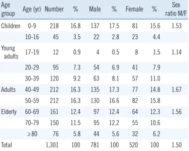
Of the 1,301 patients with urolithiasis, 781 (60%) were males and 520 (40%) were females with a sex ratio of 1.5:1. Patient’s age at presentation ranged from 6 months to 92 yr. The number of patients stratified by age and sex is shown in Table 1. The highest number of calculi in males and females was observed in the age groups 40-49 and 50-59 yr, respectively.
The upper urinary tract was most frequently affected by this condition (kidney 52.4% and ureter 29.4%). Bladder stones were noted in 18.2% of cases. A significant difference in bladder stone cases, was noted according to gender (63.2% in male vs. 36.8% in female) (P <0.001).
Children and elderly patients seemed to have a higher inci- dence of lower urinary tract stones (Fig. 2). A male predominance was pronounced in children (M/F=2.86). Prostate hyperplasia was associated with bladder stone findings in 12 elderly male patients.
A family history of stone disease was recorded for 54 patients Table 1. Distribution of urinary calculi according to age and gender
of the patients Age
group Age (yr) Number % Male % Female % Sex ratio M/F Children 0-9 218 16.8 137 17.5 81 15.6 1.53
10-16 45 3.5 22 2.8 23 4.4
Young
adults 17-19 12 0.9 4 0.5 8 1.5 1.14
20-29 95 7.3 54 6.9 41 7.9
30-39 120 9.2 63 8.1 57 11.0
Adults 40-49 212 16.3 135 17.3 77 14.8 1.67
50-59 212 16.3 130 16.6 82 15.8
Elderly 60-69 161 12.4 97 12.4 64 12.3 1.56
70-79 150 11.5 95 12.2 55 10.6
≥80 76 5.8 44 5.6 32 6.2
Total 1,301 100 781 100 520 100 1.50
%
60 50 40 30 20 10
0 ≤16 17-39
Year
40-59 ≥60
Kidney Ureter Bladder
Fig. 2. Changes in stone location according to age (N=1,301).
Table 2. Metabolic and urological status of 1,301 Tunisian patients with renal stone disease Metabolic and
anatomical defects
Stone component
Total %
COM UA COD CA Cystine MAP Calcite AU Xanthine Others
Normal 535 177 0 63 0 31 0 42 2 4 854 65.6
Metabolic abnormalities
Hypercalciuria 69 0 99 19 0 11 6 3 0 4 211 16.2
Hyperoxaluria 33 7 1 16 0 4 3 3 0 0 67 5.1
Cystinuria 0 0 0 0 21 0 0 0 0 0 21 1.6
Hyperuricosuria 6 87 0 0 0 0 0 9 2 0 104 8.0
Anatomical defects
Horseshoe kidney 3 0 1 0 0 1 0 0 0 0 5 0.4
Ureteropelvic junction
obstruction 9 0 2 6 0 1 0 0 0 0 18 1.4
Dumb kidney 2 0 0 1 0 0 0 0 0 0 3 0.2
Posterior urethral valves 2 0 0 1 0 0 0 0 0 0 3 0.2
Neuropathic bladder 0 0 0 5 0 1 0 0 0 0 6 0.5
Vesicoureteric reflux 0 2 0 4 0 1 0 2 0 0 9 0.7
Total number 659 273 103 115 21 50 9 59 4 8 1,301 100
Abbreviations: COM, calcium oxalate monohydrate; COD, calcium oxalate dihydrate; CA, carbapatite; MAP, magnesium ammonium phosphate; UA, uric acid;
AU, ammonium urate.
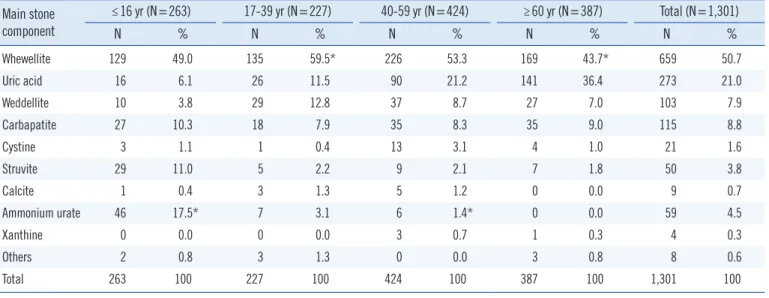
As shown in Table 3, the most frequent component of urinary stones, as determined by infrared spectroscopy, was whewellite (50.7%). However, its frequency differed with age. Whewellite was significantly more frequent in young adults (59.5%) than in the elderly (43.7%) (P <0.05). The proportion of stones with uric acid was 21.0%, and such stones were more prevalent in the el- derly than in the other age groups (P <0.02). However, ammo- nium urate stones were predominant in children (P <0.001).
Struvite-containing stones were observed in 3.8% of cases and were more frequent in children than the other age groups (P <
0.02).
According to stone location, struvite stones were more fre- quently observed in the bladder (6.8%) than in the kidneys (3.7%), whereas whewellite stones were more frequently found in the kidneys (54.1% vs. 44.1% in the bladder) (Fig. 3). Ammo- nium urate was observed in 5.1% of bladder stones, and it was noted only in children (15.8%).
The treatment procedures performed were as follows: 1) open surgical procedure was performed in 1,057 patients (81.2%); 2) stones were disintegrated with extracorporeal shock wave litho- tripsy in 185 patients (14.2%); 3) endoscopic interventions (sim- ple forceps and/or basket extraction) were used in 22 patients (1.7%); and 4) a specific treatment with D-penicillamine (urine pH in the low alkaline range) was used in 21 cystinuria patients.
However, treatment with D-penicillamine was successful in only 14 patients. The remaining 23 patients (1.8%) required no spe- cific intervention, and the majority passed the stones spontane- ously with conservative management and close follow-up.
Of the 1,301 patients, 288 (22.1%) experienced recurrences Table 3. Distribution of the main stone components according to age
Main stone component
≤16 yr (N=263) 17-39 yr (N=227) 40-59 yr (N=424) ≥60 yr (N=387) Total (N=1,301)
N % N % N % N % N %
Whewellite 129 49.0 135 59.5* 226 53.3 169 43.7* 659 50.7
Uric acid 16 6.1 26 11.5 90 21.2 141 36.4 273 21.0
Weddellite 10 3.8 29 12.8 37 8.7 27 7.0 103 7.9
Carbapatite 27 10.3 18 7.9 35 8.3 35 9.0 115 8.8
Cystine 3 1.1 1 0.4 13 3.1 4 1.0 21 1.6
Struvite 29 11.0 5 2.2 9 2.1 7 1.8 50 3.8
Calcite 1 0.4 3 1.3 5 1.2 0 0.0 9 0.7
Ammonium urate 46 17.5* 7 3.1 6 1.4* 0 0.0 59 4.5
Xanthine 0 0.0 0 0.0 3 0.7 1 0.3 4 0.3
Others 2 0.8 3 1.3 0 0.0 3 0.8 8 0.6
Total 263 100 227 100 424 100 387 100 1,301 100
*P <0.05.
%
60 50 40 30 20 10 0
Whewellite Uric acid
Weddellite Carbapatite
Cystine Struvite
Calcite Ammonium urate
Xanthine Others
Kidney Bladder
Fig. 3. Differences in kidney and bladder stone composition.
(4.1%). The most common symptom on admission was abdom- inal pain in 39% of cases. This was accompanied by colic pain in 28.2%, hematuria in 11.7%, dysuria in 9.1%, urinary tract in- fection in 3.2%, anuria in 2.8%, accidental finding in 2.8%, fe- ver in 1.9%, and dumb kidney in 1.3%.
Anatomical defects were detected in 44 patients (Table 2) and were associated with urinary tract infection in only 11 cases. Uri- nary stasis secondary to a urinary tract anomaly was noted in 8 patients (18%). Metabolic disorders (confirmed by 24-hr urine collection) were observed in 403 patients (31.0%). Two hundred and eleven patients had severe hypercalciuria, 67 had hyperox- aluria, and 21 had cystinuria (Table 2). Hyperuricosuria, detected in 104 patients, was the second most common metabolic disor- der and was more frequent in the elderly.
since an initial diagnosis of stone disease. The incidence of stone recurrence in patients who were radiographically stone free fol- lowing ESWL was reported as 40.0% (74 cases). Disease recur- rence occurred at 5 yr after the initial diagnosis based on the probable criteria.
DISCUSSION
Changes in socioeconomic conditions have generated changes in the incidence and type of urolithiasis in terms of both the site and the physicochemical composition of the calculi. Major vari- ations in worldwide occurrence of urolithiasis have been reported according to geographical areas [10]. We found that the annual incidence of new stone formation in Tunisian patients was 31.7 per 100,000 Tunisians. This rate is similar to that reported in England (22 per 100,000 inhabitants) [13] and Kuwait (23.9 per 100,000 inhabitants). However, it is significantly different from that reported in Sweden (140 per 100,000 inhabitants) [14], Italy (168 per 100,000 inhabitants) [15], and the USA (277 per 100,000 inhabitants) [16]. In Europe, urinary stones are mainly located in the upper urinary tract, and the proportion of bladder calculi does not exceed 10.0% [10, 17]. Bladder stones have also been found to be frequent in the elderly as reported by some studies [18] but not in others [17]. According to Daudon et al. [18], 40.0% of the patients they analyzed were men over 80 yr. In our study, 18.2% of the stones were from the bladder, and elderly men were the most affected (24.3%). Prostatic hyperplasia, which is considered a frequent cause of bladder outlet obstruction, is frequent in old men and could be a possible explanation for the high frequency of bladder stones in the elderly [18, 19]. Fe- males were also exposed to lower urinary tract stones (36.8%).
This suggests that other risk factors, such as changes in blad- der function associated with relaxation of smooth muscle tonic- ity in the elderly, reduce the efficiency of bladder emptying and promote urine stasis [18].
The pattern of urolithiasis in adult patients in Tunisia is not dif- ferent from that observed worldwide. Stone composition has changed substantially over the past decades with a progressive increase in the frequency of calcium oxalate stones, which rep- resents 70-80% of stones [20]. According to Daudon et al. [21], calcium oxalate stones in developing countries are mainly ob- served in North Africa and Asia Minor. In our study, calcium oxa- late stones were found in 58.6% of all cases. This rate is compa- rable to those reported in French [9], Brazilian [22], and Spanish [5] papers. As recognized worldwide, hypercalciuria was the most frequent underlying factor in calcium oxalate stones, even
though in some countries in the eastern hemisphere, hypoci- traturia has been reported as the leading cause [23]. Daudon et al. [7] used a multivariate approach based on correspondence factor analysis and confirmed the relationship between stone composition and patient age. However, some variations accord- ing to geographical areas have been reported. In 1993, Baker et al. [24] reported that in Australia, the peak frequency of calcium oxalate urolithiasis was observed in persons between 50 and 60 yr of age. In Europe, studies had revealed that calcium oxalate stones are more frequent in persons between 40 and 50 yr of age [5, 7]. In Asia, it seems that the peak frequency of calcium oxa- late stones occurs at an earlier age (30-50 yr) [8]. In our study, we found a peak frequency of calcium oxalate urolithiasis in per- sons between 16 and 39 yr of age; similar results have also been observed in Algeria [25]. The monohydrate form (whewellite) was 6.5 times more abundant than the dihydrate form (weddellite).
Weddellite calculi occurred earlier in female patients than in male patients and decreased with increasing age; this finding is similar to that of Algerian [25] and French [9] studies. Abrams [26] has reported that both vitamin D deficiency and a diminished ability to absorb dietary calcium are more prevalent in the elderly. This could explain, in part, the decrease of weddellite and whewellite frequency in older individuals. In fact, a greater proportion of di- etary calcium remains in the intestine where it is available to bind oxalate, thereby reducing oxalate absorption and the subsequent concentration of urinary oxalate [27].
Chronic diarrhea with subsequent acidosis during the first months of life and simultaneous phosphorus deficiency can ex- plain a high urine excretion of ammonium ions. Ammonium ions can form urate ammonium by combining with urate ions, which are also abundant in child urine. In our series, ammo- nium urate was observed as often in kidney stones as in bladder stones; however, a high proportion (15.8%) of pediatric bladder stones was nucleated on ammonium urate. This suggests that hyperuricosuria, low phosphorus intake, low diuresis, and chronic diarrhea in children may be factors affecting stone formation.
Struvite is the best marker of urinary tract infections by ure- ase-producing bacteria. Our study found a relatively lower rate of struvite stones (3.8%) than other stones; this rate was similar to the rates reported in Europe [7, 28], Algeria [24], and China [29]. This infection lithiasis frequency was lower than that re- ported by Najjar et al. [30] in the same area. Antibiotics abuse may be one reason for the low rate of infection lithiasis in devel- oped countries [29]. However, we think the earlier detection of urinary infections and the greater attention paid to their treat- ment in recent years could explain the lower frequency in Tuni-
sia. Contrary to the findings of other studies in France [7, 9], Al- geria [25], and China [29], we found that uric acid stone was a very common type of stone disease in our region, accounting for 21.0% of cases. However, this frequency was similar to those reported in Germany [31], the USA [32], and Australia [24].
Hyperuricosuria and low urinary pH (below 5.5) seem to be the most important risk factors for uric acid crystallization [33].
In our series, hyperuricosuria was reported in only 107 cases, thereby suggesting the presence of others risk factors such as dietary habits. Indeed, since the 1960s, these habits have changed in our country towards an increase of purine-rich food (animal protein and seafood) intake [34], which explains the as- sociated increase in uric acid excretion. As reported in industri- alized countries [5, 7-9, 24], our data shows a clear increase of uric acid stones according to age in both genders. On the basis of the well-established pH dependence of uric acid urolithiasis, the rising proportion of uric acid stones with increasing age may be due to a progressive defect in urine ammoniagenesis that manifests with aging [35]. Daudon et al. [36] have shown that diabetic patients are more likely affected by uric acid stones. In addition, recent studies have suggested that the increased prevalence of urolithiasis and recurrence is associated with obe- sity and elevated urinary excretion of calcium, uric acid, and ox- alate [37]. We now know that overeating contributes to the de- velopment of obesity and is strongly involved in the development of diabetes, kidney stones, and hypertension. These risk factors, such as obesity, diabetes, and hypertension, should be consid- ered in epidemiological investigations in order to avoid confu- sion with the influence of age on stone composition and in order to have representative results.
Some studies have shown stone recurrence rates between 26% and 53% after 10 yr [38]. Previous ESWL has been found to be a risk factor in some studies [39] but not in others [40]. In our study, we report a recurrence rate, but the implication of ESWL as a risk factor remains to be confirmed.
In conclusion, although data on kidney stone disease in Tuni- sia are scarce, our data provide an idea about its epidemiologic characteristics. Calcium oxalate stones remain the most frequent components in all age groups, even if their frequency decreases in the elderly. As reported by other studies, we found that uric acid stones increase with increasing age, and infection stones are rarely observed in adults. Analysis of these data showed that urinary stones seen in Tunisian patients are tending to evolve in the same direction as stones seen in patients in industrialized countries.
Authors’ Disclosures of Potential Conflicts of Interest
No potential conflict of interest relevant to this article was re- ported.
Acknowledgement
The authors wish to thank Professor Walid Chaouachi for his as- sistance with the English-language presentation of the manu- script.
REFERENCES
1. Trinchieri A. Epidemiology of urolithiasis. Arch Ital Urol Androl 1996;68: 203-49.
2. Ramello A, Vitale C, Marangella M. Epidemiology of nephrolithiasis. J Nephrol 2000;S(3):S45-50.
3. Robertson WG and Hughes H. Epidemiology of urinary stone disease in Saudi Arabia. In: Ryall R, Bais R, et al. eds. Urolithiasis 2. New York London: Plenum Press, 1994:453-5.
4. El-Reshaid K, Mughal H, Kapoor M. Epidemiological profile, mineral metabolic pattern and crystallographic analysis of urolithiasis in Kuwait.
Eur J Epidemiol 1997;13:229-34.
5. Costa-Bauzá A, Ramis M, Montesinos V, Grases F, Conte A, Pizá P, et al. Type of renal calculi: variation with age and sex. World J Urol 2007; 25:415-21.
6. Alaya A, Najjar MF, Nouri A. Changes in stone composition according to age in Tunisian pediatric patients. Int Urol Nephrol 2010;42:621-8. 7. Daudon M, Doré JC, Jungers P, Lacour B. Changes in stone composi-
tion according to age and gender of patients: a multivariate epidemio- logical approach.Urol Res 2004;32:241-7.
8. Koide T, Itatani H, Yoshioka T, Ito H, Namiki M, Nakano E, et al. Clinical manifestations of calcium oxalate monohydrate and dihydrate urolithia- sis. J Urol 1982;127:1067-9.
9. Daudon M, Donsimoni R, Hennequin C, Fellahi S, Le Moel G, Paris M, et al. Sex- and age-related composition of 10617 calculi analyzed by in- frared spectroscopy. Urol Res 1995;23:319-26.
10. López M and Hoppe B. History, epidemiology and regional diversities of urolithiasis. Pediatr Nephrol 2010;25:49-59.
11. Elder JS. Urinary lithiasis. In: Behrman RE, Kliegman RM, et al. eds.
Nelson textbook of pediatrics. 17th ed. Philadelphia: Saunders, 2004: 1822-6.
12. Oner A, Demircin G, Ipekçio lu H, Bülbül M, Ecin N. Etiological and clini- cal patterns of urolithiasis in Turkish children. Eur Urol 1997;31:453-8. 13. Power C, Barker DJ, Blacklock NJ. Incidence of renal stones in 18 Brit-
ish towns. A collaborative study. Br J Urol 1987;59:105-10.
14. Ahlstrand C and Tiselius HG. Renal stone disease in a Swedish district during one year. Scand J Urol Nephrol 1981;15:143-6.
15. Serio A and Fraioli A. Epidemiology of nephrolithiasis. Nephron 1999;81 (S1):S26-30.
16. Curhan GC, Rimm EB, Willett WC, Stampfer MJ. Regional variation in nephrolithiasis incidence and prevelance among United States men. J Urol 1994;151:838-41.
17. Neuzillet Y, Lechevallier E, Ballanger P, Ferriere JM, Saussine C, Doré B, et al. Urinary stones in subjects over the age of sixty. Prog Urol 2004;14:
479-84.
18. Daudon M. Évolution de la composition et de la localisation des calculs chez le sujet âgé. Feuillets de Biologie 2003;25:51-4.
19. El-Reshaid K, Mughal H, Kapoor M. Epidemiological profile, mineral metabolic pattern and crystallographic analysis of urolithiasis in Kuwait.
Eur J Epidemiol 1997;13:229-34.
20. Daudon M. Comment analyser un calcul et comment interpréter le ré- sultat. L’ Eurobiologiste 1993;203:35-46.
21. Daudon M, Bounxouei B, Santa Cruz F, Leite da Silva S, Diouf B, Ang- wafoo FF 3rd, et al. Composition of renal stones currently observed in non-industrialized countries. Prog Urol 2004;14:1151-61.
22. Silva SF, Matos DC, Silva SL, Daher Ede F, Campos Hde H, Silva CA.
Chemical and morphological analysis of kidney stones: a double-blind comparative study. Acta Cir Bras 2010;25:444-8.
23. Stitchantrakul W, Kochakarn W, Ruangraksa C, Domrongkitchaiporn S.
Urinary risk factors for recurrent calcium stone formation in Thai stone formers. J Med Assoc Thai 2007;90:688-98.
24. Baker PW, Coyle P, Bais R, Rofe AM. Influence of season, age, and sex on renal stone formation in South Australia. Med J Aust 1993;159:390-2. 25. Djelloul Z, Djelloul A, Bedjaoui A, Kaid-Omar Z, Attar A, Daudon M, et
al. Urinary stones in Western Algeria: study of the composition of 1,354 urinary stones in relation to their anatomical site and the age and gen- der of the patients. Prog Urol 2006;16:328-35.
26. Abrams SA. Calcium turnover and nutrition through the life cycle. Proc Nutr Soc 2001;60:283-9.
27. Taylor EN, Stampfer MJ, Curhan GC. Dietary factors and the risk of inci- dent kidney stones in men: new insights after 14 years of follow-up. J Am Soc Nephrol 2004;15:3225-32.
28. Leusmann DB, Blaschke R, Schmandt W. Results of 5,035 stone analy- ses: a contribution to epidemiology of urinary stone disease. Scand J Urol Nephrol 1990;24:205-10.
29. Sun X, Shen L, Cong X, Zhu H, He L, Lu J. Infrared spectroscopic anal- ysis of 5,248 urinary stones from Chinese patients presenting with the
first stone episode. Urol Res 2011;39:339-43.
30. Najjar MF, Najjar F, Boukef K, Oueslati A, Memmi J, Bechraoui T. La li- thiase infantile dans la région de Monastir étude clinique et biologique.
Le Biologiste 1986;165:31-9.
31. Strohmaier WL and Weigl A. Stone composition in upper Franconia−
unusually high percentage of uric acid lithiasis. In: Jungers P, Daudon M, eds. Renal stone disease. Amsterdam: Elsevier Science, 1997:10-11. 32. Curhan GC, Willett WC, Speizer FE, Spiegelman D, Stampfer MJ. Com- parison of dietary calcium with supplemental calcium and other nutri- ents as factors affecting the risk for kidney stones in women. Ann Intern Med 1997;126:497-504.
33. Grases F, Costa-Bauza A, Prieto RM. Renal lithiasis and nutrition.Nutr J 2006;5:23.
34. Alaya A, Nouri A, Najjar MF. Urinary stone composition in pediatric pa- tients: a retrospective study of 205 cases. Clin Chem Lab Med 2011;49: 243-8.
35. Agarwal BN and Cabebe FG. Renal acidification in elderly subjects.
Nephron 1980;26:291-5.
36. Daudon M, Jungers P. Diabetes and calculations. Feuillets de Biologie 2001;239:37-9.
37. Siener R, Glatz S, Nicolay C, Hesse A. The role of overweight and obesi- ty in calcium oxalate stone formation. Obes Res 2004;12:106-13. 38. Trinchieri A, Ostini F, Nespoli R, Rovera F, Montanari E, Zanetti G. A
prospective study of recurrence rate and risk factors for recurrence af- ter a first renal stone. J Urol 1999;162:27-30.
39. Siener R, Glatz S, Nicolay C, Hesse A. Prospective study on the efficacy of a selective treatment and risk factors for relapse in recurrent calcium oxalate stone patients. Eur Urol 2003;44:467-74.
40. Kosar A, Sarica K, Aydos K, Küpeli S, Türkölmez K, Gö üs O. Compara- tive study of long-term stone recurrence after extracorporeal shock wave lithotripsy and open stone surgery for kidney stones. Int J Urol 1999;6: 125-9.