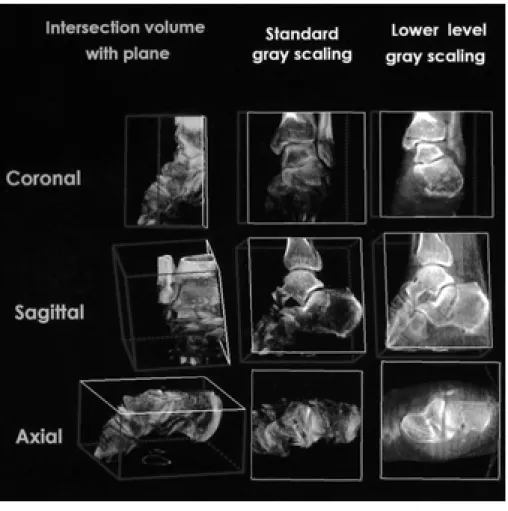
Fig. 1. Postprocessing using multipla- nar intersection with gray scale manip- ulation. After multiplanar-coronal, sagittal and axial-intersection, gray scale manipulation is performed on the intersection plane.
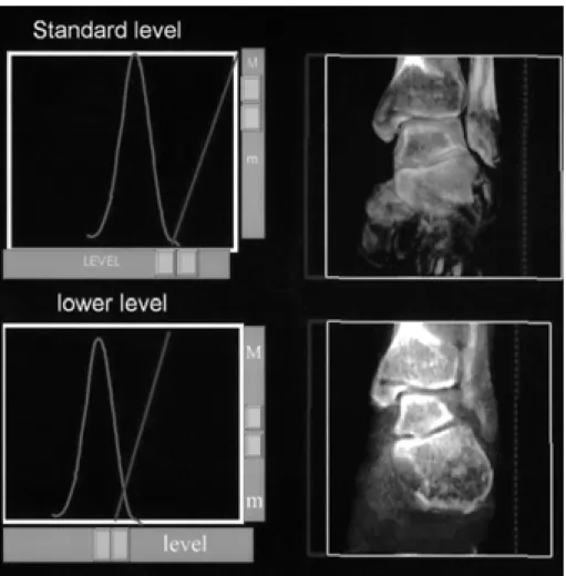
Fig. 2. Postprocessing using gray scale manipulation. The initial intersection plane shows the standard level of gray scale by auto gray scaling. The contrast resolution between the cortex and medulla on initial intersection plane is not better than that of bony algorithm on CT scan. On lowering level of gray scale, the contrast resolution between the cortex and medulla is more im- proved than that on standard level of gray scale.
A B
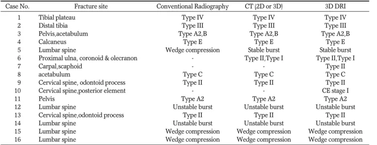
Table 1. Classification of Fractures on Conventional Radiography, CT (2D or 3D) and 3D DRI
Case No. Fracture site Conventional Radiography CT (2D or 3D) 3D DRI
01 Tibial plateau Type IV Type IV Type IV
02 Distal tibia Type III Type III Type III
03 Pelvis,acetabulum Type A2,B Type A2,B Type A2,B
04 Calcaneus Type E Type E Type E
05 Lumbar spine Wedge compression Stable burst Stable burst
06 Proximal ulna, coronoid & olecranon - Type II,Type I Type II,Type I
07 Carpal,scaphoid - - Type II
08 acetabulum Type C Type C Type C
09 Cervical spine, odontoid process Type II Type II Type II
10 Cervical spine,posterior element - - CE stage I
11 Pelvis Type A2 Type A2 Type A2
12 Lumbar spine Unstable burst Unstable burst Unstable burst
13 Cervical spine,odontoid process Type II Type II Type II
14 Lumbar spine Unstable burst Unstable burst Unstable burst
15 Lumbar spine Wedge compression Wedge compression Wedge compression
16 Lumbar spine Wedge compression Wedge compression Wedge compression
Table 2. Detection, Characterization and Comparison of Fractures on Conventional Radiography, 2D or 3D CT and 3D DRI
Case No. Conventional Radiography CT
3D DRI
2D 3D
01 + ++ + +++
02 + ++ 0 ++
03 + + 0 ++
04 ++ +++ + +++
05 + ++ + +++
06 0 + 0 +++
07 0 0 0 +
08 + ++ + +++
09 + + 0 ++
10 0 0 0 ++
11 + + 0 ++
12 + + 0 ++
13 + + ++ +++
14 + + + +++
15 + + ++ +++
16 + + ++ +++
0= poor visualization or disinterpretation
+=ambiguous visualization,++=precise visualization +++=more precise visualization)
1. Kode L, Lieberman JM, Motta AO, Wilber JH, Vasen A, Yagan R.
Evaluation of tibial plateau fractures: efficacy of MR imaging com- pared with CT. AJR Am J Roentgenol 1994;163:141-147
2. Liow RY, Birdsall PD, Mucci B, Greiss ME. Spiral computed to- mography with two- and three-dimensional reconstruction in the management of tibial plateau fractures. Orthopedics 1999;22:929- 932
3. Wicky S, Blaser PF, Blanc CH, Leyvraz PF, Schnyder P, Meuli RA.
Comparison between standard radiography and spiral CT with 3D reconstruction in the evaluation, classification and management of tibial plateau fractures. Eur Radiol 2000;10:1227-1232
4. Lawler LP, Corl FM, Fishman EK. Multi- and single detector CT with 3D volume rendering in tibial plateau fracture imaging and management. Crit Rev Comput Tomogr 2002;43:251-82
5. Anxionnat R, Bracard S, Macho J, et al. 3D angiography. Clinical interest first applications in interventional neuroradiology. J Neuroradiol 1998;25:251-262
6. Missler U, Hundt C, Wiesmann M, Mayer T, Bruckmann H.
Three-dimensional reconstructed rotational digital subtraction an- giography in planning treatment of intracranial aneurysms. Eur Radiol 2000;10:564-568
7. Fahrig R, Fox AJ, Lownie S, Holdsworth DW. Use of a C-arm sys- tem to generate true three-dimensional computed rotational an- giograms: preliminary in vitro and in vivo results. AJNR Am J Neuroradiol 1997;18:1507-1514
8. Schueler BA, Sen A, Hsiung HH, Latchaw RE, Hu X. Three-dimen- sional vascular reconstruction with a clinical x-ray angiography system. Acad Radiol 1997;4:693-699
9. Grass M, Koppe R, Klotz E, et al. Three-dimensional reconstruc- tion of high contrast objects using C-arm image intensifier projec- tion data. Comput Med Imaging Graph 1999;23:311-321
10. Anxionnat R, Bracard S, Ducrocq X, et al. Intracranial aneurysms:
clinical value of 3D digital subtraction angiography in the thera- peutic decision and endovascular treatment. Radiology 2001;218:
799-808
11. Patel NH, Hunter J, Weber TG, Routt ML Jr. Rotational imaging of complex acetabular fractures. J Orthop Trauma 1998;12:59-63 12. El-Sheik M, Heverhagen JT, Alfke H, et al. Multiplanar reconstruc-
Springer-Verlag, 1981
20. Regan W, Morrey B. Fractures of the coronoid process of the ulna.
J Bone Joint Surg Am 1989;71:1348-1354
21. Canale ST. Campbell s Operative Orthopaedics. St.Louis: Mosby, 1998:2324
22. Canale ST. Campbell s Operative Orthopaedics. St.Louis: Mosby, 1998:2711-2712
23. Anderson LD, D Alonzo RT. Fractures of the odontoid process of the axis. J Bone Joint Surg Am 1974;56(8):1663-1674
24. Leventhal MR. Fractures, dislocations, and fracture-dislocations of spine. In Canale ST. Campbell s Operative Orthopaedics. 9th ed. St.
Louis: Mosby, 1998:2704-2790
25. Murphy GA, Fractures and dislocations of foot. In Canale ST.
Campbell s Operative Orthopaedics. 9th ed. St.Louis: Mosby, 1998:
1924-1971
26. Greenspan A. Orthopedic Radiology. Philadelphia: Lippincott Williams & Wilkins, 1999:151-195
27. Scott WW Jr, Fishman EK, Magid D. Acetabular fractures: optimal
imaging. Radiology 1987;165:537-539
28. Haveri M, Junila J, Suramo I, Lahde S. Multiplanar and 3D CT of acetabular fractures. Acta Radiol 1998;39:257-264
29. Kode L, Lieberman JM, Motta AO, Wilber JH, Vasen A, Yagan R.
Evaluation of tibial plateau fractures: efficacy of MR imaging com- pared with CT. AJR Am J Roentgenol 1994;163:141-147
30. Brophy DP, O Malley M, Lui D, Denison B, Eustace S. MR imag- ing of tibial plateau fractures. Clin Radiol 1996;51:873-878 31. Kotsianos D, Rock C, Wirth S, et al. Detection of tibial condylar
fractures using 3D imaging with a mobile image amplifier (Siemens ISO-C-3D): comparison with plain films and spiral CT.
Rofo Fortschr Geb Rontgenstr Neuen Bildgeb Verfahr 2002;174:82-87 32. Calhoun PS, Kuszyk BS, Heath DG, Carley JC, Fishman EK.
Three-dimensional volume rendering of spiral CT data: theory and method. Radiographics 1999;19:745-764
33. Chiras J, Depriester C, Weill A, Sola-Martinez MT, Deramond H.
Percutaneous vertebral surgery. Technics and indications, J Neuroradiol 1997;24:45-59
Three Dimensional Digital Rotational Imaging in the Evaluation of the Fractures
1Semin Chong, M.D., Min Hee Lee, M.D.
2, Hyon Joo Kwag, M.D., Young Rae Lee, M.D., Shin-Ho Kook, M.D., Hae Won Park, M.D., Woo-Jin Moon, M.D.,
Seung Kwon Kim, M.D., Eun Chul Chung, M.D.
1Department of Radiology, Kangbuk Samsung Hospital, SungKyunKwan University School of Medicine
2Department of Diagnostic Radiology, Samsung Medical Center, SungKyunKwan University School of Medicine
Purpose:
To evaluate the usefulness and the application of three dimensional digital rotational imaging (3D DRI) by the evaluation of fractures.
Materials and Methods:
Sixteen patients with clinically diagnosed or suspicious fracture were involved in this study. The lesion or suspicious sites of all 16 cases were spines (n=7), pelvis (n=3) and so on (n=6; knee, el- bow, ankle, wrist and foot). In all cases, conventional radiography, multiplanar 2D (slice thickness/pitch=3 or 5 mm/1:1)and volume rendering 3D reconstructed single detector helical CT (HiSpeed Advantage, GE Medical Systems, Milwaukee, WIS) scans and 3D DRI (Integris V-5000,Philips Medical Systems, The Netherlands) with multiplanar intersection and gray scaling as postprocessing technique were performed. 3D DRI was eval- uated and compared with conventional radiography, multiplanar 2D CT and volume rendering 3D CT.
Results:
3D DRI provided more detail and additional information in 14 cases (88%), comparing with 2D and 3D CT scans. Two fractures were revealed only on 3D DRI other than conventional radiography and CT scans and one case was revealed on 2D CT and 3D DRI. In all cases, we could acquired more detail and additional information from 3D DRI than from 3D CT in the acquisition of 3D imaging. 3D DRI didn t change the classi- fication of fracture in 12 of 13 cases (92%),which revealed the fracture on the conventional radiography or CT.
Conclusion:
3D DRI can diagnose and evaluate the fracture rapidly and easily with anatomical and spatial res- olution by acquisition of 3D imaging with postprocessing using DRI.
Index words :