Iatrogenic vascular injuries are unusual complications of lumbar disc surgery. Their incidence is very low but because clinical manifestations may be extremely vari- able depending on the extension of trauma, is probably underestimated (1-4). Vascular injury is suspected when early signs of retroperitoneal hemorrhage appear, but may often be delayed for weeks or years due to the formation of a pseudoaneurysm or arteriovenous fistula, which may be of gradual onset and give rise to few ini- tial symptoms. Prompt diagnosis and aggressive treat- ment can improve the current mortality rate of more than 50% (1-3). We describe one case of acute hemor- rhage and aortic pseudoaneurysm caused by lumbar disc surgery in a young man.
Case Report
A 22-year-old man presented with back pain and pro- gressive sensory disturbance, as well as right lower-ex-
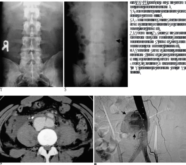
tremity motor weakness. Plain radiography revealed no definite bony abnormality but magnetic resonance imaging (MRI) demonstrated herniation of the nucleus pulposus through an annular defect at the L3-4 and L5- S1 levels. Lumbar myelography indicated the presence of an extradural defect and edematous change in the right L4 nerve root, the sleeve of which was amputated by a herniated disc at the L5-S1 level. A decompressive procedure in which focal discectomy was performed via a right-sided posterior lumbar approach was undertak- en, and after surgery the patient complained of diffuse abdominal pain. Although plain radiographs of the ab- domen depicked no ileus, bilateral psoas lines were obliterated (Fig. 1A, B). Laboratory tests showed that he- moglobin concentration and the hematocrit level had fallen to 5.3 mmol/L and 0.25, respectively, and con- trast-enhanced computed tomography (CT) of the ab- domen and pelvis, performed because vascular injury was suspected, revealed that a pseudoaneurysm abutted the infrarenal abdominal aorta and that retroperitoneal hemorrhage had occurred (Fig. 1C). Angiography of the abdominal aorta demonstrated a 3×7 cm-sized aneurys- mal sac to the right of the infrarenal abdominal aorta (Fig. 1D). The patient underwent emergency surgery during which a 3×7 cm-sized pseudoaneurysm was successfully resected by means of a prosthetic aortic
J Korean Radiol Soc 2003;48:91-93
─ 91 ─
Abdominal Aortic Pseudoaneurysm Caused by Lumbar Discectomy: Case Report1
Dong Hun Kim, M.D., Sang Won Yoon, M.D.2, Kyung Seuk Kim, M.D.3, Woo Hyuk Kim, M.D.3
Vascular injuries which occur during lumbar disk surgery, although rare, can give rise to potentially fatal complications which may be overlooked due to a broad range of clinical manifestations, and which surgeons and radiologists should be aware of. We report a recently encountered case of pseudoaneurysm of the abdominal aorta after lumbar disc surgery, and review the associated literature.
Index words : Lumbar, disc
Vascular complication Aorta, pseudoaneurysm
1Department of Radiology, Armed Forces Kwang Ju Hospital
2Department of Neurologic Surgery, Armed Forces Kwang Ju Hospital
3Department of General Surgery, Armed Forces Kwang Ju Hospital Received May 8, 2002 ; Accepted August 21, 2002
Address reprint requests to : Dong Hun Kim, M.D., Department of Radiology, Armed Forces Kwang Ju Hospital, 66 Hwajeong-dong, Seo-gu, Kwangju 502-240, Korea.
Tel. 82-62-390-5129 Fax. 82-62-381-3946
graft. After the procedure, the abdominal pain disap- peared.
Discussion
Vascular injuries are associated with vessel mobiliza- tion and retraction or laceration during spinal surgery which necessitates direct repair (1-2). The anatomical lo- cation at which spinal procedures are performed may al- so relate to the type of vascular injury involved. The most common of these is tearing of the left common iliac artery, which lies immediately anterior to the fourth lum- bar intervertebral disc and is thus particularly vulnerable.
Although the actual incidences of arteriovenous fistula, pseudoaneurysm and hemorrhage have not been pub- lished, a few isolated case reports have appeared (3-4).
Pseudoaneurysm is the least common sequela of vas- cular injury. Aortic pseudoaneurysm can manifest with- in the first few days or weeks of surgery, but may also remain clinically obscure for years. Occasionally, ab- dominal examination reveals a pulsating mass (1). The detailed cause of this is not clear, but a possible mecha- nism is that the outer layer of the aorta is injured prior to herniation of the aortic intima.
Predisposing factors which increase the risk of vascu- lar injury include degeneration or a defect of the fibrous annulus or anterior longitudinal ligament, persistent ad- hesion of the prevertebral structures to the anterior lon- gitudinal ligament, and difficult operative conditions such as reoperations, aggressive exploration, and com- plex patient positioning (5). Vascular tortuosity of the aorta and iliac arteries can increase significantly with
Dong Hun Kim, et al: Abdominal Aortic Pseudoaneurysm Caused by Lumbar Discectomy
─ 92 ─
A B
Fig. 1. A 22-year-old man with abdomi- nal aortic pseudoaneurysm.
A. Preoperative plain radiograph repre- sents no abnormality.
B. Postoperative plain radiograph shows definite obliteration of bilateral psoas lines (arrows).
C. Abdominal CT scan with contrast enhancement demonstrates saccular pseudoaneurysm (arrows) and retro- peritoneal hemorrhage (asterisks).
D. Aortogram shows typical saccular aneurysm (arrows) in the right lateral wall of infrarenal abdominal aorta.
There is inferior mesenteric artery (IMA) adjacent to aneurysm but IMA is intact.
C D
age, and may affect the degree of retraction needed to expose the spinal column.
Arterial complications are more likely to cause hemo- dynamic instability, which is evidenced initially by tachycardia and subsequently by hypotension. The clin- ical course may vary and can be divided into acute and chronic stages (2, 6-7). Acute symptoms or signs result from excessive blood loss secondary to intra-abdominal or retroperitoneal bleeding, with or without shock.
Sudden onset, such as that occurring in our case, is most often attributed to isolated laceration of a great artery, which carries a mortality rate as high as 80% (8).
The sudden onset of pain or hemodynamic instability should prompt evaluation, which should include the as- sessment of vital signs and hematocrit levels, electrocar- diography, and CT scanning of the surgically treated re- gion. The most immediate life-threatening cause or causes can thus be determined. In acute cases, angiogra- phy is required, as it is specific and precise and defines vascular topography and location and the size of the le- sion prior to preoperative appraisal. As it provides more detailed information about the relationship of injured vessels to surrouding hematoma, critical organs, and bones, CT is the preferred method for investigating complications in patients in whom the onset of symp- toms is subacute or chronic (9). In our case, we over- looked the obliteration of bilateral psoas lines, which might have been an important indicator of a retroperi- toneal or intraperitoneal lesion. However, a CT image obtained after the injection of contrast medium had the
exact appearance of an aortic pseudoaneurysm.
In conclusion, although vascular complications of spinal surgery are uncommon, the potential risk must be borne in mind, and meticulous care taken to avoid them. The radiologist should help the surgeon to rapidly and correctly establish the diagnosis and the surgical in- tervention required.
References
1. Fruhwirth J, Koch G, Amann W, Hauser H, Flaschka G. Vascular complications of lumbar disc surgery. Acta Neurochir 1996;138(8):
912-916
2. Szolar DH, Preidler KW, Steiner H, et al. Vascular complications in lumbar disk surgery: report of four cases. Neuroradiology 1996;
38(6):521-525
3. Tanaka M, Nakahara S, Tanizaki M. Aortic pseudoaneurysm in the L3-L4 disc space after lumbar disc surgery. A case report. J Bone Joint Surg Br 1998;80(3):448-451
4. Franzini M, Altana P, Annessi V, Lodini V. Iatrogenic vascular in- juries following lumbar disc surgery. Case report and review of the literature. J Cardiovasc Surg 1987;28:727-730
5. Oskouian RJ Jr, Johnson JP. Vascular complications in anterior thoracolumbar spinal reconstruction. J Neurosurg 2002;96(1):1-5 6. Ohnishi T, Neo M, Matsushita M, Komeda M, Koyama T,
Nakamura T. Delayed aortic rupture caused by an implanted ante- rior spinal device. Case report. J Neurosurg 2001;95(2Suppl):253- 256
7. Jenis LG, Leclair WJ. Late vascular complication with anterior cer- vical discectomy and fusion. Spine 1994;19(11):1291-1293 8. Smith DW, Lawrence BD. Vascular complications of lumbar de-
compression, laminectomy and foraminotomy. A unique case and review of the literature. Spine 1991;16:387-390
9. Novelline RA, Rhea JT, Rao PM, Stuk JL. Helical CT in emergency radiology. Radiology 1999;213(2):321-339
J Korean Radiol Soc 2003;48:91-93
─ 93 ─
대한방사선의학회지 2003;48:91-93
요추간반절제술후에 발생한 복부 가성 대동맥류: 증례 보고1
1국군광주병원 방사선과
2국군광주병원 신경외과
3국군광주병원 일반외과 김동훈・윤상원2・김경석3・김우혁3
요추간반 수술중 발생하는 혈관외상은 드물지만 다양한 임상증상 때문에 간과될 때는 심각한 합병증을 남긴다. 따라 서 외과의사와 방사선과의사는 증상에 따라 요추간반 수술중 발생한 혈관외상을 조기진단하여 치명적인 합병증을 예 방하는데 중점을 두어야한다. 최근 저자들이 경험한 요추간반 수술후 발생한 복부 가성 대동맥류를 문헌고찰과 함께 소개한다.