Introduction
Chronic subdural hematoma (cSDH) is a common neu- rosurgical disorder that often requires surgical interven- tion. cSDH has become an increasingly prevalent neurologi- cal disease encountered by a wide variety of both general and specialized health-care practitioners.10,16) The increas- ing incidence of this disease underscores the need for an improved understanding of both the pathophysiology of cSDH as well as available treatment options. Although the treatment goals for cSDH are well-established, several im-
portant aspects of clinical management remain controver- sial.2) cSDH is widely treated with burr hole craniostomy (BHC) with closed-system drainage.4,8,14,15,21,22) The use of closed-system drainage after BHC is thought to decrease the recurrence rate promoting brain re-expansion.11) The re- currence rate of cSDH is 2.7-37%.1-3,6,8,10,13,15,19,20) Alternative techniques have been proposed to address the problem of recurrence. For example, middle meningeal artery embo- lization has been found efficacious for refractory chronic subdural hematoma.17) An adjunct technique for recurrent cSDH treatment is implantation of an ommaya reservoir permitting repeated punctures and aspiration of subdural fluid.23) The effect of elimination of the hematoma and its capsule through craniotomy and that of evacuation of he- matoma and irrigation through burr hole craniostomy are under evaluation.2,5,9,18-20) Despite these efforts to cure the disease, there isn’t a single catheter made to evacuate the hematoma specifically for cSDH. The most widely used catheter for draining out hematoma is made for external
Effects of Newly Designed Drainage Catheter in Treating Chronic Subdural Hematoma
Bum-Soo Park, MD, Seung-Won Choi, MD, PhD, Hyon-Jo Kwon, MD, Seon-Hwan Kim, MD, Hyeon-Song Koh, MD, Jin-Young Youm, MD and Shi-Hun Song, MD
Department of Neurosurgery, School of Medicine, Chungnam National University, Daejeon, Korea
Objective: Chronic subdural hematoma (cSDH) is a common disorder that is readily surgically treated but has high recur- rence rate. This is a preliminary report to evaluate the effectiveness of a newly designed catheter compared with the con- ventional one in treating cSDH.
Methods: We conducted a retrospective study of 111 patients with unilateral chronic subdural hematoma treated by burr hole craniostomy with closed-system drainage from November 2009 to September 2012. Group A was defined as patients treated with an external ventricular drainage (EVD) catheter and B as patients treated with the new catheter. We mea- sured changes of thickness of hematoma and midline shifting in brain computed tomography (CT), amount of drainage and recurrence rate in both groups.
Results: Group A consisted of 54 and B of 57 cases. The mean duration for total removal of hematoma was 42.6±13.9 hours in group A and 30.3±11.9 hours in group B (p<0.05). The mean amount of drainage counted per six hours cumula- tively differed significantly between groups. The result (p<0.05) showed that the newly designed catheter effectively re- moved the hematoma. The total recurrence rate in group A was 11% and 3.5% in group B.
Conclusion: The study showed that the newly designed catheter effectively removed the hematoma in less time than the con- ventional one. This helps re-expand the brain block CSF from flowing into the subdural space and decrease the recurrence
rate. (Korean J Neurotrauma 2013;9:87-91)
KEY WORDS: Chronic subdural hematoma ㆍRecurrence ㆍCatheter.
Received: July 25, 2013 / Revised: September 21, 2013 Accepted: September 21, 2013
Address for correspondence: Seung-Won Choi, MD, PhD Department of Neurosurgery, School of Medicine, Chungnam National University, 282 Munhwa-ro, Jung-gu, Daejeon 301-721, Korea
Tel: +82-42-280-7361, Fax: +82-42-280-7363 E-mail: [email protected]
Korean J Neurotrauma 2013;9:87-91 http://dx.doi.org/10.13004/kjnt.2013.9.2.87
ventricular drainage (EVD). The EVD catheter we used has eight holes within two centimeters from the tip. It is de- signed to have openings placed just inside the ventricle to remove CSF. However, removing a hematoma is a different matter. Hematoma, even in its chronic form, has a solid por- tion of crust that can influence the outflow obstructing holes and disrupting the treatment process. Therefore, it is less suitable to use EVD catheter in treating cSDH. We thought the catheter for cSDH should have more holes that are more widely spread on the body of the catheter. Based on these demands, we designed a new catheter in treating cSDH. This is a preliminary report to compare the effect of the new catheter we built in treating cSDH with the conven- tional one.
Materials and Methods
The new catheter (CEM tech., Hwaseong, Korea) we de- signed has multiple bores on silicone shaft. It has eighteen openings laterally side hole of 2 mm diameter. The holes are 7 mm apart from each other and distributed within 50 mm from the tip. Although the outer diameter of the new catheter is the same as the conventional one, 4 mm, the in- ner diameter is slightly larger than that of the conventional one (2.3 mm vs. 2.2 mm)(Figure 1). The catheter passed the tension test. The EVD catheter is also made of silicone but has eight holes within 15 mm from the tip. We assessed the effects on cSDH drainage and recurrence rates.
This retrospective study included 111 unilateral cSDH patients who were treated with burr hole craniostomy and closed system drainage in our institution, from November 2009 to September 2012, consecutively. Patients with orga- nized cSDH, bilateral lesion, subacute form hematoma, mixed or septated hematoma were excluded. Burr hole craniostomies were performed at the parietal eminence with a diameter of about 2 cm. A catheters were inserted 8 cm deep from the dura aiming at glabella. No other mea- sures such as irrigation were taken. Catheters were ran-
domly selected when treating cSDH. Fifty four cases were grouped as A treated with the conventional EVD catheter and remaining 57 were grouped as B, which were treated with the newly designed catheter. Brain CT scans were tak- en one day and one week after surgery. The preoperative and postoperative thickness of hematoma and midline shifting were measured and hematoma drained from the catheter was gathered and measured every six hours and the rate of the extraction was analyzed. The rate of improve- ment was calculated by subtracting the initial value by the value after a day or a week and dividing it by the initial value.
When there was no further drainage of hematoma through the catheter, catheter was removed. Recurrence rate was eval- uated at 6 months after discharge.
The data from these studies were statistically analyzed using analysis of covariance, t-test and chi-square through SPSS version 20.0 (SPSS Inc., Chicago, IL). Null hypothe- ses of no difference were rejected if p-value were less than 0.05. Analysis of covariance (ANCOVA) evaluates wheth- er population means of a dependent variable (DV) are equal across levels of a categorical independent variable (IV), while statistically controlling for the effects of other con- tinuous variables that are not of primary interest, known as covariates (CV). Therefore, when performing ANCOVA, we are adjusting the DV means to what they would be if all groups were equal on the CV.
Results
We retrospectively analyzed clinical and imaging data of 111 patients who were randomly assigned to one of two treatments. The mean age was 65.08 (range 6-93). There were 83 (74.7%) men with 28 (25.2%) women, and left con- vexity hematoma outnumbered the right side by 62 to 49.
Variables such as age, sex, initial hematoma thickness and initial midline shifting in both groups were similar (Table 1). The mean amount of total drainage postoperatively in group A was 212.2 mL and in group B 174.2 mL. The total time consumed to evacuate hematoma and to remove the catheter was longer in group A 42.6±13.9 (range, 24-80) hours versus 30.3±11.9 (range, 24-60) hours in group B (p
<0.05).
The mean preoperative maximum thickness of hemato- ma according to the brain CT taken was 20.59±6.37 mm in group A and 20.83±4.87 mm in group B, without sig- nificant difference. The thickness a day and a week after the operation were 9.57±3.91 mm and 9.07±4.69 mm in group A and in group B 10±4.10 mm and 8.16±3.70 mm.
The improvement of the thickness over a day and a week FIGURE 1. The newly designed catheter with 18 holes within 5
centimeters from the tip (A), and external ventricular drainage (EVD) catheter used to treat chronic subdural hematoma con- ventionally (B).
B A
after surgery was 53.3±15.17%, 56.13±19.79% in group A, and 52.2±15.42%, 59.78±18.14% in group B (Figure 2). The difference between two groups was not statistically signifi- cant.
The average midline shifting checked initially was 10.74
±4.49 mm in group A and 9.71±5.18 mm in group B, with- out significant difference. The shifting a day and a week after the operation were 4.37±3.21 mm and 3.09±3.11 mm in group A and in group B 3.74±3.48 mm and 2.42±2.56 mm. The improvement of the shift over a day and a week after surgery was 60.8±26.51%, 72.66±26.32% in group A
and 67.4±28.60%, 78.65±23.74% in group B (Figure 3). The difference between two groups was not statistically signifi- cant.
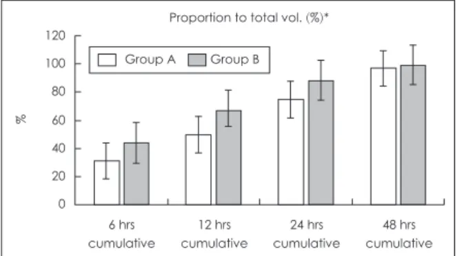
The mean cumulative amount of drainage for 6, 12 and 24 hours were 55.06±27.6 mL, 92.01±37.29 mL, 147.3±
63.21 mL in group A, and 83.9±37.8 mL, 106.35±51.69 mL, 148.95±51.42 mL in group B, respectively. In addition, the ratio of accumulative amount of hematoma checked in 6, 12, 24 hours to total drainage volume were 31.1±19.17%, 49.8±20.76%, 74.6±18.51% in group A and 44.2±24.10%, 67.2%±13.65 and 88.5±2.97% in group B respectively (Fig- Table 1. Summary of the patients characteristics between two groups
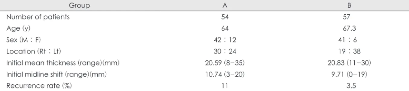
Group A B
Number of patients 54 570.
Age (y) 64 67.3
Sex (M : F) 42 : 12 41 : 60
Location (Rt : Lt) 30 : 24 19 : 38
Initial mean thickness (range)(mm) 20.59 (8-35) 20.83 (11-30)
Initial midline shift (range)(mm) 10.74 (3-20) 09.71 (0-19)0
Recurrence rate (%) 11 03.5
Group A: treatment with conventional EVD catheter, group B: treatment with newly designed catheter
FIGURE 2. Improvement rate of maximum thick- ness one day (A) and one week (B) after the sur- gery. There is no difference between groups. *im- provement rate: (1-value of 24 h after surgery/pre- operative value)×100.
90 80 70 60 50 40 30 20 10 0
POD #1 Improvement rate of depth (%)*
p=0.692
%
A
Group A Group B
90 80 70 60 50 40 30 20 10 0
POD #7 Improvement rate of depth (%)*
p=0.312
%
B
Group A Group B
FIGURE 3. Improvement rate of midline shift one day (A) and one week (B) after the surgery. There is no difference between groups. *improvement rate: (1-value of 24 h after surgery/pre-operative value)×100.
120 100 80 60 40 20
0
POD #1 Improvement rate of shift (%)*
p=0.209
%
A
Group A Group B
120 100 80 60 40 20
0
POD #7 Improvement rate of shift (%)*
p=0.210
%
B
Group A Group B
ure 4). We found significant differences in cumulative drain- age and in hematoma to drainage ratio at all time points, with results favoring Group B. The recurrence rate was 11% for group A versus 3.5% in group B, with borderline statistical significance.
Discussion
Chronic subdural hematoma is well known a curable dis- ease although its mechanism of progression and recurrence not well described.12) The spatial proportion of the brain pa- renchyma declines as atrophy proceeds.1,9) Patients with extensive brain atrophy, such as the elderly or alcoholics, are particularly vulnerable to developing SDH, due to the increased baseline stretch of the bridging vessels. Patients maintained on chronic anticoagulation are also at increased risk for SDH.1) Although the mechanism for this increased risk is incompletely understood, it has been suggested that contained, asymptomatic “microbleeds” are common in elderly people and that anticoagulants impair the ability to control these bleeds, allowing progression to symptomatic hemorrhage. cSDH may evolve from a prior traumatic acute SDH, often asymptomatic, through a series of distinct patho- logic processes.6)
Craniostomy and closed system drainage is less aggres- sive, takes less operation time and causes less complications than other methods of treatments.15) Because of its benefi- cial effects, it is widely used to treat cSDH. However, this method also has several disadvantages including brain pa- renchyma injury or neomembrane bleeding. It could also incompletely evacuate the hematoma, causing recollection.3) Catheter related infection and pneumocephalus are also po- tential problems. To address these shortcomings, attempts have been made to reduce the recurrence rate.6,15,19)
To the authors’ knowledge, there is no report concerning the effects of the drainage catheter. Multiple bores with wider
inner diameters and widely spread holes provide a larger drainage area facilitating removal of the hematoma with minimal obstruction. We think it offers significant benefits of rapid drainage of hematoma and early removal of the catheter. Effective evacuation of the hematoma allows the brain to re-expand. The other benefit of new drainage cath- eter is that partial withdrawal of the catheter is unnecessary.
The primary aim for the new catheter was to change the hemodynamics when draining out the hematoma. It was thought to promote the brain to re-expand better than the conventional one which would fill the vacant space and eventually decrease the recurrence rate. Although our re- sults showed difference in the amount of hematoma drained between the groups, recovery of midline shifting did not.
There are several reports published on the relationship be- tween postoperative drainage and brain re-expansion.11) Fukuhara et al.,7) reported the relationship between brain surface elastance and brain re-expansion after evacuation of chronic subdural hematoma.11) Patients with an enlarged subdural space or with high age have higher elastance which delays brain re-expansion. Fibrous capsule, cerebrovascular dysfunction and degenerative brain atrophy could also affect brain re-expansion.
The recurrence rate was lower when treated with the new catheter. Although the difference was not statistically sig- nificant, the new catheter took a significantly shorter time to remove a hematoma. The amount of hematoma drained, cumulatively measured every six hours and calculated as a ratio of the amount of hematoma extracted was greater in group B. Therefore, further studies with larger sample size and a longer duration could give statistically significant re- sults on brain re-expansion and the recurrence rate.
Conclusion
This study was conducted to verify the effect of the new catheter we have designed. The basic concept is that more holes that are more widely separated in the catheter to drain out cSDH, facilitate more rapid removal of the hematoma, promote brain re-expansion and eventually lower the recur- rence rate. Although the recurrence rate showed no statis- tically significant difference between the groups, it would be worthwhile to extend the research for a better under- standing of the effect of the newly designed catheter.
■ The authors have no financial conflicts of interest.
REFERENCES
1) Abouzari M, Rashidi A, Rezaii J, Esfandiari K, Asadollahi M, Ale- ali H, et al. The role of postoperative patient posture in the recur-
120 100 80 60 40 20 0
6 hrs cumulative
12 hrs cumulative
24 hrs cumulative
48 hrs cumulative Proportion to total vol. (%)*
%
Group A Group B
FIGURE 4. Mean cumulative amount of drainage in 6 hours (p<
0.05), 12 hours (p<0.05) and 24 hours (p<0.05) proportion to to- tal amount after surgery, significantly larger in group B. *propor- tion to total vol.: cumulative vol./total vol.×100.
rence of traumatic chronic subdural hematoma after burr-hole surgery. Neurosurgery 61:794-797; discussion 797, 2007 2) Ali M, Khan Z, Sharafat S, Khan KM. Craniotomy for encapsu-
lated chronic subdural haematoma. Pan Arab J Neurosurg 15:
12-14, 2011
3) Benzel EC, Bridges RM Jr, Hadden TA, Orrison WW. The single burr hole technique for the evacuation of non-acute subdural he- matomas. J Trauma 36:190-194, 1994
4) Camel M, Grubb RL Jr. Treatment of chronic subdural hematoma by twist-drill craniotomy with continuous catheter drainage. J Neurosurg 65:183-187, 1986
5) Cameron MM. Chronic subdural haematoma: a review of 114 cas- es. J Neurol Neurosurg Psychiatry 41:834-839, 1978
6) Ducruet AF, Grobelny BT, Zacharia BE, Hickman ZL, DeRosa PL, Anderson K, et al. The surgical management of chronic subdural hematoma. Neurosurg Rev 35:155-169; discussion 169, 2012 7) Fukuhara T, Gotoh M, Asari S, Ohmoto T, Akioka T. The relation-
ship between brain surface elastance and brain reexpansion after evacuation of chronic subdural hematoma. Surg Neurol 45:570- 574, 1996
8) Hamilton MG, Frizzell JB, Tranmer BI. Chronic subdural hema- toma: the role for craniotomy reevaluated. Neurosurgery 33:67-72, 9) Khalid Khanzada MA. Management of chronic subdural haema-1993
toma. JPMI 18:651-657, 2011
10) Kim JH, Kang DS, Kim JH, Kong MH, Song KY. Chronic sub- dural hematoma treated by small or large craniotomy with mem- branectomy as the initial treatment. J Korean Neurosurg Soc 50:
103-108, 2011
11) Kim JY, Hyun DK, Yoon SH, Park HS, Kim E, Park HC, et al. Pre- diction of postoperative drainage volume and brain expansion of chronic subdural hematoma: supplementary study-clinical study.
J Korean Neurotraumatol Soc 6:33-37, 2010
12) Markwalder TM. Chronic subdural hematomas: a review. J Neu- rosurg 54:637-645, 1981
13) Markwalder TM. The course of chronic subdural hematomas af-
ter burr-hole craniostomy with and without closed-system drain- age. Neurosurg Clin N Am 11:541-546, 2000
14) Markwalder TM, Steinsiepe KF, Rohner M, Reichenbach W, Markwalder H. The course of chronic subdural hematomas after burr-hole craniostomy and closed-system drainage. J Neurosurg 55:390-396, 1981
15) Miele VJ, Sadrolhefazi A, Bailes JE. Influence of head position on the effectiveness of twist drill craniostomy for chronic subdural hematoma. Surg Neurol 63:420-423; discussion 423, 2005 16) Mindiola N P-GM, Pérez-García I, Pérez-Guerra M. Chronics sub-
dural hematoma. J Med Archives 19:4-8, 2003
17) Mino M, Nishimura S, Hori E, Kohama M, Yonezawa S, Midori- kawa H, et al. Efficacy of middle meningeal artery embolization in the treatment of refractory chronic subdural hematoma. Surg Neurol Int 1:78, 2010
18) Mohamed EE. Chronic subdural haematoma treated by cranioto- my, durectomy, outer membranectomy and subgaleal suction drain- age. Personal experience in 39 patients. Br J Neurosurg 17:244- 247, 2003
19) Nakajima H, Yasui T, Nishikawa M, Kishi H, Kan M. The role of postoperative patient posture in the recurrence of chronic subdural hematoma: a prospective randomized trial. Surg Neurol 58:385- 387; discussion 387, 2002
20) Okada Y, Akai T, Okamoto K, Iida T, Takata H, Iizuka H. A com- parative study of the treatment of chronic subdural hematoma-- burr hole drainage versus burr hole irrigation. Surg Neurol 57:405- 409; discussion 410, 2002
21) Ram Z, Hadani M, Sahar A, Spiegelmann R. Continuous irriga- tion-drainage of the subdural space for the treatment of chronic subdural haematoma. A prospective clinical trial. Acta Neurochir (Wien) 120:40-43, 1993
22) Robinson RG. Chronic subdural hematoma: surgical management in 133 patients. J Neurosurg 61:263-268, 1984
23) Sato M, Iwatsuki K, Akiyama C, Kumura E, Yoshimine T. Implan- tation of a reservoir for refractory chronic subdural hematoma.
Neurosurgery 48:1297-1301, 2001