색소융모결절성 활막염(Pigmented villonodular synovitis, PVNS)은 원인이 확실하지 않은, 관절이나 건초의 활막이 융 모성 혹은 소결절성 과성장을 특징적으로 보이는 활막 병변으 로 주로 무릎이나 고관절 등을 침범한다. 축관절인 척추에 발 생한 예는 1980년 Kleinman등 (1)이 최초로 보고한 이래로 병리조직학적으로 지금까지 세계적으로 약 30예가 보고되었다 (2-7). 저자들은 요추 관절돌기관절에서 발생한 PVNS 1예를 경험하였기에 이를 보고한다.
증례 보고
44세 남자환자가 1년전부터의 하부 요통과 좌측 다리의 방 사통을 주소로 내원하였다. 환자는 7년전 교통사고에 의한 제 2 요추 압박골절로 인공 삽입물을 이용한 후방유합술을 시행 받았다. 이학적 검사상 하지직거상이 우측 70°, 좌측 60°로 감 소되어 있었으며 제 4-5번 요추이하의 운동 신경이 감소되어 있었다.
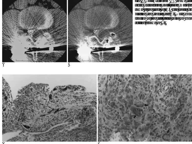
척수강조영술상 제 4-5번 요추부위에서 척수강(thecal sac) 을 누르고 있는 좌측 경막외 종괴가 있었다 (Fig. 1). 이후 시 행한 전산화단층촬영(이하‘CT’로 줄임)상 같은 부위에 경계 가 뚜렷하고 비교적 균일한 추간판보다 약간 낮은 저음영을 보 이는 난형의 경막외 연조직 종괴가 보였고 이에 연한 척추관 절돌기에 골미란이 보였다 (Fig. 2A, B).
수술 소견상 7년전에 시술한 인공 삽입물들이 완전히 풀려 그 주위로 액체 저류를 보이는 점액낭염(bursitis)의 소견을 보 이고 있었고 제 4-5번 요추의 좌측 척추 관절돌기에 연해 유 연한 피막에 둘러싸인 어두운 크림같은 종괴가 검은 색을 띠 고 있었다. 종괴는 육안적으로 완전 절제가 이루어졌으며, 수 술후 환자의 임상적 증상은 많이 호전되었고 1년 후 추적까지
증상의 재발은 보이지 않았다.
현미경 소견상 균열이나 가선양 공간(pseudoglandular space)에 의해 나뉘어진 판상의 활액막 증식이 보였고, 여러 개의 핵을 가진 거대 세포와 만성 염증 세포등 여러 가지 세 포들로 이루어져 있었으나 주된 세포는 둥글거나 다각형 모양 으로 세포질은 혈철소를 포함하여 짙은 갈색을 띠고 있어 PVNS로 확진되었다 (Fig. 3A, B).
대한방사선의학회지 2000;43:505-508
─ 505 ─
척추의 색소융모결절성활막염: 1예 보고1
성 수 옥・이 주 혁・이 정 근
색소융모결절성 활막염(PVNS)은 관절이나 건초의 활막이 융모성 혹은 소결절성 과성장을 하는 특징적인 활막 병변으로 흔히 사지관절의 윤활관절에서 발생하며 아주 드물게 축관절인 척추를 침범한다. 척추 PVNS의 예는 국내에서 아직 보고된 바 없다. 이에 저자들은 요추 관절 돌기관절에서 발생하여 경막외 종양처럼 보였던 척추의 PVNS 1예를 경험하였기에 방사선 소 견을 보고한다.
1청주성모병원 진단방사선과
이 논문은 2000년 2월 29일 접수하여 2000년 8월 25일에 채택되었음.
Fig. 1. Lumbar myelogram in oblique view shows the left- sided extradural mass(asterisk) at the level of the fourth and fifth lumbar disc space.
고 찰
PVNS는 주로 사지의 큰 관절, 특히 무릎(80%)과 고관절 (15%)에서 발생하고, 종종 손이나 발의 작은 관절을 침범할 수 있으며 건초의 활막이 관절외 PVNS의 발생 부위가 된다 (2, 8, 9). PVNS의 원인은 아직 확실하지 않아 염증성 반응, 신생물, 대사성 장애 그리고 외상에 의한 반복적인 출혈등을 원인으로 생각하고 있으며, 병리조직학적으로는 과도한 활액 세포의 증식, 대식세포 침윤, 다핵 거대세포와 혈철소(hemo- siderin) 침착이 특징이다 (10-12). 성별에 따른 발생빈도의 차이는 없으며, 흔히 젊은 연령에서 발생하여 전형적으로 20 대에 발현한다 (4, 13).
또한 PVNS는 아주 드물게 척추에서 발생하는데 경추부와 요추부가 흔히 침범되나 (41%) 모든 척추 부위에서 발생할 수 있다 (2). 척추의 PVNS는 척추궁의 가동관절(diarthrodial joint)을 둘러싸는 활액막을 침범하므로 척추관절돌기관절에서
발생한다 (2-7). Giannini등의 보고 (2)에 의하면 10예중 9예 가 어느 정도 척추 관절돌기관절을 침범하였고 1/3에서는 관 절돌기관절에 중심을 두고 있었다. 저자들의 예 역시 관절돌기 에 골미란을 보이고 있었다.
척추 PVNS의 병리학적 변화는 부속관절에서의 변화와 같 다 (9). 관절강내에서 활액막이 적갈색 비후와 융모성 돌출을 보이다가 병변이 진행함에 따라 관절 밖으로 연장되어 주위 뼈 에 골파괴성 병변을 형성하므로, 대부분 척추의 후방 성분을 침범하는 파괴적인 팽창성 골병변으로 나타나고 연조직 성분 을 동반할 수 있다 (2). Giannini등의 보고 (2)에서 9예의 파 괴성 골병변중 8예가 연조직 성분이 동반되어 있었고 그 중 5 예에서 척수강을 누르는 경막외 종괴를 보여, 4예에서 척수가 유의하게 밀리거나 변형을 보였고 1예에서는 골병변없이 추간 판의 후방에서 기시한 연조직 종괴처럼 보여 추간판 탈출과 비 슷하였다 (2). 저자들의 예에서도 관절돌기에 골미란을 동반한 경막외 종괴로 보였다.
척추의 PVNS는 융모성 표면에 국한된 활막증식, 관절낭비 성수옥 외 : 척추의 색소융모결절성활막염
Fig. 2. Post-myelogram CT shows left- sided extradural soft-tissue mass(ar- rows) at the level of L4-5 disc space dis- placing the thecal sac(A). Note bony erosion of the left facet joint on bone setting(arrows on B).
A B
A B
Fig. 3. Histology of PVNS
A. There is sheet-like synovial overgrowth pattern interrupted by pseudoglandular spaces(arrows). (H&E stain, ×100)
B. The predominant cells which show round or polygonal are ladened with hemosiderin. Note the multinucleated giant cells(ar- rows). (H&E stain, ×400)
─ 506 ─
후와 출혈이 특징인 퇴행성 변화인 과성장 활막염(hypertrophic synovitis)과 임상적으로 구별되어야 한다. 과성장 활막염은 흔 히 추간판 탈출증을 동반하며 척추 관절돌기에 비정상적인 스 트레스가 가해질 때 발생할 수 있다. 이는 또한 퇴행성 변화를 보이는 관절돌기관절에 연해 낭성 종괴를 보이는 활액낭(syn- ovial cyst)과도 감별해야 하는데 방사선학적 소견만으로는 구 분하기가 그리 용이치 않다. 척추 PVNS는 주위 뼈에 골파괴 성 병변을 일으키므로 척추 후방부를 침범하는 골종양인 골아 세포종(osteoblastoma)나 동맥류성골낭(aneurysmal bone cyst)와도 감별이 용이치 않을 수도 있다. 흔히 경막외 공간으 로 확장되어 척수를 왜곡시키거나 압박하므로 경막외 종괴(양 성; 신경초종, 드물게 경막외 수막종, 악성; 전이암)의 감별진 단에 추가될 수 있다 (2).
PVNS는 국소적으로 재발하는 경향을 보인다 (10). 병변의 완전한 수술적 제거만이 유일한 완치법이다. 따라서 처음 수술 시 종괴를 완벽히 제거하기 위해 모든 노력을 경주해야 한다.
척추 병변에서 완전절제가 불가능하였던 경우 조기 재발이 보 고되었다 (12). 방사선 치료의 역할은 아직 명확하지 않으며 방사선에 의한 이차적 육종, 관절 강직이나 상처 치유의 지연 등의 문제점이 있어 아전절제후나 국소 재발에 대해 보조적인 치료법으로 사용될 수 있다 (14).
참 고 문 헌
1. Kleinman GM, Dagi TF, Poletti CE. Villonodular synovitis in the spinal canal: case report. J Neurosurg 1980;52:846-848
2. Giannini C, Scheithauer BW, Wenger DE, Unni KK. Pigmented villonodular synovitis of the spine: a clinical, radiological, and mor- phological study of 12 cases. J Neurosurg 1996;84:592-597 3. Gezen F, Akay KM, Aksu AY, Bedu¨k A, Seber N. Spinal pigment-
ed villonodular synovitis: a case report. Spine 1996;21:642-645 4. Clark LJ, McCormick PW, Domenico DR, Savory L. Pigmented
villonodular synovitis of the spine: case report. J Neurosurg 1993;
79:456-459
5. Karnezis TA, McMillan RD, Ciric I. Pigmented villonodular syn- ovitis in a vertebra: a case report. J Bone Joint Surg(Am) 1990;72:
927-930
6. Campbell AJ, Wells IP. Pigmented villonodular synovitis of a lum- bar vertebral facet joint: a case report. J Bone Joint Surg(Am) 1982;
64:145-146
7. Bui-Mansfield LT, Youngberg RA, Coughlin W, Chooljian D. MRI of giant cell tumor of the tendon sheath in the cervical spine. J Comput Assist Tomogr 1996;20:113-115
8. Fechner RE, Mills SE. Atlas of tumor pathology. third series, fascicle 8: Tumors of the bones and joints. Washington DC: Armed Forces Institute of Pathology. 1993;282-287
9. Dorwart RH, Genant HK, Johnston WH, Morris JM. Pigmented villonodular synovitis of synovial joints: Clinical, pathologic, and radiographic features. AJR Am J Roentgenol 1984;143:877-885 10. Granowitz SP, D’Antonio J, Mankin HL. The pathogenesis and
long-term end results of pigmented villonodular synovitis. Clin Orthop 1976;114:335-351
11. Jaffe HL, Lichtenstein L, Sutro CJ. Pigmented villonodular synovi- tis, bursitis and tenosynovitis. A discussion of the synovial and bursal equivalents of the tenosynovial lesion commonly denoted as xanthoma, xanthogranuloma, giant cell tumor or myeloplaxoma of the tendon sheath with some consideration of this tendon sheath lesion itself. Arch Pathol 1941;31:731-765
12. Weidner N, Challa VR, Bonsib SM, Davis CH, Carroll TJ. Giant cell tumors of synovium(pigmented villonodular synovitis) involv- ing the vertebral column. Cancer 1986;57:2030-2036
13. Goldman AB, DiCarlo EF. Pigmented villonodular synovitis.
Diagnosis and differential diagnosis. Radiol Clin North Am 1988;26:
1327-1347
14. Bentley G, McAuliffe T. Pigmented villonodular synovitis. Ann Rheum Dis 1990;49:210-211
대한방사선의학회지 2000;43:505-508
─ 507 ─
성수옥 외 : 척추의 색소융모결절성활막염
─ 508 ─
J Korean Radiol Soc 2000;43:505-508
Address reprint requests to : Su Ok Seong, M.D., Department of Radiology, Cheongju St. Mary’s Hospital 589-5, Jujung-dong, Sangdang-gu, Cheongju, Choongchungbuk-do 360-568, Korea.
Tel. 82-43-212-5000 Fax. 82-43-212-1345 E-mail: [email protected]
Pigmented Villonodular Synovitis of the Spine: A Case Report1
Su Ok Seong, M.D., Joo-Hyuk Lee, M.D., Jeong Geun Yi, M.D.
1Department of Radiology, Cheongju St. Mary’s Hospital
Pigmented villonodular synovitis(PVNS) is a synovial lesion of joints or tendon sheaths, characterized by vil- lous and nodular overgrowth of the synovial membrane. It commonly occurs in synovial joints of the appen- dicular skeleton, particularly those of the knee and hip, but rarely affecting those of the spine. We report a case of PVNS of the lumbar spine mimicking epidural mass.
Index words :Spine, CT Spine, disease Spine, facet joints Synovitis