서론
기정맥계는 흉곽후방에 한 쌍으로 이루어지는 정맥으로 기 정맥, 반기정맥, 부반기정맥으로 이루어져 있다. 기정맥계는 복 부의 정맥 등과 연결되어 있으며, 늑간정맥, 종격동 정맥, 식도 정맥, 심막정맥 등이 유입되며, 4번째 흉추 부위에서 기정맥궁 을 형성하면서 상대정맥으로 유입된다. 기시부위, 경로, 유입되 는 정맥, 그리고 유입되는 지점에 기정맥계는 상당한 변이가 있 다(1). 기정맥계는 불완전한 판막을 가지고 있는데 판막의 위 치와 모양이 다양하며(1) 기정맥궁에 있는 판막은 CT에서 보 일 수 있다(2). 또한 발생과정에서 기정맥계, 상대정맥, 하대정 맥들이 각기 다른데서 기원하면서, 기정맥의 선천성결손, 선천 성 하대정맥의 결손과 기정맥 연장, 기정맥류와 같은 기형이 생길 수 있다(3-5). 기정맥계는 직간접적으로 상대정맥과 하 대정맥을 연결하고 있어 상대정맥이나 하대정맥의 폐색이 생기 거나 압력이 증가하면 측부순환혈관의 역할을 하면서 기정맥 과 반기정맥이 늘어난다(3).
3차원 영상과 재구성한 CT 영상들은 기정맥계의 해부학적 위치와 크기의 변화, 주변 혈관들의 변화, 동반된 혈관이나 장 기들의 기형 및 질환 등을 볼 수 있어, 기정맥계의 선천성 혹은 후천성 이상을 진단하는 데 유용하다(3, 4).
그래서 저자들은 기정맥계에 생길 수 있는 질환들을 분류하 고 CT 소견에 대해 알아보고자 한다.
본문
정상 및 정상변이
기정맥궁 판막(Azygos Arch Valve)
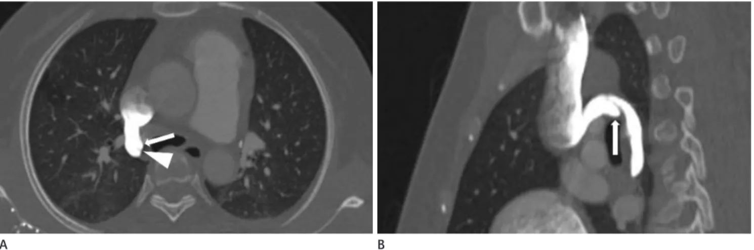
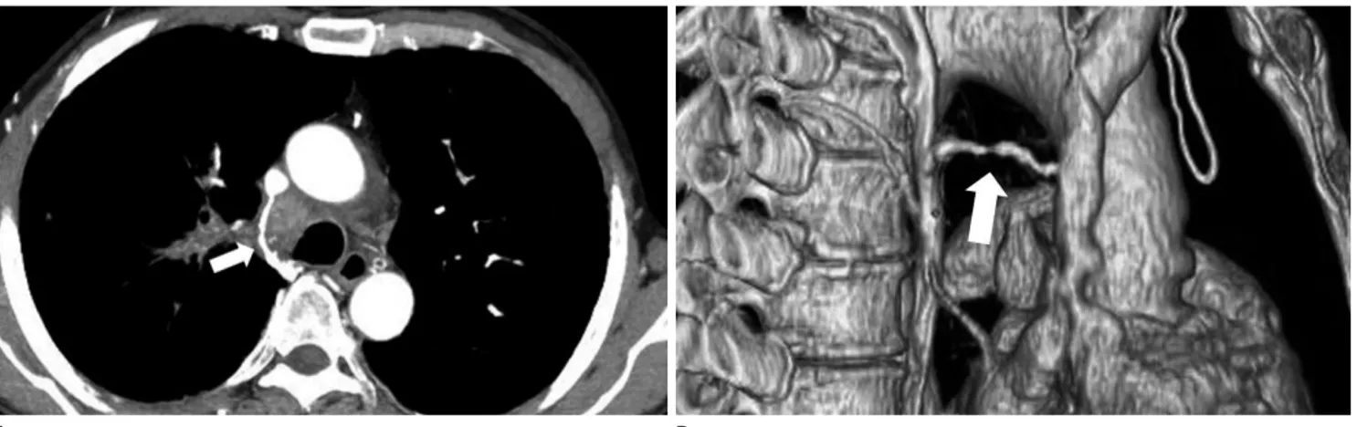
기정맥과 반기정맥은 흉강에서 판막이 있는 큰 정맥으로, 판 막은 위치에 따라 빈도가 다양하며, 상대정맥과 기정맥 사이에 는 판막이 항상 존재한다. Falla 등(1)은 기정맥 내에 물을 주 입하였을 때 기정맥 내 판막의 모양과 기능이 불완전함을 확인 하였다. 판막은 이첨판(bicuspid) 형태를 보이거나 한쪽 혹은 양측 벽에서 내피(intima)의 single fold로 보인다. CT에서 기 정맥궁으로 조영제의 역류가 있었던 154명 중 105명, 즉 68.2%에서 판막이 보였다(2). 빠른 속도로 조영제를 주입하 거나 우측 팔에서 조영제를 주입할 때 기정맥궁 판막이 잘 보 인다고 한다. 기정맥 판막은 기정맥궁의 중간부위에 있으며 판 막이 이첨판 모양으로 직접 보이기도 하지만(Fig. 1) 대부분이 판막이 있는 부위에 국소적 팽창으로 보이거나 조영제가 판막 에 남아있는 모양으로 보인다(Fig. 2). 이러한 판막들은 기능
J Korean Soc Radiol 2012;67(1):37-44
Received April 3, 2012; Accepted May 2, 2012 Corresponding author: Young Tong Kim, MD Department of Radiology, Soonchunhyang University College of Medicine, Cheonan Hospital,
31 Suncheonhyang 6-gil, Dongnam-gu, Cheonan 330-721, Korea.
Tel. 82-41-570-3513 Fax. 82-41-579-9026 E-mail: [email protected]
This work was supported in part by the Soonchunhyang University Research Fund.
Copyrights © 2012 The Korean Society of Radiology
Since three dimensional and reformatted CT images can show changes in the loca- tion and size of the azygos system, changes in the surrounding vessels, and com- bined anomalies of vessels or organs, CT is useful for diagnosing congenital and ac- quired abnormalities of the azygos system. In this article, we review CT findings in regards to various disorders involving the azygos system.
Index terms
Azygos Vein CTCongenital Anomaly Abnormalities
Azygos System on Multi-Detector Computed Tomography: Pictorial Essay
다중검출전산화단층촬영을 이용한 기정맥계 질환의 진단
Jeong Ah Hwang, MD, Young Tong Kim, MD, Sung Shick Jou, MD, Woong Hee Lee, MD
Department of Radiology, Soonchunhyang University College of Medicine, Cheonan Hospital, Cheonan, Korea
이나 형태가 불완전하기 때문에 임상적으로 의미는 없다.
기정맥 열(Azygos Fissure)
기정맥 열은 우측 후방 전주정맥의 불완전한 내측이동(in- complete medial migration)으로 인해 생기며, 정상인의 0.4~1%
에서 발생한다. 기정맥 열에 있는 기정맥은 정상적인 기정맥보 다 상부, 외측에서 상대정맥 혹은 우측 완두정맥(brachioce- phalic vein)으로 유입된다. 기정맥 열에 의해 엽이 형성되는데 이렇게 형성된 엽은 우상엽의 첨부, 후부로부터 기관지와 동맥 공급을 받기 때문에 독립적인 완전한 엽을 형성하지는 않는다.
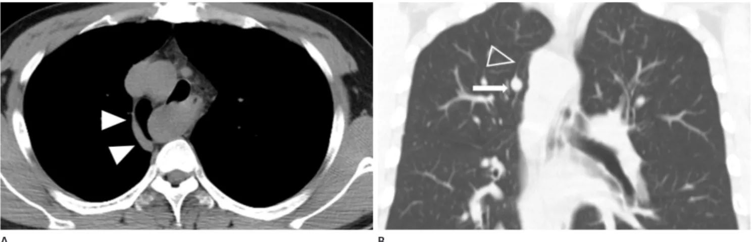
CT에서 우측 폐첨부와 연결되는 가늘고 볼록한 선으로 보이 며, 폐첨부에서는 삼각형 모양으로 보이고, 하부에 기정맥에 의해 눈물방울 모양으로 보인다(6)(Fig. 3). 기정맥열에 기흉 이 동반되기도 하며, 드물게 우측 대동맥과 동반되어 나타나면 Fig. 1. Azygos arch valve in a 48-year-old woman. Axial CT scan
shows reflux of contrast material from superior vena cava into the azygos arch. Bicuspid-shaped azygos valve (arrowhead) is at the mid- portion of azygos arch.
Fig. 2. Azygos arch valve in a 40-year-old woman.
A. Axial CT scan shows reflux of contrast material from superior vena cava into the azygos arch. Azygos arch valve is demonstrated as focal bulging (arrow) at the medial wall of distal azygos arch, and a leaflet of valve is demonstrated as a line (arrowhead).
B. Reformatted sagittal image shows a focal bulging (arrow) suggesting valve at distal azygos arch.
Fig. 3. Azygos fissure in a 36-year-old man.
A. Reformatted coronal image shows a curvilinear azygos fissure (open arrowhead) and azygos vein (arrow).
B. The 3-dimensional image in a posterior view shows azygos fissure (open arrowheads) and lobe (*).
A
A
B
B
기정맥의 선천성 결손
기정맥의 선천적 결손은 드문 기형으로 기정맥이 없으면 반 기정맥으로 혈류가 증가되어 대동맥궁상부로 늘어난 반기정맥 이 좌측 완두정맥으로 유입되면서 aortic nipple로 보일 수 있 다. CT에서는 정상적인 위치에 기정맥이 보이지 않고 약간 늘 어나 있는 반기정맥이 좌측 완두정맥으로 유입되는 것을 직접 볼 수 있다(3, 4)(Fig. 5). Falla 등(1)이 언급했던 한 개의 정 맥으로 이루어진 3형은 한 쌍이 아닌 하나로 이루어져 있다는 점에 같지만 척추전방에 한 개의 기정맥이 있으면서 상대정맥 으로 유입되기 때문에 기정맥의 결손과는 또 다른 형태이다.
선천성 하대정맥의 기형을 동반한 기정맥 연장(Azygos Continuation with Inferior Vena Cava Anomalies)
하대정맥의 기형에는 하대정맥의 전체 혹은 부분 결손, 그리 기정맥 열의 모양이 변화하기도 한다(Fig. 4).
선천성 이상
발생과정에서 기정맥은 우측 상주정맥(supracardinal vein) 에서, 기정맥궁은 전주정맥(anterior cardinal vein)에서, 반 기정맥은 좌측 상주정맥에서 기원한다. 상주정맥의 상부에서 기정맥-반기정맥이 형성되고, 중간부위는 하주정맥(subcar- dinal vein)과 합쳐져서 하대정맥을 형성하고, 하부는 요정맥 을 형성한다(3). Falla 등(1)에 따르면 100명의 기정맥계를 크게 3가지로 분류하였는데 1형은 한 쌍의 정맥이 서로 간의 연결이 없이 분리되어 있는 형, 2형은 한쌍의 정맥이 서로 연 결이 있는 형, 3형은 한 개의 정맥으로 이루어져 있는 형으로 나뉘었다. 1형과 3형이 각각 1명씩이었고 나머지 98명은 2형 이었다.
Fig. 4. Azygos fissure with right sided aortic arch in a 32-year-old man.
A. Precontrast axial scan shows laterally displaced azygos vein (arrowheads), separating azygos lobe from right upper lobe.
B. Reformatted coronal image demonstrates azygos fissure (open arrowhead) originating from right-sided aortic arch. Azygos vein (arrow) is at the inferior portion of azygos fissure.
Fig. 5. Absence of azygos vein in a 52-year-old woman.
A. Axial CT scan shows absence of azygos arch on the normal position and slightly dilated left accessory hemiazygos vein (arrow).
B. Reformatted coronal image shows the absent azygos arch on right lower paratracheal area, and slightly dilated left accessory hemiazygos vein (arrow) above aortic arch.
A
A
B
B
막강의 측부순환혈관들의 확장과 기정맥계의 확장이 보이며, 심부정맥혈전증의 원인으로 알려져 있다(8).
원인불명의 기정맥 류(Idiopathic Aneurysm of Azygos Vein) 기정맥궁의 류 형성은 드물며, 대부분이 증상이 없고 우연히 발견된다. 기정맥 류가 형성되는 기전을 정확히 알 수 없지만 기정맥계에 압력이 증가되거나 최근에 외상을 받은 병력이 없 기 때문에 선천성으로 생각하고 있다. 상대정맥과 기정맥계는 다른 부위에서 기원하는데 이들 정맥이 발생하는 과정에서 기 정맥이 기원이 되는 상주정맥과 상대정맥의 기원이 되는 전주 고 중복 하대정맥이 있다. 선천성 하대정맥 부분 결손은 발생
과정에서 하대정맥의 신장전 분절(prerenal segment)의 선행 물(precursor)인 우측 하주정맥이 간정맥과 합류의 실패로 생 겨난다. 신장정맥 이하의 하대정맥의 혈류는 기정맥과 반기정 맥을 통해 심장으로 유입된다(7). 동반되는 기형은 중복 하대 정맥, 좌측 하대정맥, 선천성 심장병, 무비증(asplenia), 다비증 (polysplenia), 복부장기의 위치 이상 등이다(Figs. 6, 7). 반기 정맥은 8번째와 9번째 흉추에서 기정맥으로 유입되거나, 부기 정맥을 통해 좌측 완두정맥 혹은 좌측 상대정맥으로 유입될 수 있다(3, 4). 하대정맥의 전체 결손은 드물며, 복벽과 후복
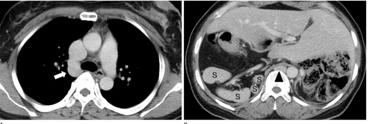
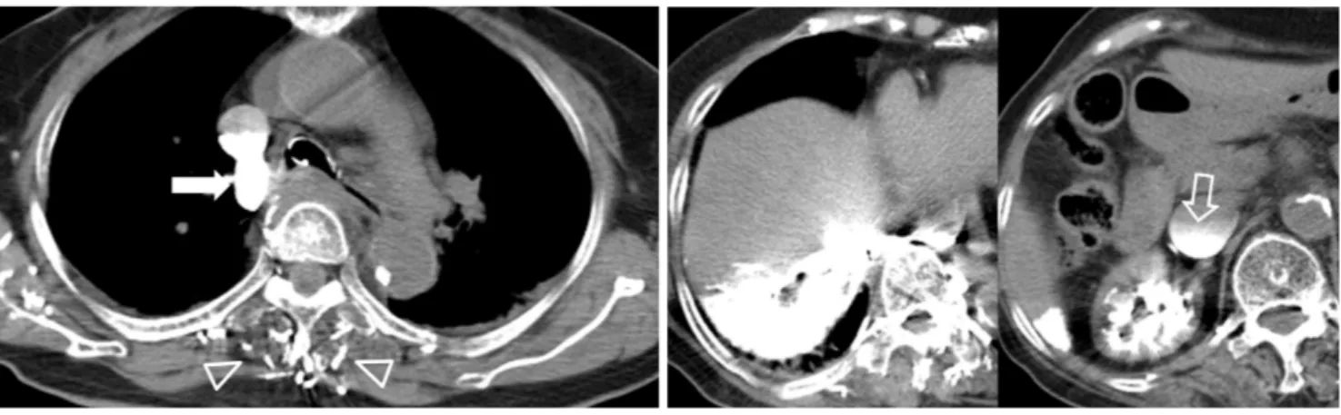
Fig. 6. Azygos continuation with interruption of inferior vena cava in a 44-year-old woman.
A. Axial CT scan at the level of carina shows dilated azygos arch (arrow).
B. Axial CT scan at the level of upper abdomen shows dilated azygos vein at the retrocrural area (arrowhead), polysplenia (S), and transverse liver.
The patient is diagnosed with heterotaxy syndrome with polysplenia and situs ambiguous.
Fig. 7. Azygos continuation with inferior vena cava anomaly in a 14-year-old girl.
A. Axial CT scan at the level of lower thoracic spines shows dilated hemiazygos vein (arrows) and slightly dilated azygos vein (arrowhead).
B. Axial CT scan at the level of kidney shows a dilated tortuous vein (open arrowheads) at the left paraaortic area, and right renal vein with anomalous course (open arrow).
C. The reformatted coronal image shows anomalous dilated tortuous vein (open arrowheads) at left paraaortic area, which drains to dilated hemiazygos vein (arrows). Also, note the anomalous course of right renal vein (open arrow) and slightly dilated collateral veins (arrowheads) in abdominal and pelvic cavities. An anomalous dilated tortuous vein at the left paraaortic area is suggestive of markedly dilated collaterals with absence of inferior vena cava or dysplastic inferior vena cava, with hemiazygos continuation.
Note.-A = aorta A
A
B
B C
측부순환혈관으로서의 기정맥계(Azygos System as Collaterals)
상대정맥 폐색
상대정맥이 폐색이 되면 기정맥계로 혈류가 증가될 수 있는 데, 폐색의 위치에 따라서 혈류의 흐름이 달라진다. 기정맥궁 이 있는 부위의 상대정맥이 폐색이 되면 기정맥계로 혈류가 증 가되고 기정맥궁의 상부에서 상대정맥이 막히면 흉벽의 측부 순환혈관들과 상늑간정맥을 통해 부반기정맥, 반기정맥, 기정 맥으로 혈류가 유입된다. 폐와 종격동의 악성종양이 상대정맥 폐색의 가장 흔한 원인이다(Fig. 9). 악성이 아닌 원인으로는 종격동섬유증, retrosternal goiter, 대동맥류, 상대정맥 혈전 등 이다(3).
하대정맥 폐색
간, 신장과 부신의 악성종양에 의한 직적적인 침범(Fig. 10), 혈전, 압박에 의해 하대정맥이 폐색이 되면 기정맥계가 측부순 환혈관으로 작용한다(3).
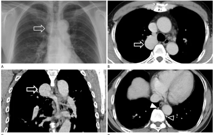
정맥이 만나는 지점이 해부학적으로 약한 부위여서 류를 형성 한다고 하였다(5). 기정맥류는 호흡에 따라 크기가 변하며 기 정맥류 내에 혈전을 동반할 수 있다. 기정맥 류는 흉부사진과 조영전 CT에서 종격동 종괴로 오인할 수 있다. 하지만 조영 후 CT에서 기정맥궁에 조영증강이 잘 되는 종괴로 보여 확진할 수 있다(Fig. 8). 또한 다른 부위의 기정맥계의 확장, 기정맥계 의 혈류나 압력을 증가시킬 수 있는 상대정맥이나 하대정맥 이 상, 간경화 등이 없는 것도 CT로 확인할 수 있다.
후천성 이상
기정맥계의 이상은 선천성 기형보다 후천적 이상이 더 많다.
기정맥은 상행요정맥, 늑골하정맥, 혹은 우측 신정맥이나 하대 정맥의 분지들과 연결되고, 반기정맥은 상행요정맥, 늑골하정 맥, 혹은 좌측 신정맥의 분지들과 연결이 된다(3). 그래서 기정 맥계는 상대정맥이나 하대정맥의 폐색이나 압력이 증가할 때 기정맥계가 측부순환혈관의 역할을 한다. 또한 드물지만 기정 맥 내에 혈전을 형성하기도 한다.
Fig. 8. Azygos vein varix in a 58-year-old man who presented as incidentally detected mediastinal mass.
A. Chest posteroanterior radiograph shows focal bulging (open arrow) of azygos arch.
B, C. Axial (B) and coronal (C) images show an aneurysmal dilation (open arrow) of distal portion of azygos arch.
D. Axial CT scan at the lower T-spines shows non-dilated azygos vein (arrowhead) and hemiazygos vein (open arrowhead). Liver was intact and inferior vena cava thrombus was not found.
C A
D B
으로 들어가지 못하고 정맥으로 역류한다. 그래서 CT 소견은 정맥으로의 진한 조영제 역류를 보인다. 하대정맥, 신정맥, 간 정맥으로 조영제의 역류, 간과 신장의 실질에 진한 조영제 역류 등을 보일 수 있다(9)(Fig. 12). 또한 하대정맥에 조영제에 의한 액체-액체층을 형성할 수 있다. 또한 심장으로 들어가지 못하 는 조영제들이 흉강 내의 측부순환혈관인 기정맥, 종격동, 심 막, 흉벽의 정맥으로 역류하게 된다. 그리고 어느 시기에 심정지 가 일어났느냐에 따라 동맥의 조영제 증강은 다양하게 보일 수 있다. 즉, 대동맥이나 좌측 심장 내에 조영제가 보일 수도 있고 대동맥과 좌측 심장에 조영제가 전혀 보이지 않을 수도 있다.
문맥고혈압(Portal Hypertension)
문맥과 전신정맥계(systemic venous system)와 연결되어 있 기 때문에 문맥의 압력이 증가하면 관상정맥, 식도정맥, 위정 맥으로 혈류가 증가하며 기정맥계를 통해 상대정맥으로 유입 된다(3)(Fig. 11).
급성심정지(Cardiac Arrest)
혈역학적으로 불안정한 환자가 CT를 시행하는 과정에 급성 심정지가 생길 수 있다. 심장이 정지하면 혈액순환이 이루어지 지 못하기 때문에 우심방의 압력이 증가하여 조영제들이 심장
Fig. 9. Tumor invasion of azygos arch in a 49-year-old man. He was confirmed to be suffering from small cell carcinoma of lung. On initial CT scan, superior vena cava was invaded and azygos arch was obliterated by metastatic lymphadenopathy.
A. Axial CT scan after chemotherapy shows luminal irregularity of azygos arch with contrast filling (arrow) surrounded by metastatic lymphade- nopathy.
B. The 3-dimensional image shows wall irregularity of azygos arch with luminal narrowing (arrow).
A B
Fig. 10. Azygos-hemiazygos dilation with tumor invasion of inferior vena cava in a 55-year-old man who was diagnosed with renal cell carcinoma of right kidney. Reformatted coronal image shows inferior vena cava (open arrow) invaded by renal cell carcinoma (M) and dilat- ed hemiazygos vein (open arrowheads).
Fig. 11. Dilated azygos vein with advanced liver cirrhosis in a 57-year- old man. Reformatted coronal image shows tortuous dilated azygos vein (arrowhead) and cirrhotic liver (L).
Fig. 12. Cardiac arrest during CT scanning in an 81-year-old woman.
A. Postcontrast axial scan shows a fluid-fluid level in superior vena cava, reflux of contrast materials in azygos arch (arrow) and veins (open ar- rowheads) in posterior chest wall.
B. Serial axial images show nonvisualized contrast materials in cardiac chambers, a fluid-fluid level in inferior vena cava (open arrow), and retro- grade opacification of hepatic veins and parenchyma of the right hepatic lobe and right kidney. Also, note dense opacification of azygos vein, hemiazygos vein and collateral veins in paraverteral areas.
A B
A B C
Fig. 13. Thrombus in azygos vein caused by central catheterization in a 44-year-old man.
A. Initial axial CT scan shows contrast filled normal azygos arch.
B. Chest radiograph obtained 8 months later shows the central venous catheter (arrow) inserted into azygos arch through left subclavian vein.
C. Axial scan obtained 5 months after central catheterization into azygos arch shows thrombosed and narrowed azygos arch (open arrow).
Fig. 14. Thrombophlebitis of azygos arch in a 56-year-old man. Three weeks ago, the patient had lobectomy of right upper lobe due to primary lung carcinoma.
A, B. Axial (A) and reformatted coronal (B) images show intraluminal thrombus and wall enhancement (arrowheads) of azygos arch. In addition, note irregular shaped low attenuation lesion (arrow) caused by postoperative infection in adjacent lung parenchyma.
A B
genital and acquired abnormalities of the azygos system.
Radiographics 1991;11:233-246
4. Demos TC, Posniak HV, Pierce KL, Olson MC, Muscato M.
Venous anomalies of the thorax. AJR Am J Roentgenol 2004;182:1139-1150
5. Gallego M, Mirapeix RM, Castañer E, Domingo C, Mata JM, Marín A. Idiopathic azygos vein aneurysm: a rare cause of mediastinal mass. Thorax 1999;54:653-655
6. Mata J, Cáceres J, Alegret X, Coscojuela P, De Marcos JA.
Imaging of the azygos lobe: normal anatomy and varia- tions. AJR Am J Roentgenol 1991;156:931-937
7. Bass JE, Redwine MD, Kramer LA, Huynh PT, Harris JH Jr.
Spectrum of congenital anomalies of the inferior vena cava: cross-sectional imaging findings. Radiographics 2000;20:639-652
8. Kondo Y, Koizumi J, Nishibe M, Muto A, Dardik A, Nishibe T.
Deep venous thrombosis caused by congenital absence of the inferior vena cava: report of a case. Surg Today 2009;
39:231-234
9. Singh AK, Gervais D, Mueller P, Shirkhoda A, Sagar P, Mc- carroll K. Cardiac arrest: abdominal CT imaging features.
Abdom Imaging 2004;29:177-179
10. Bankier AA, Mallek R, Wiesmayr MN, Fleischmann D, Kranz A, Kontrus M, et al. Azygos arch cannulation by central venous catheters: radiographic detection of malposition and subsequent complications. J Thorac Imaging 1997;12:
64-69 기정맥 내 혈전(Thrombus in Azygos Vein)
중심정맥카테터삽입술을 시행할 때 천공, 정맥혈전과 같은 합병증을 동반할 수 있다. 혈전은 30~50% 정도로 생길 수 있 으며 기전은 다인자성(multifactorial)인데, 내피손상(endothe- lial injury), 정맥혈류의 난류(turbulence), 카테터 자체의 혈전 형성(thrombogenicity) 등으로 설명하고 있다. 혈전 형성은 삽 입하는 위치에 따라 다르며 삽입되는 혈관에 따라서도 달라진 다. 중심정맥카테터의 끝이 흉곽입구의 정맥에 들어가 있으면 혈전의 빈도가 16배가량 증가한다.
기정맥의 혈전은 보고 자체가 드물다. 신장암에 의한 기정맥 의 혈전이 보고되고 있으며, 중심정맥카테터삽입술에 의해 생 기거나(Fig. 13) 정맥염에 의해 생길 수 있다(Fig. 14). Banki- er 등(10)은 중심정맥카테터삽입술을 시행할 때 1287예 중 16 예(1.2%)에서 기정맥 내로 삽관이 이루어졌으며(Fig. 13), 이 중 3예에서 정맥의 천공과 같은 합병증이 동반되었다고 한다.
좌측 정맥으로 카테터를 삽입할 때 기정맥으로 삽관될 확률이 더 높다고 하였다.
참고문헌
1. Falla A, Preston FW, Anson BJ. Classification and calibra- tion of the azygos venous system in 100 specimens. Surg- Gynecol Obstet 1963;116:405-412
2. Yeh BM, Coakley FV, Sanchez HC, Wilson MW, Reddy GP, Gotway MB. Azygos arch valves: prevalence and appearance at contrast-enhanced CT. Radiology 2004;230:111-115 3. Dudiak CM, Olson MC, Posniak HV. CT evaluation of con-
다중검출전산화단층촬영을 이용한 기정맥계 질환의 진단
황정아 · 김영통 · 조성식 · 이웅희
3차원 영상과 재구성한 CT 영상들은 기정맥계의 해부학적 위치와 크기의 변화, 주변 혈관들의 변화, 동반된 혈관이나 장기들의 기형 및 질환 등을 볼 수 있어, CT는 기정맥계의 선천성과 후천성 이상을 진단하는 데 유용하다. 저자들은 기정 맥계에 생기는 다양한 질환들의 CT 소견에 대해 알아보고자 한다.
순천향대학교 의과대학 천안병원 영상의학과학교실