This is an Open Access article distributed under the terms of the Creative Commons Attribution Non-Commercial License (http://creativecommons.org/licenses/
근관확대 및 세척 주사바늘의 근관 내 위치가 치근단 3 mm 부위의 근관 세정에 미치는 영향
문호진1*, 홍찬의2
1단국대학교조직재생공학연구소
2플란트치과병원치과보존과
Effects of canal enlargement and irrigation needle depth on the cleaning of the root canal system at 3 mm from the apex
Ho-Jin Moon1*, Chan-Ui Hong2
1Biomaterials & Tissue Engineering Lab., Dankook University, Cheonan, Korea
2Department of Conservative Dentistry, Plant Dental Hospital, Daejon, Korea
Objectives: The aim of this study was to test the hypothesis, that the effectiveness of irrigation in removing smear layer in the apical third of root canal system is dependent on the depth of placement of the irrigation needle into the root canal and the enlargement size of the canal. Materials and Methods: Eighty sound human lower incisors were divided into eight groups according to the enlargement size (#25, #30,
#35 and #40) and the needle penetration depth (3 mm from working length, WL-3 mm and 9 mm from working length, WL-9 mm). Each canal was enlarged to working length with Profile.06 Rotary Ni-Ti files and irrigated with 5.25% NaOCl. Then, each canal received a final irrigation with 3 mL of 3% EDTA for 4 min, followed by 5 mL of 5.25%
NaOCl at different level (WL-3 mm and WL-9 mm) from working length. Each specimen was prepared for the scanning electron microscope (SEM). Photographs of the 3mm area from the apical constriction of each canal with a magnification of x250, x500, x1,000, x2,500 were taken for the final evaluation. Results: Removal of smear layer in WL-3 mm group showed a significantly different effect when the canal was enlarged to larger than #30. There was a significant difference in removing apical smear layer between the needle penetration depth of WL-3 mm and WL-9 mm. Conclusions:
Removal of smear layer from the apical portion of root canals was effectively accomplished with apical instrumentation to #35/40 06 taper file and 3 mm needle penetration from the working length. (Restor Dent Endod 2012;37(1):24-28)
Key words: Apical instrumentation; Periapical periodontitis; Root canal therapy;
Scanning electron microscopy; Smear layer Received December 9, 2011;
Last Revision January 16, 2011;
Accepted January 19, 2011.
1Moon HJ, DDS, MSD, Researcher, Biomaterials & Tissue Engineering Lab., Dankook University, Cheonan, Korea
2Hong CU, DDS, MSD, PhD, President, Department of
Conservative Dentistry, Plant Dental Hospital, Daejon, Korea
*Correspondence to Ho-Jin Moon, DDS, MSD.
Researcher, Biomaterials & Tissue Engineering Lab., Dankook University, Sinbu-dong, Dongnam- gu, Cheonan, Korea 330-716 TEL, +82-10-2062-5288; FAX, +82- 41-553-5288; E-mail, alkydes@
dankook.ac.kr
서론
근관치료의주된목적은근관계의감염을제거하고재감염을방지하는것이다.1이를위하여근관계 는잔존유기물이세정되어야하며, 3차원적인밀폐를얻기위해성형되어야한다.2하지만근관형태 및크기에있어수많은변이가존재하여세정과성형의과정을어렵게한다.3특히근관계의해부학적 인구조로인해치료의난관으로작용하는부위는치근단3 mm 부위인데, 이위치에는수많은부근관, 흡수부위, 치수석, 불규칙한2차상아질, 적은수의상아세관, 미세한세관의가지등이집중적으로존 재한다. 이러한근단부의형태는그복잡한구조로인하여접근이어렵고결과적으로세척액의효과를 감소시켜성공적인치료를어렵게한다.4
근관내의기구작업시근관내에유기조직과무기질이혼합된도말층(smear layer)이형성되어세 ISSN 2234-7658 (print) / ISSN 2234-7666 (online)
http://dx.doi.org/10.5395/rde.2012.37.1.24
균의증식장소가되고상아세관내소독제와첨약의침투를방해한 다. 효과적인근관치료를위하여도말층을제거하는화학적인세척이 이루어지고있다. 차아염소산나트륨용액은현재근관치료에서가장 많이사용되고있으며유기조직및세균을제거하는데효과가있다.5,6 또한칼슘치환제인 EDTA (Ethylene Diamine Tetra-acetic Acid)는도 말층의제거에유용하게사용되는데, 차아염소산나트륨용액과의복 합적인사용으로잔존치수조직, 도말층, 전상아질(Predentin)을제거 한다.7또한도말층은미생물의부착을 증가시키므로 EDTA를활용한 근관세척은근관치료후재감염의기회를감소시켜줄수있다.8
치근단부까지세척제가침투하는데에는근관의크기, 세척바늘의 굵기, 또한세척바늘의깊이등많은요소들이영향을미친다. Chow 는과량의불용성입자를채운모의근관세척실험에서세척효과는바 늘끝에서멀어질수록그효과가떨어짐을보고하였다.9 Abou-Rass는 근관내에적용된방사선불투과성조영제를여러가지방법으로세척 하는실험을통해세척효과는근단부로세척액을전달하는바늘의위 치가중요한 역할을하며작은직경의 바늘이세척효과가뛰어남을 밝혀내었다.10 Kahn은다양하게성형한레진블록상의식염료를다양 한직경의바늘로세척하는실험을통하여근관확대와세척바늘의직 경이세척효과에영향을미침을확인하였다.11이런연구들은세척바 늘의위치가근관내부의물리적인세척에영향을미친다는결과를보 여주고있다. 이연구들은세척액으로콜로이드액이나조영제, 식염료 등을사용한모의실험으로실제의근관치료에서 감염을제거하기위 한세척결과를정확히반영하지못한다는한계를안고있다. Sedgley 는세척바늘의침투깊이를달리하여실제세균의감소를확인하였으 며깊은위치에바늘이위치할경우세균을제거하는능력이뛰어남을 보고한바있다.12몇몇연구들은깊은바늘깊이를갖는실험모델을 확립하기위해 #60에이르는치근단확대를시행하여실제빈번하게 시행되는근관치료를대표하기어려울때가있다.9,12 Boutsioukis 등이 Computational fluid dynamic model을이용한작은근단부확대, 주사 바늘의깊이및주사바늘의형태에따른연구를통해치근단확대가 세척의효율을높이며주사바늘의깊이보다는치근단에서적절한유 체의와류가더중요함을결론지었다.13-16
본연구는주사전자현미경(SEM, Scanning electron microscope)을 이용하여치근단 근관의도말층을제거하는데있어세척액의침투깊 이와각기다른근관형성정도가서로어떤영향을미치는가에대해서 알아보고자하였다.
연구 재료 및 방법 실험재료
최근 발거된하악 절치 중 치근단이완성되어있는 건전한치아 80개를실험치아로 선택하였다. 근관 세척제로5.25% NaOCl 용액 (Duksan pure chemical Co., Ansan, Korea)과 3% EDTA 용액(pH 9, Smear Clean, Nippon Shika Yakuhin Co., Yamanashi, Japan) 그리고 생리식염수(Daihan Co., Ansan, Korea)를 사용하였다. 세척 바늘로 는30 gauge needle (Max-I-Probe, Dentsply International, York, PA, USA)을이용하였다.
시편준비
#10 K-file을이용하여작업장을측정하고 #10 K-file, #15 K-file, #20 ProFile (Dentsply International) 파일로초기근관형성을하였다. 그 후80개실험치아의근관와동을개방한다음무작위로20개치아씩4 개군으로나눈다음각각 #25, #30, #35, #40 ProFile로작업장까지근 관을확대하였다. 매 기구작업마다5.25% NaOCl로근관을세척하였 다. 각군은다시10개치아씩나뉘어치근단작업장으로부터3 mm, 9 mm 두위치에서최종세척을시행하는군으로분류하였다.
최종근관세척시근단공밖으로세척액의누출을막기위해각시 편의근첨부를 green compound (Kerr, Orange, CA, USA)로 폐쇄하였 다. 모든세척에는30 gauge needle에 rubber stop을 장착하여정확 한위치에서다음과같은순서로적용하였다. 우선5.25% NaOCl 용액 을5 mL 용량으로20분간적용하였다. 3% EDTA 용액을4분간적용하 였으며, 첫1분간은1 mL의 EDTA를적용후근관내의세척액을주사 기로제거한다음, 다시2 mL의 EDTA를적용하고 #20 file로근관벽을 향한교반을시행한다음3분간기다렸다가다시5 mL의5.25% NaOCl 을 5분간적용하였다. 최종적으로5 mL의증류수로근관을세정하고 paper point로근관을건조하였으며 green compound를 제거한다음 주사전자현미경관찰시까지생리식염수에보관하였다.
SEM 관찰
실험치아의근원심부치근에 disk를이용하여홈을형성한다음, 각
치근은 chisel을 통해근원심방향으로횡절단하였다. 절단된각 시
편을2% glutaraldehyde (PBS buffer, 0.1M, pH 7.2, SIGMA Co., St.
Louis, MO, USA)로4℃에서최소한24시간고정후, Na cacodylate 완 충액(0.1M, pH 7.2, SIGMA Co)으로3회세척하였다. 그후 증가되는 농도(30%, 50%, 70%, 90%, 95%, 100%)의에틸알콜(Ethyl Alcohol Anhydrous, Carlo Erba, Italy)에10분씩처리하였고건조기에서최소 1일건조한다음, aluminium stub에 carbon tape으로시편을고정하였 다. 전도성을위해 aqueous conductive silver liquid (ProSciTech Co., Kirwan, Australia)로시편과 stub 사이를칠해주었으며 ion sputter (E-1010, Hitachi Co., Tokyo, Japan)를이용하여10 mA에서120초씩 3회 gold-palladium으로코팅하였다.
모든시편은 SEM (S-3000H, Hitachi Co., Tokyo, Japan)을이용하여 치근단 협착부(apical constriction)로부터 3 mm의 위치를확인하고 그 부위의 x250, x500, x1,000, x2,500사진을촬영하였다. Lendini의 smear layer score (Table 1)를이용하여본실험과무관한3명의보존 과전공의가각시편사진에대한도말층지수를각각맹검법으로채 점하였다.17
통계분석
근관확대간의유의성을검증하기위해 Kruskal-Wallis test를 시행 하고, 세척깊이간의유의성을검증하기위해 Mann-Whitney U test를 시행하였다. 각통계방법은95%의유의수준을사용하였다.
결과
근단부 근관확대 정도 및 세척깊이에 따른 도말층 제거 정도는 Table 2와같다.
세척바늘이작업장으로부터3 mm 짧은부위까지위치된경우에는 근단부근관확대가 증가할수록 도말층제거효과가 증가되는양상을 보이고있으나, 세척바늘이작업장으로부터9 mm 짧게삽입된경우에
는별다른효과차이를보이고있지않다. 또한세척바늘이작업장에서 3 mm 짧은부위까지삽입된경우가작업장으로부터9 mm 짧게삽입 된경우보다도말층제거효과가높게나타났다(Figures 1 and 2).
Table 3은근단부근관확대차이에따른도말층제거효과를분석한
결과로, #25까지확대한군은 #35군부터, 또한 #30군은 #35군부터 유의한차이가있었으며(p < 0.05), #25군과 #30군, #35군과 #40군 간에는유의한차이를발견할수없었다.
Table 1. Smear layer score
Score Condition of smear layer
0 no smear layer, dentinal tubuli open
1 small amount of smear layer. some dentinal tubuli open
2 homegenous smear layer covering the root canal wall, only few dentinal tubuli open 3 complete root canal wall covered by a homogenous smear layer, no dentinal tubuli open 4 heavy, nonhomogenous smear layer covering the complete root canal wall
This table was adopted from Lendini, M. (2005) The effect of high-frequency electrical pulses on organic tissue in root canals.
Int Endod J, 38, 534.
Table 2. Score of remaining smear layer according to instrumentation size and needle position from the apex in mm scale (mean ± SD, n = 10)
Size of instrumentation
#25 #30 #35 #40
Depth of needle tip
WL-3 mm 3.3 ± 0.6 3.3 ± 0.6 2.5 ± 0.4 1.9 ± 0.6 WL-9 mm 3.5 ± 0.6 3.6 ± 0.4 3 ± 0.6 2.8 ± 0.5 WL–3 mm, Needle position is 3 mm from the apex; WL–9 mm, Needle position is 9 mm from the apex.
Table 3. The statistic analysis among instrument size
#25 #30 #35 #40
#25
#30 p = 0.445
#35 p = 0.009 p = 0.009
#40 p = 0.015 p = 0.006 p = 0.383
WL–3 mm, Needle position is 3 mm from the apex; WL–9 mm, Needle position is 9 mm from the apex.
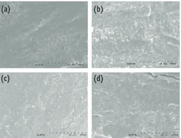
Figure 1. Representative photograph in the WL-3 mm groups. (a) #25, WL-3 mm; (b) #30, WL-3 mm; (c) #35, WL-3 mm; (d) #40, WL-3 mm. (a) and (b) presence of the smear layer on the surface, x1,000; (c) presence of debris in the dentinal tubules, x1,000; (d) removal of the smear layer from the surface, x1,000.
(a)
(c)
(b)
(d)
Figure 2. Representative photograph in the WL-9 mm groups. (a) #25, WL-9 mm; (b) #30, WL-9 mm; (c) #35, WL-9 mm; (d) #40, WL-9 mm. (a) and (b) smear layer is not removed, and tubule apertures are totally obliterated, x1,000; (c) smear layer is thin as evidenced by crack over tubule aperture, x1,000; (d) dentinal tubules are exposed, but some are blocked by smear layer, x1,000.
(a)
(c)
(b)
(d)
총괄 및 고안
근관세척액은근관내에적용된이후시간이지남에따라그효과 가떨어진다. 차아염소산나트륨용액은근관내에서시간에따라유 효염소농도가감소되어항세균효과의 감소를보이며산화, 환원능 력의감소로유기물질분해능력이떨어지게된다.5,6 EDTA 또한근관 내에서탈회가진행됨에따라 세척액내에수소이온이증가하게되 고이로인해낮아진 pH는탈회능력을감소시키는자가정체효과(self- limiting effect)를갖고있다.18따라서충분한양의세척액이적용되 기용이하도록근관이성형되며주기적인세척액의교환이이루어진 다. 하지만치근첨에서근관입구에이르는근관성형의형태는근단부 를향할수록그내부의용적이감소한다. 따라서근관내부에적용된 세척액의양은치근단부로향할수록그양이적어지게되고이에따라 세척액의효과가떨어지게된다. 본실험결과, 치근단확대가증가할 수록대부분의군은도말층이많이제거된것을확인할수있었다. 이 는증가된근단부의용적이더많은세척액을받아들일수있었기때 문으로생각된다. 하지만 #25군과 #30군은유의성있는차이가존재 하지않았는데, 이는치근단1/3근관의체적이작았고따라서유효한 세척액이더이상의도말층을제거할수없었던것으로생각된다.
근관으로세척액을전달하기위해바늘을갖는시린지가전통적으 로사용되었다. 임상에서흔히사용되는다섯가지세척바늘은 21G, 23G, 25G, 27G, 30G 로각각의외경은0.8 mm, 0.6 mm, 0.5 mm, 0.4 mm, 0.3 mm이며내경은0.490 mm, 0.317 mm, 0.232 mm, 0.184 mm, 0.133 mm이다.13본연구의예비실험에서는네 가지세척바늘(30G, 27G, 25G, 23G)을선택하여실험치아성형후각치아의근관에최대
한적용되는 needle tip의위치를기록하고최대침투깊이와평균침
투깊이를얻었다. #25집단에서, 30 gauge 세척바늘의최대적합부(근 관장으로부터평균1.5 mm, 최대2 mm)와 23 gauge 세척바늘의최 대적합부(근관장으로부터평균6.5 mm, 최대7.6 mm)에관한결과를 확인하였다. 이를토대로본실험에서는세척바늘이침투될위치를3 mm, 9 mm로설정하였고일관된세척액의적용을위해모든세척액은 첨단이측방개구형태 (closed-end and single side-openings)인30G 주 사바늘을사용하였다.
한편, Boutsioukis 등은 Computational Fluid Dynamics Model을이 용한모의실험결과측방개구형태를갖는30G 세척바늘의세척액교 환효과가바늘끝으로부터1 - 1.5 mm 위치에한정되어있다고보고 하였다.14 Gulabivala 등은이처럼 바늘끝에서제한된거리만큼은와 류가형성되어새로운세척액이교환되지만그이상의위치에서세척 액의교환은주로확산에의존하기때문에상대적으로오랜시간이 소모된다고하였다.19실험결과를살펴보면세척바늘이 작업장에서 WL-3 mm 군의경우 WL-9 mm 군보다통계적으로우수한효과를보였 는데, 이는유효한능력을갖는새로운세척제의교환이 WL-3 mm 군 에서더욱활발히이루어져근단부의도말층을제거할수있는더많 은기회가주어졌기때문으로생각된다.
또한치근단확대가서로다른 WL-3 mm 군간에서는도말층제거 능력이서로유의성있는차이를보였지만 WL-9 mm 군간의차이가 없었다는것은주목할만한사실이다. WL-3 mm와 WL-9 mm 세척군의 의미가각각30G, 23G 세척바늘의평균및 최대침투깊이를반영한 결과라는점을다시한번상기해볼때, 임상에서23G 정도의굵은세 척바늘을사용한근관세척은치근단1/3근관부위에불량한세척효
과를보임을유추할수 있다. 본실험에서근관확대의정도와세척바
늘의침투깊이의두변수는 Kahn의연구와는달리서로상호관계가
존재하지않았는데, Kahn의연구에서는세척바늘이가능한깊이위치
되었으나본실험에서는작업장으로부터특정한위치, 즉 WL-3 mm 및 WL-9 mm 군으로나누어설정한것이그원인이아닌가사료된다.11
현재 근관내의도말층을 제거하는 데에는근관을10 mL의 17% EDTA 처리후10 mL의2.5 - 5.25% 차아염소산나트륨용액으로처리 하는것이가장효과적인방법으로받아들여지고있다.2017% EDTA가 도말층의제거에효과적이기는 하지만, 칼슘치환의효과는근관벽의 세관을 과하게개방시켜관간상아질을완전히파괴시키는경우도있 다.21또한 Torabinejad 등은17% EDTA의사용이 근관의치관부1/3, 중간1/3에서과다한부식을야기함을보고한바있다.22이러한 EDTA 의과다한상아질침식은 EDTA의농도를낮춤으로서해결될수있다.21 하지만 낮은농도에서는 치환반응이 진행될수록누적되는수소이온 으로인하여산성환경이조성되고이로인해치환반응이정체된다.18 이런자가정체반응을막기위해알칼리성 pH 9로적정된 EDTA는 과
도한탈회없이도말층을제거할수있으며, sealer와근관벽간의접촉
각을보다줄이고15% EDTA 만큼이나근관약제의세관내침투가가
능하다.23본실험에서는우치를사용한 Nakashima의 연구모델에의 해 pH 9로적정된3%의 EDTA를선택하였으며치근부3 mm에바늘이 적용되고 #40으로확대된인간치아를통한예비실험을통해변형된
EDTA 세척의조건을잡고자하였다.23예비실험의결과로통상적으로
사용되는적용시간보다긴4분이설정되었으며첫1분적용뒤잔류된 차아염소산나트륨용액과 EDTA의반응을중단시키기위한시도로서
세척액이제거되고이후3분의 EDTA 수용액을다시적용하였다. 또한
EDTA가잔류되어상아세관을더욱확장시킬가능성을줄이기위해최 종적으로차아염소산나트륨수용액으로다시한번세척하였다. 하지 만예비실험에서확인된세척모델은보다작은치근단확대가이루어 진본실험에서근단부의도말층을충분하게제거하지못했으며이것 이농도에의한결과인지는각농도간의추가적인비교연구로확인 이필요할것으로판단된다.
치근단1/3근관에대한기구작업에대해다양한견해가존재한다.
Schilder는근관의형태가근관입구에서큰 직경을갖고근단협착부
에서가장작은직경을갖도록점진적으로 taper가줄어들어야한다고 하였다.2하지만이작업법은열연화충전법을위해고안되었고근관계 의 이상적인감염제거를위해개발되지는않았다.1 Ram은방사선조 영제세척실험을통하여최대한의세척을이루기위해서는 #40까지 확대되어야한다고결론지었다.24 Orstavik 등은치근단치주염이있는 치아에서 #45크기로작업하였을경우치료전에비해세균의증식이 10배나 감소되었다고주장하였다.25 하지만근관치료의 성공에대한 장기적인연구들로부터근단부의확대정도와성공률이비례하지않 음을확인할수있다.26,27본연구에서치근단확대의크기가증가하면 서더많은도말층이제거되는결과를확인하였으나오직 #30군에서
#35군으로확대하는과정만이유의성있는차이를보였다.
결론
본연구의결과를통해근단부의확대정도가크거나주사바늘의위 치가깊게삽입될수록치근단3 mm의세척효과가우수하였음을확 인하였다. 향후효과적인근관세척에영향을미치는다른인자에대한
더많은연구가필요할것으로생각된다.
References
1. Baugh D, Wallace J. The role of apical instrumentation in root canal treatment: a review of the literature. J Endod 2005;31:333-340.
2. Schilder H. Cleaning and shaping the root canal. Dent Clin North Am 1974;18:269-296.
3. Vertucci FJ. Root canal anatomy of the human permanent teeth. Oral Surg Oral Med Oral Pathol 1984;
58:589-599.
4. Verma P, Love RM. A Micro CT study of the mesiobuccal root canal morphology of the maxillary first molar tooth. Int Endod J 2011;44:210–217.
5. Zehnder M. Root canal irrigants. J Endod 2006;32:389- 398.
6. Rutala WA, Weber DJ. Uses of inorganic hypochlorite (bleach) in health-care facilities. Clin Microbiol Rev 1997;10:597-610.
7. Bystrom A, Sundqvist G. The antibacterial action of sodium hypochlorite and EDTA in 60 cases of endodontic therapy. Int Endod J 1985;18:35-40.
8. Yang SE, Bae KS. SEM study on the anaerobic bacterial adhesion to the dentin of root canal. J Korean Acad Cons Dent 2001;26:350-359.
9. Chow TW. Mechanical effectiveness of root canal irrigation. J Endod 1983;9:475-479.
10. Abou-Rass M, Piccinino MV. The effectiveness of four clinical irrigation methods on the removal of root canal debris. Oral Surg Oral Med Oral Pathol 1982;54:323-328.
11. Kahn FH, Rosenberg PA, Gliksberg J. An in vitro evaluation of the irrigating characteristics of ultrasonic and subsonic handpieces and irrigating needles and probes. J Endod 1995;21:277-280.
12. Sedgley CM, Nagel AC, Hall D, Applegate B. Influence of irrigant needle depth in removing bioluminescent bacteria inoculated into instrumented root canals using real-time imaging in vitro. Int Endod J 2005;38:97-104.
13. Boutsioukis C, Lambrianidis T, Vasiliadis L. Clinical relevance of standardization of endodontic irrigation needle dimensions according to the ISO 9,626:1991 and 9,626:1991/Amd 1:2001 specification. Int Endod J 2007;40:700-706.
14. Boutsioukis C, Lambrianidis T, Kastrinakis E. Irrigant flow within a prepared root canal using various flow rates: a computational fluid dynamics study. Int Endod J 2009;42:144-155.
15. Boutsioukis C, Lambrianidis T, Kastrinakis E, Bekiaroglou P. Measurement of pressure and flow rates during irrigation of a root canal ex vivo with three endodontic needles. Int Endod J 2007;40:504-513.
16. Boutsioukis C, Gogos C, Verhaagen B, Versluis M, Kastrinakis E, Van der Sluis LW. The effect of apical preparation size on irrigant flow in root canals evaluated using an unsteady computational fluid dynamics model. Int Endod J 2010;43:874-881.
17. Lendini M, Alemanno E, Migliaretti G, Berutti E. The effect of high‐frequency electrical pulses on organic tissue in root canals. Int Endod J 2005;38:531-538.
18. Calvo Pérez V, Medina Cárdenas ME, Sánchez Planells U. The possible role of pH changes during EDTA demineralization of teeth. Oral Surg Oral Med Oral Pathol 1989;68:220-222.
19. Gulabivala K, Ng YL, Gilbertson M, Eames I. The fluid mechanics of root canal irrigation. Physiol Meas 2010;31:R49-84.
20. Aktener BO, Bilkay U. Smear layer removal with different concentrations of EDTA-ethylenediamine mixtures. J Endod 1993;19:228-231.
21. Perez F, Rouqueyrol-Pourcel N. Effect of a low- concentration EDTA solution on root canal walls: a scanning electron microscopic study. Oral Surg Oral Med Oral Pathol Oral Radiol Endod 2005;99:383-387.
22. Torabinejad M, Khademi AA, Babagoli J, Cho Y, Johnson WB, Bozhilov K, Kim J, Shabahang S. A new solution for the removal of the smear layer. J Endod 2003;29:170- 175.
23. Nakashima K, Terata R. Effect of pH modified EDTA solution to the properties of dentin. J Endod 2005;31:
47-49.
24. Ram Z. Effectiveness of root canal irrigation. Oral Surg Oral Med Oral Pathol 1977;44:306-312.
25. Orstavik D, Kerekes K, Molven O. Effects of extensive apical reaming and calcium hydroxide dressing on bacterial infection during treatment of apical periodontitis: a pilot study. Int Endod J 1991;24:1-7.
26. Hoskinson SE, Ng YL, Hoskinson AE, Moles DR, Gulabivala K. A retrospective comparison of outcome of root canal treatment using two different protocols.
Oral Surg Oral Med Oral Pathol Oral Radiol Endod 2002;93:705-715.
27. Kerekes K, Tronstad L. Long-term results of endodontic treatment performed with a standardized technique. J Endod 1979;5:83-90.