Tissue Plasminogen Activator and Plasminogen Activator Inhibitor-1 Levels in Patients with Acute Paraquat Intoxication
To investigate the effects of reactive oxygen species (ROS) on tissue plasminogen activator (tPA) and plasminogen activator inhibitor-1 (PAI-1) plasma levels, and their possible implications on clinical outcome, we measured tPA and PAI-1 levels in 101 patients with acute paraquat (PQ) intoxication. The control group consisted of patients who ingested non-PQ pesticides during the same period. tPA and PAI-1 levels were higher in the PQ group than in the controls. PQ levels were significantly correlated with ingested amount, timelag to hospital, tPA level, and hospitalization duration. tPA levels were correlated with PAI-1, fibrin degradation product (FDP), and D-dimer. D-dimer levels were lower in the PQ group than in the controls. Univariate analysis indicated the following significant
determinants of death: age, ingested amount, PQ level, timelag to hospital, serum creatinine, lipase, pH, pCO2, HCO3-, WBC, FDP, PAI-1, and tPA. However, multivariate analysis indicated that only PQ level was significant independent factor predicting death.
In conclusion, tPA and PAI-1 levels were higher, while D-dimer levels were lower in the PQ group than in the controls, implying that ROS stimulate tPA and PAI-1, but PAI-1 activity overrides tPA activity in this setting. Decreased fibrinolytic activity appears to be one of the clinical characteristics of acute PQ intoxication.
Key Words: Tissue Plasminogen Activator; Plasminogen Activator Inhibitor 1; Paraquat;
Reactive Oxygen Species Su-Jin Seok, Su-Ji Kim, Hyo-Wook Gil,
Jong-Oh Yang, Eun-Young Lee, and Sae-Yong Hong
Department of Internal Medicine, College of Medicine, Soonchunhyang University, Cheonan, Korea
Received: 8 December 2010 Accepted: 8 February 2011 Address for Correspondence:
Sae-Yong Hong, MD
Department of Internal Medicine, Soonchunhyang University Cheonan Hospital, 8 Suncheonhyang 2-gil, Dongnam-gu, Cheonan 330-721, Korea
Tel: +82.41-570-2121, Fax: +82.41-574-5762 E-mail: [email protected]
This study was supported by a grant from the Soonchunhyang University, Korea (2010).
DOI: 10.3346/jkms.2011.26.4.474 • J Korean Med Sci 2011; 26: 474-481
INTRODUCTION
Tissue plasminogen activator (tPA) is a serine protease expressed by endothelial cells of blood vessels that catalyzes the conver- sion of plasminogen to plasmin (1). In addition to its role in the fibrinolysis system, tPA is implicated in tissue proliferation, cel- lular adhesion, and the activation of growth factors and matrix metalloproteinases (2). Insight into the regulation of the syn- thesis and release of tPA has extended markedly over the last 2 decades, including the characterization of the intracellular sig- naling pathways, promoter elements, and transcription factors involved in the transcriptional regulation of tPA (3, 4).
The tPA gene is located on chromosome 8 (5). Evidence from experimental studies indicates that reactive oxygen species (ROS) can inhibit tPA mRNA expression and synthesis, and impair the release of tPA from cultured human endothelium (6, 7). Further- more, oxidants may alter the structural characteristics of tPA, which in turn may adversely affect endothelial fibrinolytic ac- tivity (8). However, these observations were derived from in vi- tro experiments or from experimental animal models. Van Guil- der et al. observed that the antioxidant vitamin C increased the capacity of the endothelium to release tPA in overweight/obese adults (9). They concluded that ROS inhibit tPA production; how-
ever, it is not clear whether high levels of ROS suppress plasma tPA levels, leading to thrombosis in humans.
Plasminogen activator inhibitor-1 (PAI-1) is the principal in- hibitor of tPA (10). It is mainly produced by the cells lining blood vessels, but it is also secreted by other tissue types, e.g., adipose tissue. The PAI-1 gene is located on chromosome 7 (7q21.3-q22) (11). There is little information regarding the effects of ROS on the production of PAI-1 (3, 12).
Paraquat (PQ, 1,1´-dimethyl-4,4´-bipyridinium dichloride) is one of the most commonly used herbicides in the world (13-15).
In human, intentional or accidental ingestion of PQ is consid- ered to be uniformly fatal, resulting in death from multi-organ failure and cardiogenic shock within few days (16). After inges- tion of small quantities, PQ is specially taken up into and accu- mulates in the lung. Subsequent redox cycling and free radical generation triggers a neutrophil-mediated inflammatory re- sponse in the lungs, which initiates an irreversible fibrotic pro- cess that kills the majority of patients within several weeks (17).
Considering that PQ is the most frequently used chemical to produce ROS in vitro experiments, acute PQ intoxication could be a human model to study specific clinical conditions related to ROS.
The purpose of this study was to examine the plasma levels
of tPA and PAI-1 in patients with acute PQ intoxication, focus- ing on the relationship between these parameters and the clini- cal features of PQ intoxication.
MATERIALS AND METHODS
Soonchunhyang Cheonan Hospital’s Investigational Review Board approved this study, and all participants gave written in- formed consent. We enrolled 101 patients (58 males and 43 fe- males, aged 49.1 ± 16.0 yr) with acute PQ intoxication in this study. All of the patients ingested concentrated PQ (22%-23%
per volume) while committing suicide and were admitted to the Institute of Pesticide Poisoning, Soonchunhyang University Cheonan Hospital, from September 2009 to March 2010. The amount ingested was estimated from the number of swallows, where 1 mouthful was considered as 20 mL.
For the control group, we enrolled 30 patients (14 males and 16 females, aged 53.5 ± 16.6 yr) with acute non-PQ pesticide in- toxication. The control group also ingested pesticides while com- mitting suicide and were admitted to the same hospital during the same period. The frequency of ingested pesticides in the control group was as follows: organophosphate, 9 (30%); glypho-
sate, 6 (20%); glufosinate, 5 (16.7%); pyrethroid, 3 (10%); and others, 7 (23.3%). The demographic and basal laboratory find- ings for both groups of patients are summarized in Table 1.
Upon admission, the patients received the following standard- ized medical emergency procedures. Briefly, a gastric lavage was performed on all subjects who presented to the emergency room (ER) within 2 hr of PQ ingestion. For those whose intoxica- tion occurred within 12 hr before presenting to the ER, 100 g of Fuller’s earth in 200 mL of 20% mannitol was administered. He- moperfusion was initiated early and continued for 4-6 hr accord- ing to the result of the urine dithionite test. Glutathione (50 mg/
kg/24 hr) was administered intravenously for 3 additional days as an ROS scavenger. For the control group, early hemoperfu- sion was performed on the first day of hospitalization in the same manner as the PQ group and conservative therapy was followed.
For patients with organophosphate intoxication, atropine and pralidoxime (PAM) were administered.
Blood samples were collected in the ER for baseline laborato- ry findings. The PQ levels were measured by the HPLC method.
There are various ways to estimate the tPA mediated fibrino- lytic activity, such as measurement of tPA with catalytic activity and/or quantification of tPA antigen, as a free form or complex form of tPA-PAI-1. In this study, we measured tPA and PAI-1 an- tigen to observe the effect of ROS on the production and/or re- lease of tPA and PAI-1 each. Of course, tPA and PAI-1 antigen are not consistent with the activity of tPA and PAI-1. In order to compensate for the defect, we measure fibrin degradation prod- uct (FDP) and D-dimer, which are always the end-products of fibrinolysis. Besides, they represent the pre-existence of throm- bus.
To quantify tPA and PAI-1 levels, plasma samples were col- lected 3 times: the first sample was collected in the ER and the other 2 samples were taken on consecutive days at 24 hr inter- vals. tPA (Asserachrom® tPA kit; Diagnostica Stago, Asnières- Sur-Seine, France) and PAI-1 (Asserachrom® PAI-1 kit; Diagnos- tica Stago) antigen levels were measured using commercially available assay kits. tPA and PAI-1 levels were presented as the mean of these 3 measurements.
Statistical analysis
Continuous variables are presented as the mean ± standard de- viation and categorical variables as frequency (in percent). The differences between the PQ and control groups were compared using Student’s t-test for continuous variables and by the chi- squared-test or Fisher’s exact test for categorical variables. The relationship between variables was analyzed using bivariate correlation analysis. Univariate binary logistic regression analy- sis was used to verify the clinical implications of the fibrinolytic markers, e.g., tPA and PAI-1. Multivariate logistic regression anal- ysis was used to identify significant determinants of death after PQ intoxication. The results of the logistic regression analyses Table 1. The demographic and laboratory findings for the paraquat and control into
xication groups
Parameters Control group
(n = 30) Paraquat group
(n = 101) P
value
Sex (female/male), No. 16/14 43/58 0.298
Death, No. (%) 2 (6.7%) 56 (55.4%) < 0.001
Age (yr) 53.5 ± 16.6 49.1 ± 16.0 0.205
Time lag to hospital (hr) 11.9 ± 20.2 13 ± 17.6 0.798 Hospitalization duration (days) 12.2 ± 13.8 7.4 ± 6.1 0.008 Laboratory findings
AST (IU/L) 35 ± 17 43 ± 96 0.439
ALT (IU/L) 25 ± 15 28 ± 40 0.550
Urea nitrogen (mg/dL) 16.8 ± 9.6 16.7 ± 12.5 0.987 Creatinine (mg/dL) 0.9 ± 0.5 1.2 ± 1.1 0.048
Sodium (mEq/L) 143 ± 5 141 ± 7 0.042
Potassium (mEq/L) 4.0 ± 0.8 3.5 ± 0.7 0.003 Amylase (IU/L) 296 ± 306 252 ± 281 0.487 Lipase (U/L) 98 ± 192 91 ± 242 0.863
pH 7.35 ± 0.14 7.39 ± 0.08 0.039
pCO2 (mmHg) 36.1 ± 10.7 30.4 ± 7.0 0.001 pO2 (mmHg) 100.8 ± 75.8 95.7 ± 19.2 0.541 HCO3 (mEq/L) 19.6 ± 5.4 18.3 ± 5.2 0.225 WBC (× 103/µL) 14.7 ± 8.6 15.2 ± 7.5 0.769 Hemoglobin (g/dL) 14.2 ± 2.1 14.1 ± 2.1 0.936 Hematocrit (%) 42.5 ± 5.6 42.0 ± 5.7 0.712 Platelet (× 103/µL) 237 ± 72 237 ± 79 0.968 Ddimer (µg/mL) 2.1 ± 1.1 1.2 ± 1.1 0.001 FDP (µg/mL) 7.4 ± 3.3 6.3 ± 3.9 0.117 PAI1 (ng/mL) 51.3 ± 23.0 69.9 ± 25.0 0.001 tPA (ng/mL) 18.0 ± 12.7 24.8 ± 15.2 0.038 AST, Aspartate transaminase; ALT, Alanine transaminase; WBC, white blood cell count; FDP, fibrin degradation product; PAI1, plasminogen activator inhibitor1; tPA, tissue plasminogen activator.
were reported as odds ratio with 95% confidence intervals (CI).
Statistical analyses were performed using SPSS software (ver- sion 14.0; SPSS Inc., Chicago, IL, USA). All P values below 0.05
(ng/mL) P = 0.038
P = 0.001
tPA PAI-1
100 90 80 70 60 50 40 30 20 10 0
Control group PQ group
Fig. 1. Comparison of the tPA and PAI1 levels between the control and PQ intoxication groups. Note that tPA and PAI1 levels are significantly higher in the PQ group than in the control group.
were considered statistically significant.
Ethics statement
The protocol and standardized clinical format was approved by the Soonchunhyang Cheonan Hospital’s Investigational Review Board (2010-91). The informed consent was exempted.
RESULTS
Comparison of tPA, PAI-1, FDP, and D-dimer levels between the PQ and control groups
tPA (24.8 ± 15.2 vs 18.0 ± 12.7 ng/mL, respectively, P = 0.038) and PAI-1 (69.9 ± 25.0 vs 51.3 ± 23.0 ng/mL, respectively, P = 0.001) levels were significantly higher in the PQ group than in the control group (Fig. 1). D-dimer levels were significantly low- er in the PQ group than in the control group (1.2 ± 1.1 vs 2.1 ± 1.1 μg/mL, respectively, P = 0.001) (Fig. 2). However, there were no significant differences in the levels of FDP between the con- trol and PQ groups (7.4 ± 3.3 vs 6.3 ± 3.9 μg/mL, respectively, P =
(µg/mL) P = 0.001
P = 0.117
D-dimer FDP
12 10 8 6 4 2 0
Control group PQ group
Fig. 2. Comparison of the levels of Ddimer and fibvin degradation product (FDP) between the control and PQ intoxication groups. Note that the Ddimer levels are significantly lower in the PQ group than in the control group.
(µg/mL) P < 0.001
P = 0.003
tPA PAI-1
120 100 80 60 40 20 0
PQ level < 10 µg/mL PQ level ≥ 10 µg/mL
Fig. 3. Relationship between PQ levels and the levels of tPA and PAI1 in PQintoxi
cated patients. PQintoxicated patients were divided into 2 subgroups according to their PQ level: ≥ 10 µg/mL vs < 10 µg/mL.
tPA (ng/mL) PAI-1 (ng/mL)
70 60 50 40 30 20 10 0
100 90 80 70 60 50 40 30 20 10 0
Amount of PQ ingestion (mL) Amount of PQ ingestion (mL)
0 50 100 150 200 250 300 350 0 50 100 150 200 250 300 350
P < 0.001 P < 0.001
Fig. 4. Relationship between the amount of PQ ingested and the levels of tPA (A) and PAI1 (B) in PQintoxicated patients. Note that there is a positive correlation between the amount of PQ ingested and the levels of tPA and PAI1.
A B
0.117).
Relationship between tPA and PAI-1 levels and the PQ level and amount of PQ ingested in PQ-intoxicated patients
Patients with PQ intoxication were divided into 2 subgroups ac- cording to their PQ level: ≥ 10 μg/mL vs < 10 μg/mL. Patients whose PQ levels were over 10 μg/mL had significantly higher levels of tPA than those below 10 μg/mL (36.3 ± 13.0 vs 18.3 ± 11.7 ng/mL, respectively, P < 0.001) (Fig. 3). In addition, bivari- ate correlation analysis revealed a significant correlation be- tween PQ levels and the amount of PQ ingested (r = 0.281, P = 0.005), the time interval between PQ ingestion and hospital ad- mission (r = -0.286, P = 0.004), the level of tPA (r = 0.417, P = 0.001) and hospitalization duration (r = -0.419, P < 0.001).
The amount of PQ ingested demonstrated a positive relation- ship with the levels of tPA (r = 0.579, P < 0.001) and PAI-1 (r = 0.428, P < 0.001) (Fig. 4); similarly, the levels of tPA were signifi- cantly correlated with the levels of PAI-1 (r = 0.621, P < 0.001), FDP (r = 0.479, P < 0.001), and D-dimer (r = 0.312, P = 0.014).
Comparison of tPA, PAI-1, and PQ levels, the amount of PQ ingested, and blood chemistry between the deceased and survived groups for PQ intoxication
Fifty-six (55.4%) patients died in the PQ group; while 2 (6.7%) patients died in the control group. The demographic and labo- ratory findings for the survivors and non-survivors are summa- rized in Table 2. Briefly, the deceased patients were older, in- gested larger amounts of PQ, arrived at hospital later, and had higher levels of PQ, tPA, and PAI-1 than those who survived.
Univariate binary logistic regression analysis of the PQ group indicated that the following factors were significant determi- nants of death: age, amount of PQ ingested, PQ levels, time lag between PQ ingestion and admission, hospitalization duration, serum creatinine, potassium, lipase, pH, pCO2, HCO3-, white blood cell count (WBC), FDP, PAI-1, and tPA levels (Table 3).
However, multivariate binary logistic regression analysis indi-
Table 2. Demographic and laboratory findings between the survived and deceased groups of paraquatintoxicated patients
Parameters Survived group
(n = 43)
Deceased group (n = 56) P value
Sex (female/male), No. 21/22 20/36 0.189
Age (yr) 43 ± 15.3 53.6 ± 15.5 0.001
Amount of ingestion (mL) 21.9 ± 23.9 89.3 ± 90 < 0.001 PQ level (µg/mL) 0.4 ± 0.6 18.8 ± 20.5 < 0.001 Time lag to hospital (hr) 18 ± 21.4 8.4 ± 11.6 0.005 Hospitalization duration (days) 11.8 ± 6.0 4.3 ± 3.6 < 0.001 Laboratory findings
AST (IU/L) 29 ± 17 53 ± 127 0.160
ALT (IU/L) 23 ± 20 31 ± 51 0.293
Urea nitrogen (mg/dL) 15.6 ± 11.4 16.9 ± 11.3 0.570
Creatinine (mg/dL) 0.8 ± 0.6 1.4 ± 1.2 0.005
Sodium (mEq/L) 140 ± 6 141 ± 7 0.559
Potassium (mEq/L) 3.8 ± 0.5 3.2 ± 0.7 < 0.001
Amylase (IU/L) 208 ± 182 269 ± 304 0.221
Lipase (U/L) 29 ± 12 127 ± 303 0.037
pH 7.42 ± 0.04 7.36 ± 0.09 < 0.001
pCO2 (mmHg) 34.2 ± 4.8 27.4 ± 7.1 < 0.001
pO2 (mmHg) 93.4 ± 18.8 97.7 ± 19.6 0.276
HCO3 (mEq/L) 21.9 ± 2.9 15.5 ± 4.9 < 0.001 WBC (× 103/µL) 10.6 ± 4.0 18.8 ± 7.7 < 0.001
Hemoglobin (g/dL) 14.1 ± 2 14.1 ± 2.2 0.969
Hematocrit (%) 42 ± 5.5 42.1 ± 6 0.893
Platelet (× 103/µL) 239 ± 77 240 ± 79 0.949
Ddimer (µg/mL) 1.4 ± 3.8 2.9 ± 10.6 0.339
FDP (µg/mL) 4.6 ± 3 7.6 ± 4.1 < 0.001
PAI1 (ng/mL) 54.5 ± 20.8 82.4 ± 19.2 < 0.001
tPA (ng/mL) 12.6 ± 6.4 33.3 ± 13.7 < 0.001
AST, Aspartate transaminase; ALT, Alanine transaminase; WBC, white blood cell count; FDP, fibrin degradation product; PAI1, plasminogen activator inhibitor1; tPA, tissue plasminogen activator.
Table 3. Univariate binary logistic regression analysis to identify significant determi
nants of death in paraquatintoxicated patients
Variables P value OR 95% CI
Age (yr) 0.002 1.046 1.017 1.077
Amount of ingestion (mL) < 0.001 1.040 1.020 1.060
PQ level (μg/mL) < 0.001 5.270 2.384 11.649
Time lag to hospital (hr) 0.014 0.963 0.934 0.992 Hospitalization duration (days) < 0.001 0.677 0.582 0.787
Creatinine (mg/dL) 0.012 3.462 1.311 9.138
Potassium (mEq/L) < 0.001 0.236 0.110 0.506
Lipase (U/L) 0.005 1.050 1.015 1.087
pH < 0.001 1.9 × 10⁶ 1.4 × 10⁹ 0.003
pCO2 (mmHg) < 0.001 0.827 0.755 0.905
HCO3 (mEq/L) < 0.001 0.642 0.533 0.774
WBC (× 103/µL) < 0.001 1.0003 1.0001 1.0004
FDP (µg/mL) < 0.001 1.266 1.110 1.443
PAI1 (ng/mL) < 0.001 1.064 1.032 1.097
tPA (ng/mL) < 0.001 1.212 1.094 1.344
WBC, white blood cell count; FDP, fibrin degradation product; PAI1, plasminogen activator inhibitor1; tPA, tissue plasminogen activator.
Table 4. Multivariate binary logistic regression analysis to verify significant determi
nants of death in paraquatintoxicated patients. Values of tPA and PAI1 were adjusted by age, sex, time interval between PQ ingestion and hospital arrival, PQ levels, amount of PQ ingested and serum creatinine levels
Variables P value OR 95% CI
Amount of ingestion (mL) 0.456 1.037 0.943 1.140
PQ level (µg/mL) 0.014 16.052 1.747 147.520
Potassium (mEq/L) 0.850 1.362 0.055 33.742
Lipase (U/L) 0.859 1.002 0.980 1.025
pH 0.307 4.7 × 10¹¹ < 0.001 1.2 × 1034
pCO2 (mmHg) 0.541 0.894 0.623 1.281
HCO3 (mEq/L) 0.835 1.044 0.693 1.573
WBC (× 103/µL) 0.349 1.0003 0.9996 1.001
FDP (µg/mL) 0.835 1.081 0.521 2.242
PAI1 (ng/mL) 0.997 0.801 < 0.001 8.7 × 1057
tPA (ng/mL) 0.991 16.827 < 0.001 1.1 × 10219
WBC, white blood cell count; FDP, fibrin degradation product; PAI1, plasminogen activator inhibitor1; tPA, tissue plasminogen activator.
cated that only PQ levels, from these factors, were a significant independent factor predicting death (Table 4).
DISCUSSION
Coagulation and fibrinolysis proceed concomitantly, since a thrombus is formed on a damaged vessel wall and leads to the release of tPA from the intact endothelium nearby the throm- bus (18). Consequently, tPA activates the conversion of plas- minogen to plasmin, inducing the so-called “thrombus-specific fibrinolysis (19)”. Venous thrombosis has many different etiolo- gies and occurs when several risk factors are present simultane- ously (20). Risk factors in patients with acute pesticide intoxica- tion, in addition to the traditional ones, include prolonged bed rest (21), placement of central venous catheters (22), and he- moperfusion (23). This is the reason why we recruited a control group consisting of patients with non-PQ pesticide intoxication who underwent similar treatment modalities as the PQ group,
including bed rest, a central venous catheter for the extracorpo- real elimination of poisons, and/or the intravenous administra- tion of a large volume of fluid.
Contrary to the previous reports (6, 7, 9), we observed signifi- cantly higher levels of tPA and PAI-1 in the PQ group than in the control group (Fig. 1). Furthermore, the levels of tPA and PAI-1 were significantly higher in patients whose PQ levels were high- er than 10 μg/mL (Fig. 3). We cannot explain this discrepancy, but in the previous reports (6, 7), the results were described from not in vivo but in vitro experiments. In another previous study (9), they reported decreased levels of tPA after the injection of vitamin C in human subjects. With this result, they concluded that ROS may inhibit tPA production. However, their theory is open to criticism because the function of vitamin C is compli- cated in ROS formation, not scavenging ROS but stimulate ROS production in some situation.
In our results, there was a significant correlation between the levels of tPA and/or PAI-1 with the amount of PQ ingested (Fig. 4).
A B
C D
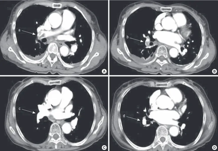
Fig. 5. Pulmonary artery computerized tomography angiogram from a PQintoxicated patient who suffered pulmonary artery thrombosis. A 64yrold woman swallowed a mouthful of a 24.5% PQ solution. Her plasma PQ level was 0.08 µg/mL at 15 hr after PQ ingestion. On day 9 of admission, the patient complained of right sided chest pain and dyspnea. Arterial blood gas analysis indicated: pH, 7.459; PaO2, 46.3 mmHg; and PaCO2, 37.9 mmHg. Ddimer was elevated at 2.9 (0–0.5) µg/mL. A chest Xray showed an increased density along the apex of the right lung. (A) obtained at 9 days after PQ ingestion. Note the thrombus in the pulmonary artery and interlobar pulmonary artery (arrow a, b). A subcutaneous injection of low molecular weight heparin was started on the 9th day and warfarin was started on the 10th day. (B) obtained at 20 days after PQ ingestion.
The thrombus was completely resolved (arrow c, d). The patient made a full recovery from this condition.
a
c
d b
A B
C D
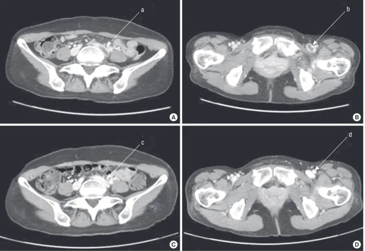
Fig. 6. Low extremity venogram computerized tomography from a PQintoxicated patient who suffered deep vein thrombosis in the leg. A 50yrold woman swallowed 2 mouth
fuls of a 24.5% PQ solution. (A) On day 21 after admission, the patient complained of left leg swelling and pain, and a deep vein thrombosis was suspected. Ddimer was elevated at 25.6 µg/mL. (B) Deep vein thrombosis was noted in the left deep venous system (arrow a, b). (C, D) Obtained at 63days after PQ ingestion. The extent of the thrombus was significantly reduced (arrow c, d).
a
c
b
d
The plasma PQ level at a given time is a function of the amount ingested and the time lag after ingestion; therefore, the PQ level does not represent the severity of intoxication in our study be- cause the patients arrived at the ER with different time lags after ingestion. However, it has been our clinical observation that no patients survive with a plasma PQ level > 10 μg/mL during the admission period. This observation led us to divide the PQ group according to their PQ levels: greater than and lower than 10 μg/
mL.
This raises the question of whether these relationships are due to PQ itself or an ROS effect generated by PQ. ROS are formed primarily in cells; however, direct measurement of ROS forma- tion in cells is very difficult in the clinical setting. Instead, sever- al indirect methods have been developed to measure free radi- cals and their metabolites, e.g., malondialdehyde (MDA; the fi- nal product of lipid peroxidation) (24) and hydroperoxides (ROOH; intermediate products of lipid peroxidation) (25). Re- cently, we reported that neither cross-sectional nor sequential measurements of plasma MDA provided reliable data on ROS formation in patients with acute PQ intoxication (24). Given the
lack of a reliable marker for ROS production, it is difficult to find a relationship between ROS levels and other parameters in acute PQ intoxication.
The plasma levels of PQ reach a peak level at 1.5 hr after PQ ingestion and decrease so rapidly that its levels are undetect- able after 24 hr in the majority of patients; therefore, plasma PQ levels observed in the ER are not comparable with those from the subsequent days. Contrary to the PQ levels, the 3 measure- ments of tPA and PAI-1 levels taken in this study were compara- ble in each patient; the mean coefficient of variation was 0.42 ± 0.22 for tPA (range 0.08-1.02) and 0.39 ± 0.30 for PAI-1 (range 0.00-1.31). These results suggest that it is not the mere level of PQ that affects tPA and PAI-1 levels, but some other factor(s) ac- tivated or stimulated by PQ; therefore, we consider that ROS gen- erated by PQ trigger the endothelium to release tPA and PAI-1.
D-dimer levels were lower in the PQ group than in the con- trol group (Fig. 2). The presence of D-dimer indicates the pre- existence of fibrin formation and the concurrent formation of plasmin by tPA (26). Coagulation and fibrinolysis are on-going physiological processes that account for the presence of D-di-
mer. Fibrin formation is the result of the imbalance between coagulation and anticoagulation activities in a pathological en- vironment. Therefore, the possibility exists that some thrombo- genic factor such as tissue factor could have been activated dur- ing PQ intoxication; however, changes in coagulation/antico- agulation activity were not the focus of the current study. In the PQ group, tPA and PAI-1 levels were higher, but D-dimer levels were lower than in the control group, indicating that, in this set- ting, PAI-1 activity overrode tPA activity.
Taken together, these results indicate that thrombosis should be a common complication in patients with acute PQ intoxica- tion, although only 2 of the 101 PQ patients suffered from pul- monary artery thrombosis or deep vein thrombosis (Figs. 5, 6).
However, bearing in mind that venous thrombosis is often clini- cally silent, we believe that there must have been more patients with subclinical thrombosis complications in the PQ group.
In order to verify the clinical implications of the tPA and PAI-1 levels, we performed univariate and multivariate binary logistic regression analyses. Univariate binary logistic regression analy- sis of the PQ group showed that age, amount of PQ ingested, plasma PQ levels, time lag after PQ ingestion, tPA levels, and PAI-1 levels were significant determinants of death (Table 3).
However, multivariate binary logistic regression analysis indi- cated that only PQ levels were a significant independent factor predicting death (Table 4).
We have few limitations in this study. First, we had better es- timate the tPA mediated fibrinolytic activity with both tPA anti- gen and tPA activity. Furthermore, parameters of coagulation, such as a tissue factor and its inhibitors would have enhanced the significance in the interpretation of the change in both tPA and PAI-1 levels. However, in practice, it was hard to obtain blood samples frequently in large amount because of their critical con- dition. The other question posed is the lack of quantitative mea- surement of ROS.
As we described in the introduction, it was impossible to mea- sure ROS directly and only surrogate markers were available in clinical practice. Even with these limit, our observations imply that ROS stimulate the production of tPA and PAI-1, but PAI-1 activity overrides tPA activity in this setting. Decreased fibrino- lytic activity due to increased PAI-1 activity appears to be one of the clinical characteristics of acute PQ intoxication.
In conclusion, the levels of tPA and PAI-1 were higher, but D- dimer levels were lower in the PQ group than in the control group.
REFERENCES
1. Collen D, Lijnen HR. Tissue-type plasminogen activator: a historical per- spective and personal account. J Thromb Haemost 2004; 2: 541-6.
2. Zorio E, Gilabert-Estellés J, España F, Ramón LA, Cosín R, Estellés A.
Fibrinolysis: the key to new pathogenetic mechanisms. Curr Med Chem 2008; 15: 923-9.
3. Nagamine Y. Transcriptional regulation of the plasminogen activator inhibitor type 1 with an emphasis on negative regulation. Thromb Hae- most 2008; 100: 1007-13.
4. Wu WS. The signaling mechanism of ROS in tumor progression. Cancer Metastasis Rev 2006; 25: 695-705.
5. Nusbaum C, Mikkelsen TS, Zody MC, Asakawa S, Taudien S, Garber M, Kodira CD, Schueler MG, Shimizu A, Whittaker CA, Chang JL, Cuomo CA, Dewar K, FitzGerald MG, Yang X, Allen NR, Anderson S, Asakawa T, Blechschmidt K, Bloom T, Borowsky ML, Butler J, Cook A, Corum B, DeArellano K, DeCaprio D, Dooley KT, Dorris L 3rd, Engels R, Glöckner G, Hafez N, Hagopian DS, Hall JL, Ishikawa SK, Jaffe DB, Kamat A, Kudoh J, Lehmann R, Lokitsang T, Macdonald P, Major JE, Matthews CD, Mauce- li E, Menzel U, Mihalev AH, Minoshima S, Murayama Y, Naylor JW, Ni- col R, Nguyen C, O’Leary SB, O’Neill K, Parker SC, Polley A, Raymond CK, Reichwald K, Rodriguez J, Sasaki T, Schilhabel M, Siddiqui R, Smith CL, Sneddon TP, Talamas JA, Tenzin P, Topham K, Venkataraman V, Wen G, Yamazaki S, Young SK, Zeng Q, Zimmer AR, Rosenthal A, Birren BW, Platzer M, Shimizu N, Lander ES. DNA sequence and analysis of human chromosome 8. Nature 2006; 439: 331-5.
6. Shatos MA, Doherty JM, Stump DC, Thompson EA, Collen D. Oxygen radicals generated during anoxia followed by reoxygenation reduce the synthesis of tissue-type plasminogen activator and plasminogen activa- tor inhibitor-1 in human endothelial cell culture. J Biol Chem 1990; 265:
20443-8.
7. Eberhardt W, Beck KF, Pfeilschifter J. Cytokine-induced expression of tPA is differentially modulated by NO and ROS in rat mesangial cells. Kidney Int 2002; 61: 20-30.
8. Feng YH, Hart G. In vitro oxidative damage to tissue-type plasminogen activator: a selective modification of the biological functions. Cardiovasc Res 1995; 30: 255-61.
9. Van Guilder GP, Hoetzer GL, Greiner JJ, Stauffer BL, DeSouza CA. Acute and chronic effects of vitamin C on endothelial fibrinolytic function in overweight and obese adult humans. J Physiol 2008; 586: 3525-35.
10. Dellas C, Loskutoff DJ. Historical analysis of PAI-1 from its discovery to its potential role in cell motility and disease. Thromb Haemost 2005; 93:
631-40.
11. Asselbergs FW, Pattin K, Snieder H, Hillege HL, van Gilst WH, Moore JH. Genetic architecture of tissue-type plasminogen activator and plas- minogen activator inhibitor-1. Semin Thromb Hemost 2008; 34: 562-8.
12. Kruithof EK. Regulation of plasminogen activator inhibitor type 1 gene expression by inflammatory mediators and statins. Thromb Haemost.
2008; 100: 969-75.
13. Seok SJ, Gil HW, Jeong DS, Yang JO, Lee EY, Hong SY. Paraquat intoxi- cation in subjects who attempt suicide: why they chose paraquat. Korean J Intern Med 2009; 24: 247-51.
14. Lee KH, Gil HW, Kim YT, Yang JO, Lee EY, Hong SY. Marked recovery from paraquat-induced lung injury during long-term follow-up. Korean J Intern Med 2009; 24: 95-100.
15. Gil HW, Kang MS, Yang JO, Lee EY, Hong SY. Association between plas- ma paraquat level and outcome of paraquat poisoning in 375 paraquat poisoning patients. Clin Toxicol (Phila) 2008; 46: 515-8.
16. Kim YT, Jou SS, Lee HS, Gil HW, Yang JO, Lee EY, Hong SY. The area of ground glass opacities of the lungs as a predictive factor in acute para- quat intoxication. J Korean Med Sci 2009; 24: 636-40.
17. Hong SY, Yang JO, Lee EY, Lee ZW. Effects of N-acetyl-L-cysteine and glu-
tathione on antioxidant status of human serum and 3T3 fibroblasts. J Korean Med Sci 2003; 18: 649-54.
18. Collen D, Lijnen HR. Thrombolytic agents. Thromb Haemost 2005; 93:
627-30.
19. Murray V, Norrving B, Sandercock PA, Terént A, Wardlaw JM, Wester P.
The molecular basis of thrombolysis and its clinical application in stroke.
J Intern Med 2010; 267: 191-208.
20. Lijfering WM, Rosendaal FR, Cannegieter SC. Risk factors for venous thrombosis- current understanding from an epidemiological point of view. Br J Haematol 2010; 149: 824-33.
21. Kahn SR, Shrier I, Kearon C. Physical activity in patients with deep ve- nous thrombosis: a systematic review. Thromb Res 2008; 122: 763-73.
22. Ortel TL. Acquired thrombotic risk factors in the critical care setting. Crit Care Med 2010; 38(2 Suppl): S43-50.
23. Gil HW, Kim SJ, Yang JO, Lee EY, Hong SY. Clinical outcome of hemo- perfusion in poisoned patients. Blood Purif 2010; 30: 84-8.
24. Gil HW, Seok SJ, Jeong DS, Yang JO, Lee EY, Hong SY. Plasma level of malondialdehyde in the cases of acute paraquat intoxication. Clin Toxi- col (Phila) 2010; 48: 149-52.
25. Kim JH, Gil HW, Yang JO, Lee EY, Hong SY. Effect of glutathione admin- istration on serum levels of reactive oxygen metabolites in patients with paraquat intoxication: a pilot study. Korean J Intern Med 2010; 25: 282-7.
26. Thachil J, Fitzmaurice DA, Toh CH. Appropriate use of D-dimer in hos- pital patients. Am J Med 2010; 123: 17-9.
AUTHOR SUMMARY
Tissue Plasminogen Activator and Plasminogen Activator Inhibitor-1 Levels in Patients with Acute Paraquat Intoxication
Su-Jin Seok, Su-Ji Kim, Hyo-Wook Gil, Jong-Oh Yang, Eun-Young Lee, and Sae-Yong Hong
Paraquat (PQ), a toxic herbicide, is highly reactive to oxygen. Here, in PQ-intoxicated patients, we investigated the plasma levels of both tissue plasminogen activator (tPA) and plasminogen activator inhibitor-1 (PAI-1), and their possible implications on clinical outcome. We observed that the levels of both tPA and PAI-1 were higher, while D-dimer levels were significantly lower in the patients with PQ-intoxication than in the control group. Univariate analysis indicated the following significant determinants of death: age, ingested amount, PQ level, time lag to hospital, serum creatinine, lipase, pH, pCO2, HCO3-, WBC, FDP, PAI-1, and tPA. However, multivariate analysis indicated that only PQ level was significant independent factor predicting death. Our findings imply that PQ-induced ROS might stimulate tPA and PAI-1, but PAI-1 activity overrides tPA activity in this setting. Decreased fibrinolytic activity appears to be one of the clinical characteristics of acute PQ intoxication.