INTRODUCTION
A family history of colorectal cancer is an important deter- minant of an individual’s risk for the disease. The highest degree of familial risk is provided by the dominantly inher- ited syndromes of colorectal cancer, hereditary non-polypo- sis colorectal cancer (HNPCC) and familial adenomatous polyposis (FAP), which constitute 1% to 3% of all colorec- tal cancers, respectively. In these syndromes, the probabili- ties of an un- treated gene carrier developing colorectal can- cers are about 80% and 100%, respectively (1, 2). An addi- tional 15% of individuals with colorectal cancer have other affected family members, but their family histories do not fulfill the criteria for either FAP or HNPCC, and they may not appear to follow a recognizable pattern of inheritance (3). These families, categorized as having familial colorectal cancer (4), are at increased risk of developing colorectal can- cer, but the familial risk, which is likely to have a genetic component, is difficult to define.
Susceptibility to HNPCC is caused by mutations in one of the genes in the DNA mismatch repair (MMR) gene sys- tem. The majority of germline mutations have been detect- ed in hMLH1 and hMSH2, whereas germline mutations in
hMSH6, hPMS1, and hPMS2 seem to be rare (5). MMR gene mutations are accompanied by a mutator phenotype, which is also caused by hMLH1-promoter methylation even if not sufficient for complete gene inactivation. Tumors arising in carriers of MMR gene mutations exhibit a characteristic phe- notype termed microsatellite instability (MSI), which is char- acterized by alterations in the length of simple repetitive mic- rosatellite sequences found throughout the genome. MSI fre- quency seems to be directly proportional to the hereditary basis of a tumor, with frequencies ranging from 15% in spo- radic colorectal cancer to 85% in HNPCC, with suspected HNPCC being intermediate (6, 7).
Recent studies have demonstrated that colorectal tumors with high-level sporadic microsatellite instability (MSI-H) share several clinicopathological features with HNPCC tumors (8). In contrast to HNPCC, however, the molecular and clin- ical characteristics of familial colorectal cancer have not been clearly defined. We found that familial colorectal cancer can be comprehensively explained as multiple occurrences of col- orectal and accompanying cancers, inherited by dominant or recessive transmission (9). The risk associated with family history varies greatly according to the age of onset of colorec- tal cancer in the family members, the number of affected rel-
In Ja Park*, Hee Cheol Kim*,�, Yong Sik Yoon*, Chang Sik Yu*, Se Jin Jang*,�, Jin Cheon Kim*,�
Departments of Surgery*and Pathology�, University of Ulsan College of Medicine and Asan Medical Center;
Laboratory of Cancer Biology & Genetics�, Asan Institute for Life Sciences, Seoul, Korea
Address for correspondence Jin Cheon Kim, M.D.
Department of Surgery, University of Ulsan College of Medicine and Asan Medical Center, 388-1 Poongnap-2-dong, Songpa-gu, Seoul 138-736, Korea Tel : +82.2-3010-3489, Fax : +82.2-474-9027 E-mail : [email protected]
*This study was supported by a Korea Research Foundation Grant (KRF-2004-014-E00063).
S91
Clinicopathological Characteristics of Colorectal Cancer with Family History: an Evaluation of Family History as a Predictive Factor for Microsatellite Instability
To determine whether family history of cancer may be a risk factor for the mutator phenotype in colorectal cancer, we recruited 143 consecutive colorectal cancer patients with a family history of accompanying cancers not meeting the Amster- dam criteria. Microsatellite instability (MSI) at 5 markers, hMLH1-promoter methy- lation, and expression of mismatch repair (MMR) proteins (hMLH1, hMSH2, hMSH6, hMPS1, and hPMS2) were determined. Among the relatives of familial colorectal cancer patients, colorectal cancer was the most common tumor type. Of the pro- band colorectal cancers, 26 (18.2%) showed high-level MSI (MSI-H); 47 additional tumors with mutator phenotype (32.9%) were identified by hMLH1-promoter methy- lation and/or loss of MMR protein expression. Mutator phenotype was associated with right-sided colon cancer and the type of accompanying cancer. Family histo- ry, which was differentially quantified according to the degree of relatives and the type of accompanying cancers, effectively discriminated MSI-H from microsatellite stable (MSS) and low-level microsatellite instability (MSI-L) and mutator phenotypes.
Our findings indicate that familial colorectal cancer may be associated with multi- ple occurrences of colorectal or accompanying cancers and that family history could be correlated with microsatellite instability.
Key Words : Familial Colorectal Cancer; Family History; Mutator Phenotype; Ncrosatellite Instability
Received : 15 June 2006 Accepted : 15 December 2006
atives, the closeness of the genetic relationship, and whether cancers have occurred across generations (10). Various crite- ria have provided screening recommendations for patients with a family history of colorectal tumors (11-14). Most of these criteria, however, include a family history of colorectal cancer but omit other tumor types, and thus do not consid- er the frequency of their occurrence and their genetic impli- cations.
We have formulated a familial impact value (FIV), in which the number of alleles shared between subjects was found to vary by their degree of relationship and the differential asso- ciation of colorectal cancer and other accompanying cancers in an ancestry (9). This value could be determined by a sim- ple calculation, suggesting it may be used efficiently as a risk index in familial colorectal cancer. Moreover, we found that FIV differed significantly between MSI-H and MSI-L/MSS tumors. This study was done to confirm our previous results, and to evaluate the clinical usefulness of FIV for selecting relatives of patients with familial colorectal cancer who require genetic testing.
MATERIALS AND METHODS Patients
Of the 1250 colorectal cancer patients treated at the Asan Medical Center (Seoul, Korea) between November 2003 and June 2005, we recruited 143 consecutive patients (87 men, 56 women; mean age, 56 yr; range, 28-89 yr) with a family history of accompanying cancers. Detailed family histories were obtained through questionnaires and through interviews with patients and their relatives. The questionnaire included cancer history in first- and second-degree relatives and con- tained questions regarding their age at diagnosis, type of can- cer, hospital at which the diagnosis had been made, current age, and current status. Patients with HNPCC meeting the Amsterdam criteria, patients with familial adenomatous poly- posis, and patients with a vague family history were exclud- ed. All solid cancers were included as accompanying cancers, except for cancers associated with viral infection, such as pri- mary liver and uterine cervix cancers. Hospital records were used to confirm family history. Patients treated with preop- erative radiotherapy were also excluded because of possible alteration of tumor DNA.
Histologically identified normal and tumor samples were freshly obtained from each patient. This study was conduct- ed prospectively, with the approval of the Institutional Review Board for Human Research, and all patients provided writ- ten informed consent.
Detection of MSI and hMLH1-promotor methylation MSI was determined by PCR, using primers amplifying
the microsatellite markers BAT25 and BAT 26 for mono- nucleotide repeats and D5S346, D2S123, and D17S250 for di-nucleotide repeats (7). Tumors were scored as MSI-H (i.e., high MSI) when ≥2 markers showed instability; MSI-L (i.e., low MSI) when 1 marker showed instability; and microsatel- lite stable (MSS) when none of the assayed markers showed MSI.
Methylation of the hMLH1 promoter region in tumor DNA was determined by methylation-specific PCR, as described previously (15), using 1 g of genomic DNA that had been denatured with NaOH and treated with sodium bisulfite.
Primer sequences were selected to cover the region upstream of the hMLH1 promoter, i.e., nucleotides -716 to -602. Pri- mers for unmethylated DNA were 5′-TTTTGATGTAGA- TGTTTTATTAGGGTTGT-3′(sense) and 5′-ACCACCT- CATCATAACTACCCACA-3′(antisense), whereas those for methylated DNA were 5′-ACGTAGACGTTTTATTAGG- GTCGC-3′(sense) and 5′-CCTCATCGTAACTACCCGCG- 3′(antisense). Ten L of each PCR reaction product were load- ed directly onto nondenaturing 6% polyacrylamide gels, which were stained with ethidium bromide and visualized under UV illumination. DNA from the colon cancer cell line SW48, which is completely methylated at the hMLH1-promoter region, was used as a positive control, whereas DNA from normal tissue was used as a negative control.
Immunohistochemical staining
Immunohistochemical staining for hMLH1, hMSH2, hMSH6, hPMS1, and hPMS2 proteins was performed in all 143 familial colorectal cancers as described previously (9), using diluted monoclonal antibodies to hMLH1 (G168-15;
BD PharMingen, San Diego, CA, U.S.A.), hMSH2 (G219- 1129, BD PharMingen), hMSH6 (clone 44, BD Transduc- tion Laboratories, San Jose, CA, U.S.A.), hPMS1 (sc-615, Santa Cruz Biotechnology Inc., Santa Cruz, CA, U.S.A.), and hPMS2 (sc-618, Santa Cruz Biotechnology). Samples were also stained with diluted monoclonal antibody to P53 (clone DO-7, Dako, Glostrup, Denmark). Normal colonic epithe- lium and lymphocytes, which exhibit strong nuclear staining for hMLH1, hMSH2, hPMS1, hPMS2, and hMSH6, were used as positive controls. The percentage of positively stained cells in a sample was divided into two grades for MMR pro- teins (i.e., negative expression for ≤10% and positive expres- sion for >10% nuclear staining) and into four grades for p53 (<10%, 10-<30%, 30-<50%, and ≥50%).
Calculation of FIV
The FIV score reflecting the severity of familial risk of colorectal cancer was based on the results of a meta-analysis (16) and suspected HNPCC criteria proposed by the Korean Hereditary Tumor Registry (17). Each first-degree relative with colorectal or HNPCC-associated cancer was scored as 4
points, each second-degree relative with colorectal or HNPCC- associated cancer was scored as 3 points, each first-degree relative with another accompanying cancer was scored as 2 points, and each second-degree relative with another accom- panying cancer was scored as 1 point. Points were doubled for an affected relative under 50 yr of age. FIV for each pa- tient was calculated as the sum of colorectal and accompa- nying cancers in each family multiplied by the relative degree.
FIVtwas defined as the sum of all accompanying cancers, and FIVcas the sum of HNPCC-associated cancers, accord- ing to the Amsterdam criteria.
Statistical methods
The Altman’s nomogram, assuming a putative MSI-H incidence of 15-40%, was used to determine the sample size to obtain an 80% power to detect MSI-H in familial colorectal cancer. Cross-table analysis using the Fisher’s exact test compared the respective mutator phenotype with MSI, hMLH1-promoter methylation, mismatch repair protein expression, and clinicopathologic variables. FIV was com- pared with MSI using the unpaired Student’s t test. All anal- yses were performed using SPSS software (ver. 11, SPSS Inc., Chicago, IL, U.S.A.), and the significance level was set at 5%.
RESULTS
Characteristics of family history and clinicopathological features
There were 206 colorectal and other cancers present in the first- and/or second-degree relatives of the 143 colorectal can- cer patients in this study. The 206 cancers consisted of 80 colorectal cancers, 69 gastric cancers, 9 HNPCC-associated cancers (small bowel, urinary tract, and endometrial cancers),
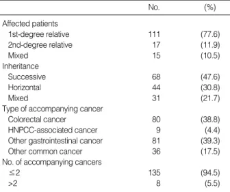
12 other digestive cancers (biliary, pancreatic, and esophageal cancers), and 36 other common cancers (lung, breast, and ovari- an cancers). Among the 143 probands, 111 (77.6%) had at least one affected first-degree relative, 15 (10.5%) had at least one affected second-degree relative, and 15 (10.5%) had affect- ed first- and second-degree relatives (Table 1).
Correlation of MSI status, hMLH1-promoter methylation, MMR protein expression, and mutator phenotypes with clinicopathological parameters
Of the 143 colorectal cancers, 26 (18.2%) were MSI-H and 3 (2.1%) were MSI-L. Younger age at onset, right-sided colon cancer, and mucinous cancer showed significant asso- ciations with MSI-H (p=0.05-0.001) (Table 2). The number of patients with MSI-H tumors was significantly higher in families affected with colorectal or HNPCC-associated can- cers than in those with other common cancers (p<0.001)
hMLH1-promoter methylation was observed in 26 patients (18.2%). Eleven of 26 MSI-H tumors (42.3%) showed hM- LH1-promoter methylation compared with 15 of 117 MSS/
MSI-L tumors (12.8%; p<0.001).
No. (%)
Affected patients
1st-degree relative 111 (77.6)
2nd-degree relative 17 (11.9)
Mixed 15 (10.5)
Inheritance
Successive 68 (47.6)
Horizontal 44 (30.8)
Mixed 31 (21.7)
Type of accompanying cancer
Colorectal cancer 80 (38.8)
HNPCC-associated cancer 9 (4.4)
Other gastrointestinal cancer 81 (39.3)
Other common cancer 36 (17.5)
No. of accompanying cancers
≤2 135 (94.5)
>2 8 (5.5)
Table 1.Family history of patients with familial colorectal cancer
MSI-H (n=26) MSS/MSI-L
(n=117) p
Sex 0.09
Male 12 (46.2) 75 (64.1)
Female 14 (53.8) 42 (35.9)
Age (yr) 0.05
≤50 13 (50.0) 35 (29.9)
>50 13 (50.0) 82 (70.1)
Mean CEA (ng/mL) 4.1 4.8 0.5
Location of primary tumor 0.001
Right colon 12 (46.1) 19 (7.7)
Left colon 6 (23.1) 28 (23.9)
Rectum 4 (15.4) 65 (55.6)
Multiple 4 (15.4) 5 (4.3)
Differentiation 0.05
Well differentiated 2 (7.7) 11 (9.4) Moderately differentiated 16 (61.5) 94 (80.3) Poorly differentiated 3 (11.5) 2 (1.7)
Mucinous 5 (19.3) 8 (6.8)
AJCC stage
I/II/III/IV 3/15/5/3 23/36/45/13 0.11
Synchronous cancer 4 (15.4) 10 (8.5) 0.29
Inheritance 0.07
Successive 10 (38.5) 58 (49.6)
Horizontal 6 (23.0) 38 (32.5)
Mixed 10 (38.5) 21 (17.9)
Affected patients 0.11
1st-degree relatives 21 (80.8) 90 (76.9) 2nd-degree relatives 0 (0) 25 (21.4)
Mixed 5 (19.2) 2 (1.7)
Table 2.Comparison MSI-H and MSS/MSI-L cancers (%)
MSI-H, microsatellite-high frequency; MSS, microsatellite stable; MSI-L, microsatellite-low frequency; CEA, carcinoembryonic antigen; AJCC, American Joint Committee for Cancer.
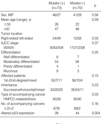
Of the 143 colorectal cancer patients, 73 (51%) exhibited mutator phenotype, defined as a tumor with MSI-H, hMLH1- promoter methylation, or loss of MMR protein expression.
This phenotype showed significant associations with right- sided colon cancer, lower expression of altered P53 protein, and a greater number of accompanying HNPCC-associated cancers (Table 3).
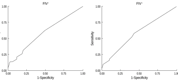
FIV as a function of MSI status and mutator phenotype
FIVtwas significantly higher in patients with MSI-H (6.9
±4.7) than in those with MSS/MSI-L (3.6±2.4) tumors when any categories of accompanying cancers were included (p<0.001). At a cut-off value of 3.5, FIVtshowed 88.5% sensi- tivity and 54% specificity for MSI status, whereas FIVcshow- ed 73% sensitivity and 60% specificity (Fig. 1). FIV was linearly correlated with the presence of the mutator pheno- type, but this correlation was weaker than that with MSI status. At a cut-off value of 2.5, FIVtshowed 64.4% sensi- tivity and 47.1% specificity for detection of mutator pheno- type, whereas, at a cut-off value of 1.5, FIVcshowed 57%
sensitivity and 57.1% specificity (Fig. 2).
Classification according to the revised Bethesda guidelines
Of our 143 patients, 84 (58.7%) met the revised Bethesda guidelines, whereas 49 (41.3%) did not (18). Patients meet- ing the revised Bethesda guidelines showed a higher inci- dence of MSI-H tumors (23.8% vs. 10.2%; p=0.04) and pro- moter methylation (21.4% vs. 13.6%; p=0.04) than those not meeting the guidelines. However, loss of mismatch pro- tein expression did not differ significantly between these two groups. Mean FIVt(5.3±3.7 vs. 2.7±1.3; p=0.002) and FIVc (4.0±4.2 vs. 1.2±1.8; p<0.001) were significantly higher in patients meeting the revised Bethesda guidelines than those not meeting the guidelines.
DISCUSSION
MSI-H has been shown to have a dominant impact on the
Mutator (+) (n=73)
Mutator (-)
(n=70) p
Sex, M/F 46/27 41/29 0.58
Mean age (range), yr 0.59
≤50 26 22
>50 47 48
Tumor location
Right-sided/ left-sided 24/49 12/58 0.03
AJCC stage 0.37
I/II/III/IV 9/30/25/8 17/21/23/8
Differentiation 0.25
Well differentiated 6 7
Moderately differentiated 54 58
Poorly differentiated 4 1
Mucinous 8 3
Affected patients 0.15
1st-/2nd-degree/mixed 55/7/11 56/10/4
Inheritance 0.23
Successive/horizontal/mixed 33/20/20 35/24/11
Type of accompanying cancer 0.03
HNPCC-related/others 45/28 30/40
No. of accompanying cancers 0.16
≤2/>2 67/6 68/2
Altered p53 expression 28 44 0.004
Table 3.Comparison of clinicopathologic characteristics regard- ing mutator phenotype
Sensitivity
1.00
0.75
0.50
0.25
0
0 0.25 0.50 0.75 1
1-Specificity FIVt
Sensitivity
1.00
0.75
0.50
0.25
0
0 0.25 0.50 0.75 1
1-Specificity FIVc
Fig. 1.Association between the familial impact value (FIV), a quantification of family history according to the type of cancer and the rela- tionship of the affected relatives, and MSI status on receiver operating characteristic (ROC) curves. FIVt, all accompanying cancers; FIVc, colorectal and HNPCC-associated cancers.
global molecular phenotype in colorectal cancer. We found that 18.2% of the tumors in this study were MSI-H, simi- lar to the proportion in our previous study (9) and within the range of 12% to 18% reported in sporadic colorectal cancers (19, 20), but significantly lower than that reported in patients with HNPCC and suspected HNPCC (7, 21, 22). The rela- tively high incidence of MSI-H tumors might have been due to the higher incidence of distal cancer in Korea than in West- ern countries and to the high rate of rectal cancer in our pati- ent cohort.
We found that over 70% of the accompanying cancers in first- and second-degree relatives of color cancer patients were gastric and colorectal cancers, whereas 4% were HNPCC- associated cancers. Gastric, ovarian, urinary tract, small bowel, hepatobiliary, and skin cancers have been found to be associ- ated with HNPCC, with brain tumors and prostate cancers occasionally reported in HNPCC-kindreds (23, 24). When all accompanying cancers were included, together with col- orectal and HNPCC-associated cancers, FIV showed high correlation with MSI status, suggesting that, in patients with familial colorectal cancer, most solid cancers should be con- sidered as associated cancers.
In contrast to the Amsterdam criteria, the revised Bethes- da guidelines included the variable type of HNPCC-related cancers and constituted a useful approach for identifying pa- tients at risk for HNPCC. Introducing the MSI determina- tion as an initial screening test for colorectal cancers enables the molecular detection of HNPCC in large populations.
Both the MSI test and immunostaining have been shown to be highly effective for selecting patients who should be test- ed for hMSH2/hMLH1 germline mutations (25).
Although the revised Bethesda guidelines were formulat- ed to detect HNPCC patients, they did not include many patients at risk for familial colorectal cancer. We found that
58.7% of our patients met the revised Bethesda guidelines;
in patients not fulfilling these criteria, however, testing for MSI-H tumors should not be neglected. Moreover, patients not fulfilling the revised Bethesda criteria showed similar findings of immunohistochemical staining for MMR pro- tein expression as patients fulfilling the criteria. Therefore, some patients not meeting the revised Bethesda guidelines may have familial colorectal cancer.
Of our familial colorectal cancer patients, 51% had a muta- tor phenotype. In HNPCC, molecular mechanisms other than the MMR pathway suggest the possibility of shared environ- mental carcinogens and the presence of HNPCC phenocopies exclusive of unidentified MMR alterations (26, 27). In agree- ment with our previous study (9), we found that the num- ber of patients with mutator phenotype did not differ with respect to the inheritance pattern of accompanying cancers, i.e., successive or horizontal generation. These results suggest that, in familial colorectal cancer, associations among horizon- tal generations would be similar to those among successive generations. Although the mutator could be correlated with family history, expressed as FIV, this correlation was weaker than that between MSI and FIV, suggesting that family his- tory should first be considered for determining the MSI status.
During the past decade the genetic etiology of all of the high-penetrance inherited colon cancer syndromes has been determined. Genetic testing to confirm the diagnosis and to test asymptomatic relatives has become a part of standard care for persons and families with these syndromes. For clin- icians, the most difficult aspect of genetic testing may be to know when and whom to test. Familial clustering of colorec- tal cancer is generally recognized, even when cases are not part of defined genetic syndromes such as FAP and HNPCC.
The strength of the relationship between colorectal cancer and family history varies according to the age at diagnosis
Sensitivity
1.00
0.75
0.50
0.25
0.000.00 0.25 0.50 0.75 1.00
1-Specificity FIVt
Fig. 2.Association between the familial impact value (FIV) and mutator phenotype on receiver operating characteristic (ROC) curves. FIVt, all accompanying cancers; FIVc, colorectal and HNPCC-associated cancers.
Sensitivity
1.00
0.75
0.50
0.25
0.000.00 0.25 0.50 0.75 1.00
1-Specificity FIVc
in the index case, the type of relative, and the number of rel- atives affected.
Although the increased risk of colorectal cancer is associ- ated with a family history of the disease (16), it is difficult to quantify the level of risk in a clinically meaningful way.
Our use of FIV assumed that the number of alleles shared between subjects varied by the degree of relationship and the differential association with colorectal and other accompany- ing cancers. This value, which differed significantly between MSI-H and MSI-L/MSS tumors, could be determined by a simple calculation, suggesting it may be used as a risk index in familial colorectal cancer. The current standard for assess- ing for DNA mismatch repair competency is molecular MSI testing (28). Screening by family history is simpler, suggest- ing it may be the primary approach for identifying families at risk. Since FIV showed high sensitivity for the MSI status, it might be suitable for screening candidates for MSI testing.
One potential weakness of this study is the lack of verifica- tion of family history. To rule out any possibility of sporadic colorectal cancers, we attempted to complete a pedigree for each patient by interviewing patients and their family mem- bers. A family history of solid cancers, as recorded in medi- cal records, is often inaccurate and may reduce the value of the scoring system. Another aspect of family history that lim- its its value as a risk predictor is the size of the family. Small families are less likely to show the presence of an inherited trait because there are fewer people at risk, a limitation that applies to the use of family history in any context. Here we did not record the family size, but perhaps this should be a routine part of future family history documentation. Anoth- er potential limitation to our study may arise from inaccu- rate results of immunohistochemical staining or methylation- specific PCR. In the present study, we found that 40.1% of non-MSH-H tumors had mutator phenotypes (47/117), and 13% of MSS/MSI-L cancers showed hypermethylation. This suggested the possibility of environmental effects might have altered the position of the amplified fragment within the hMLH1 promoter and its correlation to gene silencing. In addition, we did not perform direct hMLH1/hMSH2 germ- line mutation analysis. The aim of this study, however, was to evaluate the usefulness of FIV for selecting patients requir- ing genetic testing in familial colorectal cancer.
We found that familial colorectal cancer may be associated with multiple occurrences of colorectal and accompanying cancers. About half of the patients did not exhibit a mutator phenotype, indicating that a molecular genetic mechanism other than MMR alterations remains to be identified in famil- ial colorectal cancer. FIV, which can be determined by sim- ple calculations, well reflected the MSI status and may be used to identify familial colorectal cancer patients with a mutator phenotype. Even in the absence of mutator pheno- type status, this information may identify familial colorectal cancer patients requiring genetic testing for MMR genes.
REFERENCES
1. Bulow S. Results of national registration of familial adenomatous polyposis. Gut 2003; 52: 742-6.
2. Lin KM, Shashidharan M, Thorson AG, Ternent CA, Blatchford GJ, Christensen MA, Watson P, Lemon SJ, Franklin B, Karr B, Lynch J, Lynch HT. Cumulative incidence of colorectal and extracolonic cancers in MLH1 and MSH2 mutation carriers of hereditary non- polyposis colorectal cancer. J Gastrointest Surg 1998; 2: 67-71.
3. McGrath DR, Spigelman AD. Hereditary colorectal cancer: keep- ing it in the family--the bowel cancer story. Intern Med J 2002; 32:
325-30.
4. Abdel-Rahman WM, Peltomaki P. Molecular basis and diagnostics of hereditary colorectal cancers. Ann Med 2004; 36: 379-88.
5. Peltomaki P, Vasen HF. Mutations predisposing to hereditary non- polyposis colorectal cancer: database and results of a collaborative study. The International Collaborative Group on Hereditary Non- polyposis Colorectal Cancer. Gastroenterology 1997; 113: 1146-58.
6. Pedroni M, Tamassia MG, Percesepe A, Roncucci L, Benatti P, Lan- za G Jr, Gafa R, Di Gregorio C, Fante R, Losi L, Gallinari L, Scor- cioni F, Vaccina F, Rossi G, Cesinaro AM, Ponz de Leon M. Mic- rosatellite instability in multiple colorectal tumors. Int J Cancer 1999;
81: 1-5.
7. Boland CR, Thibodeau SN, Hamilton SR, Sidransky D, Eshleman JR, Burt RW, Meltzer SJ, Rodriguez-Bigas MA, Fodde R, Ranzani GN, Srivastava S. A National Cancer Institute Workshop on Micro- satellite Instability for cancer detection and familial predisposition:
development of international criteria for the determination of micro- satellite instability in colorectal cancer. Cancer Res 1998; 58: 5248- 57.
8. Shia J, Ellis NA, Paty PB, Nash GM, Qin J, Offit K, Zhang XM, Markowitz AJ, Nafa K, Guillem JG, Wong WD, Gerald WL, Klim- stra DS. Value of histopathology in predicting microsatellite insta- bility in hereditary nonpolyposis colorectal cancer and sporadic colorectal cancer. Am J Surg Pathol 2003; 27: 1407-17.
9. Kim JC, Lee KH, Ka IH, Koo KH, Roh SA, Kim HC, Yu CS, Kim TW, Chang HM, Gong GY, Kim JS. Characterization of mutator phenotype in familial colorectal cancer patients not fulfilling Ams- terdam Criteria. Clin Cancer Res 2004; 10: 6159-68.
10. Slattery ML, Levin TR, Ma K, Goldgar D, Holubkov R, Edwards S.
Family history and colorectal cancer: predictors of risk. Cancer Causes Control 2003; 14: 879-87.
11. Winawer S, Fletcher R, Rex D, Bond J, Burt R, Ferrucci J, Ganiats T, Levin T, Woolf S, Johnson D, Kirk L, Litin S, Simmang C; Gas- trointestinal Consortium Panel. Colorectal cancer screening and surveillance: clinical guidelines and rationale-Update based on new evidence. Gastroenterology 2003; 124: 544-60.
12. Helm JF, Sandler RS. Colorectal cancer screening. Med Clin North Am 1999; 83: 1403-22.
13. Bradshaw N, Holloway S, Penman I, Dunlop MG, Porteous ME.
Colonoscopy surveillance of individuals at risk of familial colorec- tal cancer. Gut 2003; 52: 1748-51.
14. Church JM. A scoring system for the strength of a family history of colorectal cancer. Dis Colon Rectum 2005; 48: 889-96.
15. Esteller M, Levine R, Baylin SB, Ellenson LH, Herman JG. MLH1 promoter hypermethylation is associated with the microsatellite in- stability phenotype in sporadic endometrial carcinomas. Oncogene 1998; 17: 2413-7.
16. Johns LE, Houlston RS. A systematic review and meta-analysis of familial colorectal cancer risk. Am J Gastroenterol 2001; 96: 2992- 3003.
17. Park JG, Vasen HF, Park KJ, Peltomaki P, Ponz de Leon M, Rodri- guez-Bigas MA, Lubinski J, Beck NE, Bisgaard ML, Miyaki M, Wijnen JT, Baba S, Lynch HT. Suspected hereditary nonpolyposis colorectal cancer: International Collaborative Group on Hereditary Non-Polyposis Colorectal Cancer (ICG-HNPCC) criteria and results of genetic diagnosis. Dis Colon Rectum 1999; 42: 710-6.
18. Umar A, Boland CR, Terdiman JP, Syngal S, de la Chapelle A, Ru- schoff J, Fishel R, Lindor NM, Burgart LJ, Hamelin R, Hamilton SR, Hiatt RA, Jass J, Lindblom A, Lynch HT, Peltomaki P, Ramsey SD, Rodriguez-Bigas MA, Vasen HF, Hawk ET, Barrett JC, Freed- man AN, Srivastava S. Revised Bethesda Guidelines for hereditary nonpolyposis colorectal cancer (Lynch syndrome) and microsatel- lite instability. J Natl Cancer Inst 2004; 96: 261-8.
19. Ionov Y, Peinado MA, Malkhosyan S, Shibata D, Perucho M. Ubiq- uitous somatic mutations in simple repeated sequences reveal a new mechanism for colonic carcinogenesis. Nature 1993; 363: 558-61.
20. Thibodeau SN, Bren G, Schaid D. Microsatellite instability in can- cer of the proximal colon. Science 1993; 260: 816-9.
21. Vasen HF, Watson P, Mecklin JP, Lynch HT. New clinical criteria for hereditary nonpolyposis colorectal cancer (HNPCC, Lynch syn- drome) proposed by the International Collaborative group on HN- PCC. Gastroenterology 1999; 116: 1453-6.
22. Samowitz WS, Curtin K, Lin HH, Robertson MA, Schaffer D, Ni- chols M, Gruenthal K, Leppert MF, Slattery ML. The colon cancer burden of genetically defined hereditary nonpolyposis colon cancer.
Gastroenterology 2001; 121: 830-8.
23. Wang Q, Lasset C, Desseigne F, Saurin JC, Maugard C, Navarro C, Ruano E, Descos L, Trillet-Lenoir V, Bosset JF, Puisieux A. Preva- lence of germline mutations of hMLH1, hMSH2, hPMS1, hPMS2, and hMSH6 genes in 75 French kindreds with nonpolyposis colorec- tal cancer. Hum Genet 1999; 105: 79-85.
24. Soravia C, van der Klift H, Brundler MA, Blouin JL, Wijnen J, Hu- tter P, Fodde R, Delozier-Blanchet C. Prostate cancer is part of the hereditary non-polyposis colorectal cancer (HNPCC) tumor spec- trum. Am J Med Genet A 2003; 121: 159-62.
25. Pinol V, Castells A, Andreu M, Castellvi-Bel S, Alenda C, Llor X, Xicola RM, Rodriguez-Moranta F, Paya A, Jover R, Bessa X; Gas- trointestinal Oncology Group of the Spanish Gastroenterological Association. Accuracy of revised Bethesda guidelines, microsatel- lite instability, and immunohistochemistry for the identification of patients with hereditary nonpolyposis colorectal cancer. JAMA 2005;
293: 1986-94.
26. Moslein G, Tester DJ, Lindor NM, Honchel R, Cunningham JM, French AJ, Halling KC, Schwab M, Goretzki P, Thibodeau SN. Mic- rosatellite instability and mutation analysis of hMSH2 and hMLH1 in patients with sporadic, familial and hereditary colorectal cancer.
Hum Mol Genet 1996; 5: 1245-52.
27. Charbonnier F, Olschwang S, Wang Q, Boisson C, Martin C, Bui- sine MP, Puisieux A, Frebourg T. MSH2 in contrast to MLH1 and MSH6 is frequently inactivated by exonic and promoter rearrange- ment in hereditary nonpolyposis colorectal cancer. Cancer Res 2002;
62: 848-53.
28. Lindor NM, Burgart LJ, Leontovich O, Goldberg RM, Cunningham JM, Sargent DJ, Walsh-Vockley C, Petersen GM, Walsh MD, Le- ggett BA, Young JP, Barker MA, Jass JR, Hopper J, Gallinger S, Bapat B, Redston M, Thibodeau SN. Immunohistochemistry versus microsatellite instability testing in phenotyping colorectal tumors. J Clin Oncol 2002; 20: 1043-8.