INTRODUCTION
Both the newly available powerful immunosuppressive agents and the steady increase of the number of organ trans- plantion have increased the incidence of posttransplantation lymphoproliferative disorders (PTLDs) in recent years. Most PTLDs are B-cell derived and are closely linked to Epstein- Barr virus (EBV). It is estimated that 14% of PTLDs are of T-cell origin and over 60 cases of posttransplant T-cell malig- nancies have been reported so far (1-3). The most common type of PTLDs of T-cell origin is peripheral T-cell lymphoma, and in contrast to the B-cell PTLDs, the association with EBV is less constant in posttransplant T-cell lymphomas. We here- in present the clinical, histological, immunophenotypic, and molecular features of a focal Ki-1-positive, anaplastic large cell lymphoma kinase (ALK)-negative T-cell lymphoma that occurred in a patient receiving long-term immunosuppres- sive therapy after renal transplantation.
CASE REPORT
A 47-yr-old male patient received a cadaveric renal trans- plant in July 1991 at the age of 40 yr for chronic renal failure of 10 yr. His immunosuppression regimen consisted of aza- thioprine, prednisolone, and oral cyclosporine. In May 1997, splenomegaly was noted on ultrasonography. In January 1998, he presented with a general ache, mild fever, recurrent para- nasal sinusitis, and enlargement of multiple right cervical lymph nodes which measured up to 1.5 cm in diameter.
Physical examination revealed enlarged tonsils with a whitish plaque. A computerized tomography scan revealed numer- ous enlarged bilateral cervical lymph nodes and variable-sized multiple masses in the spleen without definite abnormality in the liver. A cervical node biopsy was performed and the diagnosis of a malignant T-cell lymphoma was made. The dosage of cyclosporine was adjusted by monitoring the whole blood cyclosporine level and was in the range of 125 ng/mL.
He received whole neck radiation therapy with 4.14 Gy from January 1998 to February 1998. Laboratory studies
Hye Kyung Lee, Hee Jung Kim, Eun Hee Lee, Suk Young Kim*, Tae In Park�, Chang Suk Kang�, Woo-Ick Yang�
Department of Pathology and Internal Medicine*, Daejon St. Mary’s Hosptial, Catholic University College of Medicine, Daejon; Department of Pathology, KyungPook National University College of Medicine�, Daegu;Department of Pathology, St. Mary’s Hosptial, Catholic University College of Medicine�, Seoul;
Department of Pathology, Yonsei University College of Medicine�, Seoul, Korea
Address for correspondence Hye Kyung Lee, M.D.
Department of Pathology, Daejon St. Mary’s Hospital, Catholic University of Korea College of Medicine, 520-2 Taeheung 2-dong, Chung-gu, Daejon 301-723, Korea
Tel : +82.42-220-9610, Fax : +82.42-252-9722 E-mail : [email protected]
*This work was supported by a grant 2002 from Catholic Medical Center Research Fund.
272
Epstein-Barr Virus-Associated Peripheral T-Cell Lymphoma involving Spleen in a Renal Transplant Patient
The incidence of posttransplantation lymphoproliferative disorders (PTLDs) has increased in recent years. Although rare, various types of T-cell lymphoma have been reported and their association with Epstein-Barr virus (EBV) has been com- pared with B-cell PTLDs. We report a case of splenic peripheral T-cell lymphoma occurring in a 47-yr-old male patient 7 yr after renal allograft transplantation. The spleen showed sinusoidal proliferation of focal CD30 positive, large, atypical lym- phoid cells. Positivity for CD3 and cytolytic granule-associated proteins was also demonstrated in the tumor cells, while anaplastic large cell lymphoma kinase (ALK) and CD8 were not expressed. Strong nuclear signals for EBV mRNA were noted by EBER1 in situ hybridization. A molecular genetic study demonstrated a rearrangement of the gamma T-cell receptor gene. To our knowledge, this case is unique in terms of a posttransplant T-cell lymphoma that shows focal CD30, cytolytic granule-associated proteins, and EBV positivity.
Key Words : Lymphoproliferative Disorders; Lymphoma, T-Cell; Spleen; Herpesvirus 4, Human; Anti- gens, CD30; Antigens, CD3
Received : 8 March 2002 Accepted : 3 June 2002
showed the followings; hemoglobin, 9.0 g/dL; hematocrit, 25%; WBC count, 1.2×10⁹/L; and platelet count, 6×10⁹/L.
Examination of peripheral blood smear showed microangio- pathic hemolytic anemia; burr cells, helmet cells, along with immature granulocyte precursors and toxic granules of mature granulocytes. Bone marrow biopsy showed occasional hemo- phagocytic histiocytes without atypical lymphocytes. Results of the serologic study for virus were as follows: Epstein-Barr virus viral capsid antigen (VCA) IgG: >1:160, VCA IgM:
negative, EBNA: positive, EBV DNA PCR: positive, EBV early antigen (EA)-DR IgG: positive, EA-DR IgA: negative, and EA-DR IgM: positive. There were a diffuse pattern of EA-D both in the nucleus and cytoplasm and a restricted pattern of EA-R in the cytoplasm. Other viral test were cyto- megalovirus IgG/IgM (+/-), herpes virus IgM: negative, hepatitis C virus antibody: negative, human immunodefi- ciency virus antibody: negative, and anti-platelet antibody:
negative. In May 1998, a diagnostic and therapeutic splenec- tomy was performed because of a diagnostic impression of thrombotic thrombocytopenic purpura. After splenectomy, he suffered a postoperative intracerebral hemorrhage and died. Autopsy was not performed.
Methods of Laboratory Investigations
Routine morpholoic studies were done on 4- m tissue sections fixed in formalin, and stained with hematoxylin and eosin. Immunohistochemical studies were performed on for- malin-fixed, paraffin-embedded sections of the spleen using a labeled streptavidin-biotin method (Universal LSAB kit, DAKO, Carpinteria, CA, U.S.A.). All antibodies were ob- tained from commercial sources. The primary antibodies were CD3, CD20, CD30, CD43, CD45RO, CD68, ALK (Dako, Carpinteria, CA, U.S.A.), CD4, CD5, CD8, CD56, granzyme B (Novocastra, Newcastle-upon-Tyne, U.K.), TIA-1 (Im- munotech, Miami, FL, U.S.A.), and TCR / (T cell Diag- nostics, Cambridge, MA, U.S.A.). We performed the in situ hybridization to detect and localize EBV genomes using a biotinylated 30-base oligonucleotide probe complementary to a portion of the EBER1 gene, which is a gene of the EBV genome that is actively transcribed (up to 107copies/cell) in latently infected cells.
Serologic tests for IgG and IgM antibodies to EBV-VCA (BION, Illinois, U.S.A.), EBNA (BION, Illinois, U.S.A.) and IgG, IgA, and IgM antibodies to EBV EADR (Granbio, California, U.S.A.) were performed by immunofluorescent antibody assay. The patient’s blood was screened for EBV DNA using the Bam W primers and 35 cycles of PCR amplification. The sequences of the primers and probe used were as follows:
TC60 primer: CCAGAGGTAAGTGGACTT TC61 primer: GACCGGTGCCTTCTTAGG TC62 probe: TTCTGCTAAGCCCAAC
For genotyping, DNA was extracted from the paraffin-
embedded spleen. The V and J regions of the TCR- chain primer were used for TCR- gene rearrangement amplifica- tion using GeneAmp 9600 (4). Nested PCR was first per- formed with VAand JAprimers and the second round was performed with V and J primers. The PCR products were denatured and then renatured on ice for 20 min. The prod- ucts were eletrophoresed on a 15% polyacrylamide gel at 12℃ and developed with silver staining. The sequences of primers for TCR (5′�3′) were as follows:
VA: 5′-GAAGCTTCTAGCTTTCCTGTCTC-3′ JA: 5′-CGTCGACAACAAGTGTTGTTCCAC-3′ V : 5′-AGGGTTGTGTTGGAATCAGG-3′ J : 5′-CGTCGACAACAAGTGTTGTTCCAC-3′ Pathologic findings
The cervical lymph node showed diffuse effacement of the structure by medium-sized lymphocytes having irregular nuclear outlines as well as immunoblasts (Fig. 1). The atypical lymphoid cells demonstrated positivity for CD3 and TIA-1, focal positivity for CD30, and negative reaction to CD20 and CD79a on the immunohistochemical study. The spleen mea- sured 20×10×5 cm and weighed 550 g. Cut sections re- vealed multifocal, poorly defined pinkish-brown nodular mass- es measuring up to 7 cm in diameter (Fig. 2). Histologic sec- tions of the spleen revealed a diffuse red pulp infiltrate of large lymphoid cells with a near-total effacement of the white pulp structure. The tumor tissue was diffusely infiltrated by large-sized lymphoid cells with irregular, often multilobated vesicular nuclei with one or more prominent nucleoli and ex- tensive pale to basophilic cytoplasms (Fig. 3A). Mitotic figures were frequently encountered. In the congested red pulp, there were also numerous banal-looking histiocytes showing hemo- phagocytosis (Fig. 3B). The large atypical lymphoid cells demonstrated an aberrant T-cell immunophenotype (CD3+, CD4‐ , CD5‐ , CD8‐ , CD43+, and CD45RO+) in the immunohistochemical studies. The tumor cells demonstrated cytoplasmic positivity for TIA-1, CD30, and granzyme B (Fig. 4). Neither ALK nor TCR / expression was observed.
The hybridization signal for the EBV EBER1 RNA probe was detected in more than 95% of the large atypical neoplastic cells in both cervical lymph node and the spleen (Fig. 5).
PCR DNA analysis using the splenic tissue showed a clonal rearrangement of the TCR- chain gene (Fig. 6). DNA study and immunostain of the lymph nodes were not done.
DISCUSSION
PTLDs are a heterogenous group of lymphoid proliferative disorders that are distinguished by specific histological, phe- notypic, and genotypic features. Non-Hodgkin’s lymphoma is the most common lymphoma that occurs in transplant recipients (TRs) accounting for 21% of all malignant neo-
plasias. Most of these lymphomas are classified as large-cell lymphomas, the great majority of which are of the B-cell type. However, non-B-cell lymphomas also occur in TRs, with 14% being of T-cell origin (1-3). Like T-cell lymphomas that occur in immunocompetent patients, the majority are
peripheral T-cell lymphomas, not otherwise specified. These T-cell lineage lymphomas, like most peripheral T-cell lym- phomas (PTCL), commonly have either a T-helper (CD4+) or a T-cytotoxic (CD8+) phenotype and express the / T-cell receptor heterodimer. CD8 is more commonly expressed by / T cells, but CD8 can also be expressed by some / T- cell lymphomas. Interestingly, our patient showed a chain
Fig. 1.Lymphoma cells are intermediate to large in size and have oval-shaped nuclei with irregular contour, coarse chromatin, and moderate amount of pale cytoplasm (H&E, ×200).
Fig. 2.Cut section of the spleen reveals multifocal ill-defined red- brown nodular masses (arrows).
1 2 3 4 5 6 7 8 9 10 11 12 13
Fig. 3.The splenic red pulp is diffusely infiltrated with an abnormal population of lymphoid cells showing irregular large nuclei (A) (H&E, ×200). Some histiocytes showed erythrophagocytosis (B) (H&E, ×400).
A B
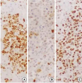
Fig. 4.Immunohistochemical demonstration of TIA-1 (A), CD30 (B), and granzyme B (C) in the tumor tissue (PAP, ×100).
A B C
TCR gene rearrangement with an expression of TIA-1 and granzyme B, indicating activated cytotoxic T-cells pheno- type without definite immunoreactivity of CD8. Recently, several studies have shown that the expression of these cyto- toxic proteins in PTCL tumors is associated with an extran- odal site, a T gamma delta-cell phenotype, CD30 expres- sion, and anaplastic feature (5). Rothenberg et al. (6) men- tioned an immune reaction of cytotoxic T lymphocytes in an EBV-induced PTLD. The cytotoxic T cells predomi- nantly expressed TCR rather than TCR and mediated non-major histocompatibility-restricted cytotoxicity against EBV-infected cells. Under these circumstances, although we can not apply this suggestion to our case definitely, we think that the cytolytic granule-associated proteins may suggest cytotoxic T lymphocyte differentiation in spite of the nega- tive immunoreactivity of CD8. Also, EBV is known to induce the CD30 expression in EBV-transformed cell lines; there- fore EBV-associated posttransplant lymphomas may prove to be CD30+.
As differential diagnosis, we considered the possibility of anaplastic large cell lymphoma (ALCL) and hepatosplenic T-cell lymphoma. Ki-1-positive ALCL is a subtype of PTCLs showing CD30 immunoreactivity and only a few cases of Ki-1-positive B-cell lymphoma have been reported as PTLD.
However, with the focal CD30 expression, it might be im- proper to diagnose the present patient as ALCL without showing ALK positivity. We also considered the possibility of hepatosplenic T-cell lymphoma. The findings of the chain TCR gene rearrangement, the expression of TIA-1, and the negative reactions for both CD4 and CD8 have been described but a positive expression of CD30 and granzyme
B are not features of hepatosplenic T-cell lymphoma.
Lymphoproliferative disorders occurring in association with immunosuppression are unique. Of concern is the role of EBV in the pathogenesis of these EBV-associated PTLDs. Com- pared with B-cell PTLDs, T-cell PTLDs show a looser asso- ciation with EBV and more often monoclonality. EBV infec- tion was believed to be limited to B lymphocytes, follicular cells of lymph nodes and tonsils, and epithelial cells of the pharynx and cervix. B lymphocytes are known to be infected via the C3d receptor (CD21), which is expressed constantly in benign and malignant B cells. However, recent reports have shown that CD21 and EBV may be present in some malignant T-cells. Medeiros et al. (7) suggested that EBV may be involved in the transformation of low grade T cells proliferation to high grade lesions, and Huh et al. (8) described the high incidence of EBV in peripheral T cell lymphomas in Koreans. However, the exact mechanism of how the EBV get into the T-cells is still unclear. Because most B-cell PTLDs are primary EBV infections, a lack of previous EBV infection is a risk factor of B-cell PTLDs. Conversely, chronic EBV infection was assumed to be the cause of EBV-associated T- cell lymphoma in the previous reports. All 10 cases of EBV- associated T-cell lymphomas reported by Su et al. (9). had evidence of previous infection, and none of them had an ele- vated level of IgM class anti-VCA. In our patient, IgM and IgG EA-DR were positive. This suggests reactivation of a past EBV infection. We could not find a report of T-cell PTLD with IgM positivity in the literature.
Clinically, at the time of diagnosis, most of the T-cell PTLD cases had fully developed T-cell malignant neoplasms. The long-term survival of patients with T-cell PTLD is generally poor, as was shown in the present case and in others. Most of the patients died within 1 yr from the diagnosis. In view of high mortality rate of this lymphoma, early diagnosis is critical to the patients’ outcome.
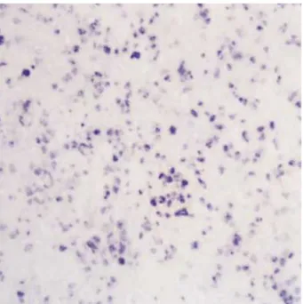
Fig. 5.Diffuse positivity for EBV on in situ hybridization using the EBV EBER1 RNA probe (×100).
Fig. 6.Polymerase chain reaction (PCR) analysis for T-cell recep- tor V-gamma-J-gamma reveal a clonal V-gamma-J-gamma rear- rangement (lane T). Lane N is the negative control and lane P is the positive control.
P T N
To our knowledge, this is the first case report of a post- transplant T-cell lymphoma involving spleen, which is asso- ciated with EBV positivity, gamma TCR gene rearrangement, cytolytic granule-associated proteins, and CD30 positivity.
REFERENCES
1. Frizzera G. Atypical lymphoproliferative disorders. In: Knowles DM editor, Neoplastic hematopathology. Lippincott Williams & Wilkins 2001; 569-622.
2. Craig FE, Gulley ML, Banks PM. Posttransplantation lymphoprolif- erative disorders. Am J Clin Pathol 1993; 99: 265-76.
3. Hanson MN, Morrison VA, Peterson BA, Stieglbauer KT, Kubic VL, McCormick SR, McGlennen RC, Manivel JC, Brunning RD, Litz CE.
Posttransplant T-cell lymphoproliferative disorders: an aggressive, late complication of solid-organ transplantation. Blood 1996; 88: 3626-33.
4. Weiss LM, Wood GS, Nickoloff BJ, Sklar J. Gene rearrangement studies in lymphoproliferative disorders of skin. Adv Dermatol 1988;
3: 141-60.
5. Kanavaros P, Boulland ML, Petit B, Arnulf B, Gaulard P. Expression of cytotoxic proteins in peripheral T-cell and natural killer-cell (NK) lymphomas: association with extranodal site, NK or T gamma delta phenotype, anaplastic morphology and CD30 expression. Leuk lym- phoma 2000; 38: 317-26.
6. Rothenberg ME, Weber WEJ, Longtine JA, Hafler DA. Cytotoxic gamma-delta T lymphocytes associated with an Epstein-Barr virus- induced posttransplantation lymphoproliferative disorder. Clin Immunol Immunopathol 1996; 80: 266-72.
7. Medeiros LJ, Jaffe ES, Chen Y-Y, Weiss LM. Localization of Epstein- Barr viral genomes in angiocentric immunoproliferative lesions. Am J Surg Pathol 1992; 16: 439-47.
8. Huh J, Cho K, Heo DS, Kim JE, Kim CW. Detection of Epstein-Barr virus in Korean peripheral T-cell lymphoma. Am J Hematol 1999;
60: 205-14.
9. Su IJ, Hsieh HC, Lin KH, Uen WC, Kao CL, Chen CJ, Cheng AL, Kadin ME, Chen JY. Aggressive peripheral T-cell lymphomas con- taining Epstein-Barr viral DNA: a clinicopathologic and molecular analyisis. Blood 1991; 77: 799- 808.