Baicalein and Baicalin from the Radix of Scutellaria baicalensis Georgi Inhibits Oxidative DNA Damage and Apoptosis via
its Antioxidant Activity
Nellie Ann S. Garcia1,2 and Hyung Jin Jeong2*
1University of Philippines, Manila, Philippines
2School of Bioresources, Andong National University, Andong 760-749, Korea
Abstract - In this study, we evaluated and compared the protective effects of two major constituents, baicalein and baicalin, against oxidative DNA and cell damages caused by hydroxyl radical. Antioxidant properties were evaluated using DPPH and hydroxyl radicals scavenging assays and Fe2+ chelating assay. φX 174 RFI plasmid DNA and intracellular DNA migration assay were used to evaluate the protective effect against oxidative DNA damage. Also, MTT and lipid peroxidation assays were used to evaluate their protective effects against oxidative cell damage. Both baicalein and baicalin prevented intracellular DNA and cells from oxidative damage caused by hydroxyl radical via antioxidant activities. Baicalein demonstrated a stronger antioxidant activity in scavenging DPPH radicals and chelating Fe2+ while baicalin scavenged hydroxyl radicals more efficiently. The differences in the level of baicalein and baicalin pose a different pathological pathway for each. The antioxidant activity of baicalin was due to its ability to scavenge hydroxyl radical whilst baicalein was a stronger Fe2+ chelator.
Further investigation to compare the molecular mechanisms of antitumor activities of baicalein and baicalin is vital to anticancer research.
Key words - Reactive oxygen species (ROS), Oxidative DNA damage, Oxidative cell death, Lipid peroxidation
*Corresponding author. E-mail : [email protected]
Introduction
Scutellaria baicalensis Georgi is one of the most widely used traditional Chinese herbal medicines. Its roots have been used for anti-inflammation, anti-cancer, antiviral and antibac- terial infections of the respiratory and the gastrointestinal tract, cleaning away heat, moistening aridity, purging fire, detoxi- fying toxicosis, reducing the total cholesterol level and decrea- sing blood pressures (Li, 2004). Two of its active compounds, the flavonoids baicalin and baicalein, have been extensively studied for their antioxidant activities. Flavonoids have been reported to be ideal candidates for reducing oxidative stress, since they possess free radical scavenging (Rice Evans, 2004) and metal ion chelating properties (More et al, 1993), and increase the expressions of certain antioxidant proteins (Cai and Wei, 1996; Sudheesh et al, 1999). Most interest has been devoted to the antioxidant activity of flavonoids, which is due to their ability to reduce free radical formation and to scavenge
free radicals. The capacity of flavonoids to act as antioxidants in vitro has been the subject of several studies in the past years (Pietta, 2000).
Oxidative DNA damage is mediated by reactive oxygen species (ROS) and is thought to play an important role in muta- genesis, aging and carcinogenesis (Barja, 2004). Aging and aging-related diseases might be due to the long term effects of oxidative damage to the cells and tissues of the body that arises primarily as a result of aerobic metabolism (Balaban et al., 2005; Wickens, 2001). Lots of research have clearly shown that many reactive oxygen species (ROS), such as hydrogen peroxide (H2O2), superoxide radical (O2), and hydroxyl radical (OH), would damage nearby structures including DNA, proteins or lipids (Barja, 2004; Benzie, 2000; Bokov et al., 2004; Yin and Chen, 2005). Radical scavenging antioxidants are particul- arly important in antioxidative defense in protecting cells from the injury of free radicals. Among DNA damage causing cancer development, approximately 80% of the damage is caused by the reactive oxygen species (ROS) such as hydrogen peroxide (H2O2), singlet oxygen (1O2), and hydroxyl radical (OH) (Ghosal
et al., 2005). Cancer pathogenesis is a multi-step process involving mutations in critical genes required for maintaining cellular homeostasis and the clonal expansion of these mutated cells (Armitage and Doll, 1954). The foremost is the ability to induce DNA damage that can cause to mutation if replication proceeds without proper repair. Oxidative DNA damage can lead to mutations and be suspected to be a major cause of cancer (Schwarz et al., 1984). Furthermore, persistent oxidative DNA damage can alter signaling cascades and gene expression, induce or arrest transcription, and increase replica- tion errors and genomic instability, all of which have been described in the progression of cancer development (Powell et al., 2005). Of the ROS, hydroxyl radical is the most reactive oxygen radical formed via Fenton reaction in living systems.
In general, this radical is considered to be a harmful byproduct of oxidative metabolism, which can cause molecular damage in living system, and also play a critical role in initiating and catalyzing a variety of radical reactions in the presence of oxy- gen (Livingstone, 2001).
In this study, protective effects of two major flavonoids from the radix of S. baicalensis Georgi were investigated on oxida- tive damage induced by hydroxyl radical in non-cellular system and cellular systems.
Materials and Methods
Chemical reagents
Mouse skin fibroblast cell line, NIH 3T3, was purchased from American Type Culture Collection (ATCC, Manassas, VA, USA) and the mediums used for the cell growth were purchased from GIBCO BRL Co. (NY, USA). All chemicals, baicalein and baicalin and the DPPH (1,1diphenyl-2-picryl hydrazyl) were obtained from Sigma Chemicals Co. (St. Louis, USA). φX-174 RF I plasmid was purchased from New Engl- and BioLabs (County Road Ipswich, MA)
Sample preparation and Identification of baicalein and baicalin by LC/MS
One kilogram of dried Scutellaria baicalensis Georgi radix was ground and extracted with 800 ml of 80% methanol with shaking for 24 hours. The methanol-soluble fraction was then
filtered, concentrated by a vacuum evaporator, and fractioned in a separating funnel with ethyl acetate. The ethyl acetate frac- tion was separated, evaporated by a vacuum evaporator, pre- pared aseptically, and kept in refrigerator (- 80℃) for further assays. For identification of baicalein and baicalin, the LC/MS system as an API-2000 LC/MS/MS system (Applied Biosys- tems, USA) was used, which has the power and performance of triple quadrupole mass spectrometry. The MS/MS was cou- pled with the HPLC for the detection of analytes. The following conditions were used for the system: (1) detector (DF): -200;
(2) CEM: 1900; (3) Polarity: negative; (4) scan type: Q1; (5) ion spray voltage: 5500; and (6) ion source gas: 15. Methanol and water (1:1) were used as the solvents. The injection volume was maintained at 10.0 µl at a flow rate of 10 µl/min. Data integration was performed with Analyst 1.4.1 software version (Applied Biosystems, USA).
DPPH radical scavenging activity
The antioxidant activities of baicalein and baicalin were evaluated first by monitoring its ability in quenching the stable free radical DPPH (Hus et al., 2006). Reaction mixture containing 40 μl of test samples of baicalein and baicalin (4 mg/ml dissolved in DMSO) and 760 µl of 300 μM DPPH ethanol solution in micro tube were incubated at 37℃ for 30 min and absorbance was measured at 515 nm according to the increasing concentrations of the test samples. The DPPH quen- ching ability was calculated from the log-dose inhibition curve.
All determination was carried out in triplicate.
Hydroxyl radical scavenging assay
Hydroxyl radical scavenging ability was measured according to a literature procedure (Smirnoff and Cumbes, 1989) with a few modifications. Hydroxyl radical was generated from fenton reaction between 1.5 mM FeSO4 and 6 mM H2O2 (1.4:1, v/v) at 37℃ for 30 min before the assay and detected by their ability to hydroxylate salicylate. The reaction mixture (1 ml) contained 760 µl of hydroxyl radical, 40 µl of varying concen- trations of baicalein and baicalin and 200 µl of sodium salicy- late (20 mM). After a reaction for 1 hour at 37℃, the absor- bance of the hydroxylated salicylate complex was measured at 562 nm. Hydroxyl radical scavenging ability was calculated from the log-dose inhibition curve. All determination was car-
ried out in triplicate.
Fe2+-chelating activity assay
This assay was measured according to a literature procedure (Hus et al., 2006) with a few modifications. The reaction mix- ture (800 µl) contained 120 µl of 2 mM FeCl2, 40 µl of varying concentrations of baicalein and baicalin and 640 µl of distil- led water. The mixture was shaken vigorously and left at room temperature for 5 min. After 5 min, 200 µl of 5 mM ferrozine was added and mixed. The absorbance of the Fe2+-ferrozine complex was measured at 562 nm. Fe2+-chelating activity assay was calculated from the log-dose inhibition curve. All determination was carried out in triplicate.
φX-174 RF I plasmid DNA cleavage assay
Conversion of the supercoiled form of plasmid DNA to the open-circular and further linear forms has been used as an index of DNA damage (Jung and Surh, 2001). For DNA cleavage assay by hydroxyl radical and ferrous iron, reaction mixtures (90 µl) contained 10 µl of φX-174 RF I plasmid DNA, 4 μl of varying concentrations of baicalein and baicalin, 76 µl of hydroxyl radical generated from Fenton reaction between 250 µl of 1.5 mM FeSO4 and 175 µl of 6 mM H2O2. The mix- tures were incubated at 37℃ for 30 min. After 30 min, 10 µl of a solution containing 50% glycerol (v/v), 40 mM EDTA and 0.05% bromophenol blue was added to stop the reaction and the reaction mixtures was electrophoresed on 1% agarose gel. The DNA in the gel was visualized and photographed under ultraviolet light after ethidium bromide staining.
Intracellular DNA damage assay
This assay was carried out according to literature procedure (Cho et al., 2008) with some modifications. NIH 3T3 cells (2×
106) were cultured in 6-well plates for 24 hours at 37℃ in an incubator with a humidified atmosphere of 5% CO2. After 24 hours, the cells were treated with the varying concentrations of baicalein and baicalin for 30 min and then added with 1.5 mM FeSO4 and 6 mM H2O2 (1.4:1, v/v) for 1 hour. After 1 hour, each cell was harvested and then the supernatant was discarded. Each cell was resuspended with 20 μl of lysis buffer containing 50 mM Tris-HCl, pH 8.0, 10 mM EDTA, 0.5% SDS and 0.5 mg/ml proteinase K by pipetting cells to
ensure complete lysis and then incubated at 55℃ for 60 min.
After 60 min, each cell was centrifuged, 5 μl of RNase A was added to the supernatant, and each cell was incubated at 55℃
for 60 min. After 60 min, each cell was spun briefly to remove any further cell debris and collect the supernatant. Each lysate was heated at 70℃ for a few minutes and mixed with 10 μl of loading buffer (50% glycerol (v/v), 40 mM EDTA and 0.05%
bromophenol blue). the reaction mixtures was electrophoresed on 2% agarose gel, and the DNA in the gel was visualized and photographed under ultraviolet light after ethidium bromide staining.
Cell viability assay
NIH 3T3 (5×10³ cells/well) were cultured in a 96-well plate at 37℃ for 24 hours. After 24 hours, the varying concent- rations of baicalein and baicalin were treated to correspon- ding wells, and then incubated at 37℃ for 30 min. After 30 min, 10 µl of hydroxyl radical generated by fenton reaction between 1.5 mM FeSO4 and 6 mM H2O2 (1.4:1, v/v) was appli- ed to each well and then incubated at 37℃ for 24 hours. After 24 hours, 50 µl of MTT solution (1 mg/ml) was treated to each well for 4 hours, then the supernatant was removed and 100 µl DMSO was injected to each well. The absorbance was mea- sured with a microplate reader at 570 nm.
Lipid peroxidation assay
This assay was carried according to literature procedure (Kang et al., 2008) with some modifications. The NIH 3T3 cells were cultured in a 6-well plate at 2×10⁶ cells/well for 16 hours.
Sixteen hours after plating, the cells were treated with the varying concentrations of baicalein and baicalin for 30 min.
After 30 min, 1 mM H2O2 and FeSO4 were added to the plate and then the cells were incubated for 12 hours. The cells were then washed with cold phosphate-buffered saline (PBS), harve- sted, and homogenized in an ice-cold 1.15% KCl. One hundred microliter of the cell lysate was mixed with 0.1 ml of 8.1%
sodium dodecylsulfate, 0.75 ml of 20% acetic acid (adjusted to pH 3.5), and 0.75 ml of 0.8% thiobarbituric acid (TBA). The mixtures were made up to a final volume of 4 ml with distilled water and heated to 95℃ for 2 hours. After cooling to room temperature, 2.5 ml of an n-butanol/pyridine mixture (15:1, v/v) was added and the mixtures were shaken. After centrifu-
Baicalein, MW 270
Fig. 1. LC/MS chromatogram of extracts containing baicalein purified from ethyl acetate fraction from the radix of Scutellaria baicalensis Georgi.
Baicalein, MW 446
Fig. 2. LC/MS chromatogram of extracts containing baicalin purified from ethyl acetate fraction from the radix of Scutellaria baicalensis Georgi.
gation at 1000ⅹg for 10 min, the supernatant fractions were isolated and the absorbance was measured spectrophotomet- rically at 532 nm.
Statistical analysis
The series of experiments were performed as three or more independent examination with at least three replicates for each sample. Data were expressed as means ±S.D. Statistical comparison was performed using Student’s t-test.
Results
Identification of baicalein and baicalin by LC/MS
Liquid chromatography coupled with mass spectrometry (LC/MS) is a powerful tool for the rapid identification of chemi- cal constituents in plant extracts (Han et al., 2006). Fig. 1 and 2 show the LC/MS chromatograms of baicalein and baicalin
from the radix of S. baicalensis Georgi.
Antioxidant activities of baicalein and baicalin
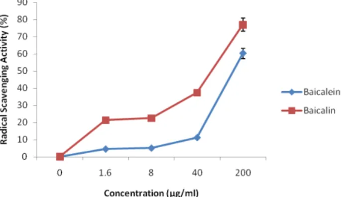
The antioxidant activities of baicalein and baicalin were evaluated by DPPH radical scavenging assay (Fig. 2), hydro- xyl radical scavenging assay (Fig. 3) and Fe2+ chelating assay (Fig. 4). The results show a dose-dependent effects in scaven- ging DPPH and hydroxyl radicals as well as in chelating the Fe2+. Baicalein showed higher antioxidant activities in scaven- ging the DPPH radical and chelating the Fe2+ while baicalin was more effective in removing the hydroxyl radical. Baicalein scavenged DPPH by 6.91% at 0.32 μg/ml, 31.27% at 1.6 μg/ml, 87.11% at 8 μg/ml, 87.16% at 40 μg/ml and 87.72% at 200 μg/ml, respectively, while baicalin scavenged DPPH by 3.21% at 0.32 μg/ml, 14.18% at 1.6 μg/ml, 60.16% at 8 μg/ml, 88.97% at 40 μg/ml and 89.19% at 200 μg/ml. Moreover, baicalein’s IC50
value was two-fold higher than baicalin at 2.9 for baicalein and
Fig. 3. DPPH radical scavenging activity of baicalein and baicalin purified from the extracts of the radix of Scutellaria baicalensis Georgi. The absorbance values were converted to scavenging effects (%) and data plotted as the means of replicate scavenging effect (%) against extract concentration in μg baicalein or baicalin per ml reaction volume.
Fig. 4. Hydroxyl radical scavenging activity of baicalein and baicalin purified from the extracts of the radix of Scutellaria baicalensis Georgi. The absorbance values were converted to scavenging effects (%) and data plotted as the means of replicate scavenging effect (%) against extract concentration in μg baicalein or baicalin per ml reaction volume.
Fig. 5. Fe2+ chelating activity of baicalein and baicalin purified from the extracts of the radix of Scutellaria baicalensis Georgi.
The absorbance values were converted to scavenging effects (%) and data plotted as the means of replicate scavenging effect (%) against extract concentration in μg baicalein or baicalin per ml reaction volume.
7.5 for baicalin. As for hydroxyl radical scavenging assay, baicalein removed hydroxyl radical by approximately 4.53%
at 1.6 μg/ml, 5.08% at 8 μg/ml, 11.21% at 40 μg/ml and 60.28%
at 200 μg/ml, respectively. Baicalin, on the other hand, remo- ved hydroxyl radical by approximately 21.35% at 1.6 μg/ml, 22.57% at 8 μg/ml, 37.34% at 40 μg/ml and 77.09% at 200 μ g/ml, respectively. In Fe2+ chelating assay, baicalein chelated Fe2+ ions by approximately 3.11% at 1.6 μg/ml, 15.83% at 8 μg/ml, 35.88% at 40 μg/ml and 81.47% at 200 μg/ml, respectively while baicalin chelated Fe2+ ions by approximately 2.62% at 1.6 μg/ml, 3.37% at 8 μg/ml, 14% at 40 μg/ml and 26% at 200 μg/ml.
Protective effects of baicalein and baicalin on oxidative DNA damage in non-cellular and cellular system
Protective effects of baicalein and baicalin from the radix of Scutellaria baicalensis Georgi on oxidative DNA damage was evaluated by the plasmid DNA cleavage assay using φ X-174 RFI plasmid DNA in non cellular system and intracel- lular DNA migration in the cellular system. In the plasmid DNA cleavage assay (Fig. 6A and 7A), induction of single strand breaks to supercoiled double stranded plasmid DNA leads to formation of open circular DNA, while the formation of a linear form of DNA is indicative of double strand breaks (Li and Trush et al., 1993). Fig. 6A and 7A show the gel elect- rophoretogram of the cleavage of the plasmid DNA by hydro- xyl radical. As observed in the figures, the plasmid DNA was mainly of the supercoiled form in the absence of hydroxyl ra- dical (untreated group). When in addition of hydroxyl radical without baicalein or baicalin, the supercoiled DNAs were converted into an open circular form. In the presence of hyd- roxyl radical, however, addition of baicalein inhibited the conversion at 17% at 1.6 μg/ml, 29% at 8 μg/ml, 52% at 40 μg/ml and 60% at 200 μg/ml while baicalin inhibited the con- version of the supercoiled form into the open-circular or linear form by 15% at 1.6 μg/ml, 43% at 8 μg/ml, 68% at 40μg/ml and 72% at 200 μg/ml, respectively. DNA migration assay is a sensitive biomarker of the DNA damage. In DNA migration assay (Fig. 6 and 7), both baicalein and baicalin inhibited
Fig. 6. Protective effects of baicalein purified from the radix of Scutellaria baicalensis Georgi against oxidative DNA cleavage using cleavage of φX-174 RF I plasmid DNA in the non-cellular system (A) and DNA migration in the cellular system induced by hydroxyl radical (B). Contents of the conversion from supercoiled form to open circular form were using the software Un-SCAN-IT gel Version 5.1 (Silk Scientific, Inc.).
Fig. 7. Protective effects of baicalin purified from the radix of Scutellaria baicalensis Georgi against oxidative DNA cleavage using cleavage of φX-174 RF I plasmid DNA in the non-cellular system (A) and DNA migration in the cellular system induced by hydroxyl radical (B). Contents of the conversion from supercoiled form to open circular form were using the software Un-SCAN-IT gel Version 5.1 (Silk Scientific, Inc.).
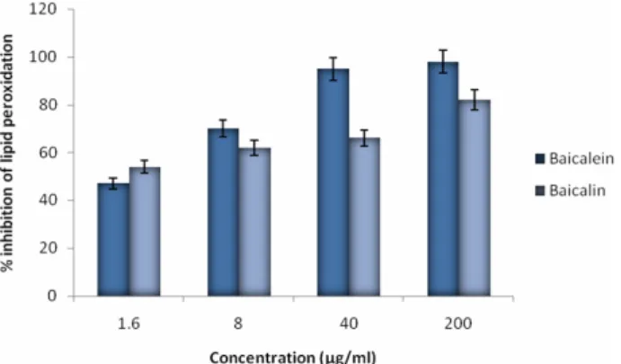
Fig. 8. Inhibitory effect of the baicalein and baicalin on lipid peroxidation. The effects of the flavonoids on the inhibition of lipid peroxidation were evaluated by measuring the amount of TBARS formation.
Fig. 9. Inhibitoryeffect of baicalein and baicalin from oxidative cell death. The viability of NIH 3T3 cells against the treatment of hydroxyl radical was evaluated by MTT assay.
DNA migration induced by hydroxyl radical in a dose- dependent manner.
Protective effects of baicalein and baicalin from the radix of Scutellaria baicalensis Georgi on oxidative cell damage
The effect of baicalein and baicalin on the oxidative cell damage was evaluated by lipid peroxidation assay (Fig. 8) and MTT assay (Fig. 9). In lipid peroxidation assay, baicalein inhi- bited lipid peroxidation by 30% at 1.6 µg/ml, 48% at 8 µg/ml, 55% at 40 µg/ml and 59% at 200 µg/ml while baicalin inhibi- ted peroxidation by 27% at 1.6 µg/ml, 42% at 8 µg/ml, 48% at 40 µg/ml and 52% at 200 µg/ml. In MTT assay, the cells treated with hydroxyl radical alone induced cell death by approxi- mately 50% compared with the untreated cells (control), while the addition of baicalein and baicalin in the presence of
hydroxyl radical inhibited oxidative cell death in a dose- dependent manner. Hydroxyl radical can react with a number of molecules including proteins, membrane lipids and DNA.
Oxidation of lipids induced by the hydroxyl radical can gene- rate products, such as malondialdehyde and unsaturated aldehy- des, that can bind to DNA to generate mutagenic adducts (Chaud- hary et al., 1994).
Discussion
POxidative stress causes various forms of tissue damage and inflammation, and plays an important role in the develop- ment of several degenerative changes in cells and tissues which ultimately lead to several degenerative disorders. Bodily de- fenses are not completely efficient in preventing on-going oxi- dative damage to DNA, lipids and proteins. Dietary antioxi- dants, vitamins, flavonoids, plant phenolics and herbal formula- tions are very essential in protecting against oxidative stress (Weiss & Landauer, 2000). Among DNA damage causing can- cer development, approximately 80% of the damage is caused by ROS such as hydrogen peroxide (H2O2), single oxygen (1O2) and hydroxyl radical (OH) (Ghosal, 2005).
Scutellaria baicalensis is one of the most popular and multi- purpose herb used in China traditionally for treatment of infl- ammation, hypertension, cardiovascular diseases and bacterial and viral infections. Accumulating evidence demonstrate that Scutellaria baicalensis possesses potent anticancer activities (Li-Weber, 2008).
In this study, we give a stronger background on the anti- cancer properties of Scutellaria baicalensis by evaluating its’
cancer chemopreventive property through inhibition of oxida- tive DNA damage and apoptosis caused by H2O2 and OH.
More specifically, we tested two of the major flavonoids from the radix of Scutellaria baicalensis called baicalein and bai- calin, in non-cellular and cellular systems. The principal find- ings from the above described experiments may be stated as follows; (I) baicalein and baicalin could scavenge DPPH free radical, hydroxyl radical and intracellular ROS, and could chelate Fe2+; (II) baicalein and baicalin could inhibit the strand scission in φX-174 RF I plasmid DNA mediated by hydroxyl radical, inhibit DNA migration mediated by hydro- xyl radical, inhibit oxidative cell death, and inhibit lipid pero-
xidation in cell membranes caused by hydroxyl radical.
Furthermore, it can be noted from our results that one of the flavonoids has a stronger activity than the other in some assays. Baicalein scavenged DPPH radical more efficiently and chelated Fe2+ on a much higher level than baicalin. The anti- fenton activity may be due to a combined effect of chelation and radical scavenging activities of baicalein. Also, the strong chelation of iron by baicalein appears to be in general agree- ment with the observations that flavonoids with an “iron- binding motif” can chelate iron under a number of different conditions such as buffer, pH and solvent (Guo, 2007).On the other hand, baicalin scavenged hydroxyl radical better. It also showed a consistently more favorable result as a better hydroxyl radical scavenger by inhibiting the strand scission φX-174 RF I plasmid DNA. However, baicalein inhibited lipid peroxi- dation in cell membranes better than baicalin, and showed a slightly higher activity in inhibiting oxidative cell death caused by H2O2. The differences in the level of antioxidant and free radical scavenging activities of baicalein and baicalin pose a different mechanism of action or pathological pathway between them. Further investigation to compare the molecular mecha- nisms of antitumor activities of the baicalein and baicalin is vital to anticancer research. The results in this study indicate that the radix of S. baicalensis Georgi possesses a spectrum of antioxidant and DNA-protective properties and showed addi- tional evidence that both baicalein and baicalin possess potent anticancer properties.
Acknowledgement
This work was supported by a support work for training problem solving human resources from Korea research founda- tion and by the second stage of BK21 grants from Ministry of Education and Human Resources Development and GB- Regional Innovation Research Program (2008), Korea
Literature Cited
Armitage, P. and Doll, R. 1954. The age distribution of cancer and a multi-stage theory of carcinogenesis. Br. J. Cancer 8:
1-12.
Balaban, R.S., S. Nemoto and T. Finkel, 2005. Mitochondria,
oxidants and aging, Cell 120: 483-495.
Barja, G. 2004. Free radicals and aging, Trends Neurosci. 27:
595-600.
Barreto, R., S. Kawakita, J. Tsuchiya, E. Minelli, K. Pavasu- thipasit, A. Helmy and F. Marotta. 2005. Metal-induced oxidative damage in cultured hepatocytes and hepatic lysoso- mal fraction: beneficial effect of a curcumin/absinthium compound. Chinese J. Digest. Dis. 6: 31-36.
Benzie, I.F.F. 2000. Evolution of antioxidant defence mecha- nisms. Eur. J. Nutr. 39: 53-61.
Bokov, A., A. Chaudhuri and A. Richardson. 2004. The role of oxidative damage and stress in aging, Mech. Ageing Dev.
125: 811-826.
Cai, Q and H. Wei. 1996. Effect of dietary genistein antioxi- dant enzyme activities in SENCAR mice. Nutr. Cancer 25:
1-7.
Chaudhary, A.K., M. Nokubo, G.R. Reddy, S.N. Yeola, J.D.
Morrow, I.A. Blair and L.J. Marnett. 1994. Detection of endogenous malondialdehyde-deoxyguanosine adducts in human liver. Science 265: 1580-1582.
Cho, E.S., K.W. Lee and H.J. Lee. 2008. Cocoa procyanidins protect PC12 cells from hydrogen-peroxide-induced apoptosis by inhibiting activation of p38 MAPK and JNK. Mutat. Res.
640: 123-130.
Esposito E., D. Rotilio, V. Di Matteo, C. Giulio, M. Cacchio and S. Algeri. 2002. A review of specific dietary antioxidants and the effects on biochemical mechanisms to neuro- degenerative processes. Neurobiol. Aging 23: 719-735.
Esterbauer, H., 1982. In: McBrien, D.C.H., Slater, T.F. (Eds), Free Radicals, Lipid Peroxidation and Cancer. Academic Press, New York, pp. 101-128.
Ghosal, D., M.V. Omelchenko, E.K. Gaidamakova, V.Y.
Matrosova, A. Vasilenko, A. Venkateswaran, M. Zhai, H.M. Kostandarithes, H. Brim, K.S. Makarova, L.P. Wackett, J.K. Fredrickson and M.J. Daly. 2005. How radiation kills cells: survival of Deinococcus radiodurans and Shewanella oneidensis under oxidative stress. FEMS Microbiol. Rev.
29: 361-375.
Grisham, M.B. 1992. Reactive oxygen metabolism. In: Gris- ham M.B., ed. Reactive metabolites of oxygen and nitrogen in biology and medicine. Austin: RG Landers Company. pp.
39.
Guo, M., C.A. Perez, Y. Wei, E. Rapoza, G. Su, F. Bou- Abdallah and N.D. Chasteen. Dalton Trans. (2007), pp. 4951- 4961.
Han, J., M. Ye, M. Xu, J.Sun, B.Wang and D. Guo. 2006. Cha- racterization of flavonoids in the traditional Chinese herbal medicine-Hangqin by liquid chromatography coupled with electrospray ionization mass spectrometry. J. Chrom. B 848:
355-362.
Hus, B., I.M. Coupar and K. Ng. 2006. Antioxidant activity of hot water EtOAC extract from the fruit of the Doum palm.
Hyphaene thebaica. Food Chem. 98: 317-328.
Jung, Y and Y. Surh. 2001. Oxidative DNA damage and cytoto- xicity unduced by copper-stimulated redox cycling of salsolinol, a neurotoxic tetrahydroisoquinoline alkaloid.
Free Radic. Biol. Med. 30: 1407-1417.
Kang, K.A., K.H. Lee, S.W. Chae, R. Zhang, M.S. Jung, Y.K.
Lee, S.Y. Kim, H.S. Kim, H.G. Joo, J.W. Park, Y.M. Ham.
L.H. Lee and J.W. Hyun. 2005. Eckol isolated from Ecklonia cava attenuates oxidative stress induced cell damage in lung fibroblast cells. FEBS Lett. 579: 6295-6304.
Kang, K.A., R. Zhang, M.J. Piao, D.O. Ko, Z.H. Wang, B.J.
Kim, J.W. Park, H.S. Kim, D.H. Kim and J.W. Hyun. 2008.
Protective effect of irisolidone, a metabolite of kakkalide, against hydrogen peroxide induced cell damage via antioxidant effect. Bioorgan. Med. Chem. 16: 1133-1141.
Li, H.B., Y. Jiang and F. Cheng. 2004. Separation methods used for Scutellaria baicalensis active components, J.
Chrom. B 812: 277-290.
Li, Y. and M.A. Trush. 1993. Oxidation of hydroquinone by copper: chemical mechanism and biological effects. Arch.
Biochem. Biophys. 300: 346-355.
Lloyd, R.V., P.M. Hanna and R.P. Mason. 1997. The origin of the hydroxyl radical oxygen in the Fenton reaction. Free Radic. Biol. Med. 22: 885-888.
Morel, I, G. Lescoat, P. Cogrel, O. Segen, N. Pasdeloup, P.
Brissot, P. Cillard amd J. CillardJ. 1993. Antioxidant and iron- chelating activities of the flavonoids catechin, quercetin and diosmetin on iron-loaded rat hepatocyte cultures.
Biochem. Pharmacol. 45: 13-19.
Moriwaki, H., M.R. Osborne and D.H. Phillips. 2008. Effect of mixing ions on oxidative DNA damage mediated by a Fenton- type reduction. Toxicol. in Vitro. 22, 36-44.
Namba, T.1993. Coloured Illustrations of Wakan-Yaku, Hoiku- sha, p. 152.
Pietta, P.G. 2000. Flavonoids as antioxidants. J. Nat. Prod. 63:
1035-1042.
Powell, C.L., J.A. Swenberg and I. Rusyn. 2005. Expression of base excision DNA repair genes as a biomarker of oxidative DNA damage. Cancer Lett. 229: 1-11.
Schwarz, S.M., G. Peres, W. Kunz, G. Furstenberger, W.
Kittstein and F. Marks. 1984. On the role of superoxide anion radicals in skin tumour promotion. Carcinogenesis. 5:
1663-1670.
Smirnoff, N. and Q.J. Cumbes. 1989. Hydroxyl radical sca- venging activity of compatible solutes. Phytochemistry. 28:
1057-1060.
Stohs, S.J. and D. Bagchi. 1995. Oxidative mechanism in the toxicity of metal ions. Free Radic. Biol. Med. 18: 321-336.
Sudheesh S., C. Sandaya, A. Sarah Koshy and N.R. Vijayala- kshmi. 1999. Antioxidant activity of flavonoids from Solanum melongena. Phytother .Res. 13: 393-396.
Vaca, C.E., J. Wilhelm and M. Harms-Ringdahl. 1988. Inte- raction of lipid peroxidation products with DNA. A review.
Mutat. Res. 195: 137-149.
(Received 7 April 2009 ; Accepted 8 December 2009)