This is an Open Access article distributed under the terms of the Creative Commons Attribution Non-Commercial License (http: //creativecommons.org/licenses/by- nc/4.0/) which permits unrestricted non-commercial use, distribution, and reproduction in any medium, provided the original work is properly cited.
© 2021 THE KOREAN SOCIETY OF MYCOLOGY.
Accepted: March 11, 2021 Revised: March 10, 2021 Received: February 01, 2020 https://doi.org/10.4489/KJM.20210008 Kor. J. Mycol. 2021 March , 49(1): 81-86
OPEN ACCESS pISSN : 0253-651X eISSN : 2383-5249
RESEARCH ARTICLE
목본식물 잎에서 분리된 Phyllosticta 속의 국내 미기록종 내생균
박혁
1, 이종철
1, 권주희
1, 이향범
2, 엄안흠
1,*1
한국교원대학교 생물교육과,
2전남대학교 농업생명과학대학 농생명화학과
Unrecorded Endophytic Fungi Belonging to Genus Phyllosticta Isolated from Leaves of Woody Plants
Hyeok Park
1, Jong-Chul Lee
1, Ju-Hui Gwon
1, Hyang Burm Lee
2, and Ahn-Heum Eom
1,*1
Department of Biology Education, Korea National University of Education, Cheongju 28173, Korea
2
Department of Agricultural Biological Chemistry, College of Agriculture & Life Sciences, Chonnam National University, Gwangju 61186, Korea
*
Corresponding Author: [email protected]
ABSTRACT
We isolated endophytic fungi from Smilax china and Cryptomeria japonica. These fungal strains were identified based on their morphological characteristics and phylogenetic analyses of their internal transcribed spacer, large subunit rDNA, and translation elongation factor 1-α DNA. Among them, we discovered two fungi belonging to the genus Phyllosticta, which have not been previously recorded in Korea. We have described these two fungal strains, Phyllosticta ericarum 19E458 and Phyllosticta philoprina 19E012 in this study.
Keywords: Endophytic fungi, Phyllosticta ericarum, Phyllosticta philoprina
서론
Phyllosticta Pers. 속은 자낭균류에 속하며, 병자각(pycnidium)에서 나오는 투명한 원통형의 분생 포자(conidia)를 형성하는 것이 특징이다[1,2]. Phyllosticta에 속하는 종은 300종 이상이며[3], 이 중 에는 간혹 Guignardia 혹은 Leptodothiorella 속의 무성 세대(anamorph)로 알려졌던 종들도 존재한다 [1]. Phyllosticta 속의 균류들은 식물 내생균(endophytic fungi), 부생균(saprophytic fungi), 식물 병원 균 등 다양한 형태로 살아가고 있으며[3-5], 국내에서도 Phyllosticta에 속하는 종들이 내생균[6-8]
혹은 식물 병원균[9]으로 보고된 바 있다. 본 연구에서는 목본식물의 잎에서 내생균으로 분리된
Phyllosticta 속의 국내 미기록종 2종에 대해 보고하고자 한다.
재료 및 방법
숙주 식물로는 전남 여수시 금오산의 청미래덩굴(Smilax china L.)과, 경남 통영시 한산도의 삼 나무(Cryptomeria japonica (Thunb. ex L. f.) D. Don)를 선정하였다. 병반 없이 깨끗한 식물 잎을 채 취하여 폴리에틸렌 백에 담아 24시간 이내에 실험실로 운반하였다. 잎은 증류수로 깨끗이 세척 한 후 1%의 NaClO 용액에 3분, 70% EtOH 용액에 2분 처리하여 표면살균 하고[10], 적당한 크기 로 잘라 potato dextrose agar (PDA) 배지 위에 올려놓았다. 배지를 25℃ 암소(dark side)에서 3일 이 상 배양하면서 균사가 뻗어 나오면 멸균된 메스로 잘라 새로운 PDA배지로 계대 배양하여 단일 종을 순수 분리하였다. 확보된 균주는 PDA배지와 더불어 malt extract agar (MEA) 배지에서 7일 간 배양하여 해부현미경 및 광학현미경 상에서 형태적 특성을 관찰하였다. 또한 DiaStar
TM10X
Direct Lysis Buffer (SolGent., Daejeon, Korea)의 protocol에 따라 균사에서 genomic DNA를 추출하 고, ITS1F와 ITS4 프라이머[11]를 이용하여 rDNA의 internal transcribed spacer (ITS) 영역을 증폭하 여 균주를 분자생물학적으로 동정하였다. 보다 정확한 동정을 위해 rDNA의 large subunit (LSU) 영 역을 LR0R과 LR16 프라이머[12]를 이용하여 증폭하였고, EF1-688F와 EF1-1251R 프라이머[13]
를 이용하여 translation elongation factor 1-α DNA를 증폭하였다. PCR이 끝난 DNA는 1.5% agarose gel에서 20분간 전기 영동을 진행하여 각각의 염기서열 단편 크기를 확인한 후 염기서열 분석을 의뢰하였다(SolGent, Daejeon, Korea). 분석된 염기서열은 미국 국립생물정보센터(NCBI) 상에서 BLAST하여 각각 유사도를 확인하였고, MEGA7 프로그램[14] 상에서 여러 영역의 DNA 염기서 열을 이용하여 phylogenetic tree를 작성하였다. 동정된 미기록종 균주는 국립생물자원관(NIBR) 에 기탁하였으며, BLAST 및 계통수 작성에 이용된 DNA 염기서열은 NCBI에 등록하여 GenBank
accession number를 부여 받았다.
결과 및 고찰
Phyllosticta ericarum Crous, Persoonia 28: 161 (2012) [MB#800377]
여수 금오산의 청미래덩굴 잎에서 분리된 균주이다. PDA 배지에서 7일간 배양된 균총의 직경
은 36-39 mm 정도이며, 균총의 앞면은 전체적으로 회색을 띠나 가장자리에 흰색의 균사들이 불
규칙하게 분포하고 뒷면은 전체적으로 어두운 회록색 혹은 올리브색을 띠나 앞면과 마찬가지로
가장자리에 흰색의 균사들이 보인다. 균총의 고도는 배지에서 살짝 융기 되어 있으며, 균총의 가
장자리는 전체적으로 둥근 형태이나 간혹 불규칙한 경우도 있다(Fig. 1A and 1E). MEA 배지에서
7일간 배양된 균총의 직경은 29-32 mm 정도로 PDA 배지에서 배양한 것보다 천천히 자라고, 앞면
은 베이지색의 영양 균사가 배지에 납작 붙어 있고 그 위를 연한 올리브색의 공중균사가 덮고 있
는 형태이다. 뒷면은 중앙부는 짙은 국방색을 띠고, 중간 부분에 살짝 노란빛이 감돌며 가장자리
는 밝은 흰색 혹은 베이지색을 띤다(Fig. 1B and 1F). 병자각은 검은색 혹은 회갈색의 구형이고, 병
자각이 열리면서 분생포자경이 발달되어 나오며 그로부터 분생포자가 형성된다(Fig. 1I). 분생포
자는 무색 투명한 원통형이며 바깥쪽의 점액질 층(mucoidal layer)과 안쪽의 포자벽이 뚜렷이 구
분되고, 끝부분에 분생포자경과 연결된 균사가 흔적으로 남아있는 경우도 있다. 분생포자의 크
Specimen examined. Keumosan mountain, Yeosu-si, Jeollanamdo, Korea, 34˚35 ´28.52˝N, 127˚47´57.13˝E, July 16, 2019, Phyllosticta ericarum, isolated from leaf of Smilax china, strain 19E458,
NIBRFG0000506771, GenBank No. MW521142.
Notes: P. ericarum은 2012년에 Crous에 의해 최초로 발표되었으며, 남아프리카 공화국의 진달래 속 식물(Erica gracilis) 잎에서 최초 분리되었다[15]. Saraca 속[16], Dioscorea 속[17] 등의 식물에서 내생균으로 분리된 기록이 존재한다. 점액질의 바깥층을 가진 원통형의 분생포자는 원 기재문에 기술된 것과 일치하였다 (Table 1). ITS 염기서열은 P. ericarum KR025424.1과 99.04%, LSU 염기서 열은 P. ericarum NG_042678.1과 99.67%, TEF 염기서열은 P. ericarum KR025452.1과 99.30%의 일치 도를 보였으며, 모두 같은 계통을 형성하였다(Fig. 2).
Phyllosticta philoprina (Berk. & M.A. Curtis) Wikee & Crous, Studies in Mycology 76 (1): 23 (2013) [MB#805660]
한산도의 삼나무 잎에서 분리된 균주이다. PDA 배지에서 7일간 배양된 균총의 직경은 17-19
mm 정도이고, 균총의 앞면은 연한 국방색 혹은 올리브색이며 뒷면은 전체적으로 검은색이나 가
장자리에 하얀색의 띠가 둘러싸고 있다. 균총의 고도는 배지에서 볼록 솟아 있고, 가장자리는 매
Fig. 1. Cultural characteristics of three endophytic fungal strains. Colonies of Phyllosticta ericarum
19E458 grown for 7 days on potato dextrose agar (PDA) (A: Surface, E: Reverse) and malt extract
agar (MEA) (B: Surface, F: Reverse), pycnidium (I) and conidia (J); Colonies of Phyllosticta philoprina
19E012 grown for 7 days on PDA (C: Surface, G: Reverse) and MEA (D: Surface, H: Reverse),
pycnidium (K) and conidia (L) (Scale bars: I, K=100 μm, J, L=10 μm).
우 불규칙한 형태이다(Fig. 1C and 1G). MEA 배지에서 7일간 배양된 균총의 직경은 20-21 mm 정 도이며, 앞·뒷면 모두 검은색과 베이지색, 그리고 흰색이 불규칙하게 섞여 있다. 균총의 고도는 배지에 납작 붙어 있고 가장자리는 불규칙한 형태이다(Fig. 1D and 1H). 황갈색 또는 적갈색의 병 자각으로부터 얇고 긴 분생포자경이 뻗어 나오며, 분생포자경으로부터 구형 혹은 원통형의 무색 투명한 분생포자가 형성된다(Fig. 1K). 분생포자는 주로 분생포자경에 붙어 있는 상태로 관찰되 고, 어린 분생포자에서는 바깥쪽의 점액층이 관찰되지만 성숙하면서 탈락되는 경우가 많다. 분 생포자의 크기는 (13.41-)18.10(-22.09)×(10.72-)13.10(-15.76) μm 정도이다(Fig. 1L).
Fig. 2. Neighbor-joining phylogenetic tree based on the three combined DNA regions, internal transcribed spacer (ITS), large subunit (LSU) and translation elongation factor (TEF) DNA sequences.
Test of phylogeny was 1,000 replicated with a bootstrap method. Botryosphaeria dothidea denotes an outgroup. The fungal strains isolated in this study are in bold.
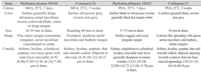
Table 1. Morphological characteristics of endophytic fungal species isolated in this study.
Strain Phyllosticta ericarum 19E458 P. ericarum [15] Phyllosticta philoprina 19E012 P. philoprina [1]
Colony MEA, 25℃, 7 days MEA, 25℃, 3 weeks PDA, 25℃, 7 days PDA, 25℃, 2 weeks
Color Surface generally beige, olivaceous aerial mycelium;
reverse yellowish-khaki, white or beige margin
Surface olivaceous grey;
reverse iron-grey Surface khaki to olivaceous; reverse
generally black but margin white A surface greenish black, reverse iron-grey
Size 36-39 mm in diam. Reaching 60 mm in diam. 17-19 mm in diam. 30 mm in diam.
Shape Flat, entire margin (sometimes irregular), aerial mycelium
concentrated in center
Erumpent, moderate aerial
mycelium and feathery margin Surface rugged, convexed,
irregular margin Colonies flat, spreading with sparse aerial mycelium, and feathery,
lobate margins Conidia Solitary, hyaline, cylindrical,
aseptate, two-layer spore wall, outer layer mucoidal, (6.92- )8.06(-9.59)×(5.42-)6.23(-7.08)
μm in diam.
Solitary, hyaline, aseptate, thin and smooth walled, ellipsoid or obovoid, (8-)9-10(-12)×(6-)7
μm in diam.
Solitary, subglobose to cylindrical, hyaline, mucoidal outer layer generally disappear in mature
conidia, (13.41-)18.10(- 22.09)×(10.72-)13.10(-15.76) μm
Solitary, hyaline, aseptate, thin and smooth-walled, ellipsoid, tapering towards a narrow truncate base, mucoid appendage, (10-)11-13(-
14)×(6-)8(-9) μm
Specimen examined. Hansando Island, Tongyoung-si, Gyeongsangnam-do, Korea, N34˚46´16.45˝, E128˚30´25.83˝, April 5, 2019, Phyllosticta philoprina, isolated from conifer leaf of Cryptomeria japonica, strain 19E012, NIBRFG0000506586, GenBank No. MW521141.
Notes: P. philoprina 는 1876년에 유성 세대인 Sphaeria philoprina로 최초 보고되었으며[18], van der Aa는 자낭 포자의 크기 외에는 종을 구분할 수 있는 형태적 특성이 없음을 근거로 이 종을 Guignardia philoprina 로 재조합하고 무성세대명을 Phyllosticta concentrica로 분류하였다[2]. 그러나 Wikee et al.은 2013년에 처음 미국에서 수집된 S. philoprina의 표본과 유럽에서 수집된 표본이 분 자생물학적으로 서로 다른 계통을 형성함을 근거로 Phyllosticta philoprina를 독립된 종으로 주장 하였다[1]. 본 연구에서 분리된 내생균 균주는 Slippers et al.에 의해 2013년 보고된[19] P. philoprina CBS 174.77 균주와 확실히 같은 계통을 형성함(Fig. 2)을 근거로 P. philoprina로 동정하였다 (Table 1). ITS 염기서열은 P. philoprina AB095507.1과 99.66%, LSU 염기서열은 P. philoprina KF766340.1과 99.84%, TEF 염기서열은 P. philoprina KF966401.1과 99.67%의 일치도를 보였다.
적요
전남 여수 금오산의 청미래덩굴 잎과 경남 통영 한산도의 삼나무 침엽을 표면살균하여 내생균 을 분리하였다. 분리된 균주는 internal transcribed spacer (ITS), large subunit rDNA (LSU), translation elongation factor 1-α DNA (TEF)의 염기서열을 이용한 계통분석 및 형태적 특성을 이용하여 동정 하였다. 본 연구에서 Phyllosticta 속에 속하는 두 종의 국내 미기록종 내생균이 확인되었으며, 확인 된 종은 Phyllosticta ericaum과 Phyllosticta philoprina이다. 두 종의 내생균 균주에 대해 형태적 특성 및 계통분석의 결과를 보고하고자 한다.
ACKNOWLEDGEMENT
This work was supported by a grant from the National Institute of Biological Resources (NIBR), funded by the ministry of Environment (MOE) of the Republic of Korea (NIBR201902202).
REFERENCES