247
강원 삼척 및 경북 영주의 담수지역에서 발굴된 수생균류 3 종의 국내 최초 보고
문혜연* · 고재덕 · 오유선 · 정남일
국립낙동강국립생물자원관 담수생물조사연구실 균류자원조사부
New Records of Three Aquatic Fungi Isolated from Freshwater in Samcheok and Yeongju, Korea
Hye Yeon Mun*, Jaeduk Goh, Yoosun Oh and Namil Chung
Fungal Resources Research Division, Freshwater Bioresources Research Bureau, Nakdonggang National Institute of Biological Resources, Sangju 37242, Korea
ABSTRACT : Three aquatic fungi were isolated from samples of freshwater-deposited plant litter and foam collected in Samcheok, Gangwon-do, and Yeongju, Gyeongsangbuk-do, Korea. Based on their morphological characteristics and a phylogenetic analysis of the internal transcribed spacer (ITS) rDNA region, the three isolates NNIBRFG329, NNIBRFG339, and NNIBRFG19 were confirmed as aquatic fungi: Articulospora tetracladia, Margaritispora aquatica, and Aquanectria penicillioides. These species were known as aquatic fungi but neither species has been previously reported in Korea.
KEYWORDS : Aquatic fungi, Aquanectria penicillioides, Articulospora tetracladia, Margaritispora aquatica
서 론
수생 불완전사상균(aquatic fungi = aquatic hyphomyce- tes)은 수생태계를 중심으로 생활사를 진행하는 다양한 종 류의 균류를 총칭하는 것으로서, 1942년 영국의 Ingold [1]
에 의해서 처음 정의되었다. 이들 균류는 용존산소가 풍부 한 청정수 하천에 침전되어 있는 분해중인 나뭇잎이나 유 기물에서 주로 발견된다. 이들은 유기물을 분해하여 2차대 사산물을 생산하는 등 생태계에 양분과 탄소원 순환에 중 요한 매개체 역할을 한다[2]. 우라늄 등의 중금속으로 오염
된 하천에서는 중금속을 흡착시키는 등 환경정화 역할도 하고 있다[3]. 현재까지 약 300종 이상의 수생균류가 알려 져 있으며, 대부분 자낭균문(Ascomycota)으로서 크게 Sor- dariomycetes (~11 spp.), Dothideomycetes (~10 spp.), Pe- zizomycetes (1 sp.), Orbiliomycetes (3~5 spp.), Leotiomy- cetes (>75 spp.)에 속하는 종들이 차지하고 있다[4]. 하지만 국내에서는 수생균류에 대한 보고나 연구가 거의 이루어지 지 않고 있는 실정이다.
본 연구에서는 3종의 수생균류, Leotiomycetes에 속하는 Articulospora tetracladia, Margaritispora aquatica, 그리고 Sordariomycetes에 속하는 Aquanectria penicillioides를 국 내의 담수환경에서 발굴하였으며, 이를 국내 미기록종으로 보고하고자 한다.
재료 및 방법
시료 채집
강원도 삼척시 소한천, 경상북도 영주시 죽계천의 담수지 역에서 침전식물체 및 포말 시료를 채집하였다. 담수 포말 은 병에 담은 후 물은 버리고 사용하며, 담수 침전식물체는 폴리에틸렌 봉투에 부식된 식물 잎을 담아서 채집하였다.
시료는 수분이 있는 상태에서 냉장보관하면서 실험에 사용
*Corresponding author E-mail: [email protected] Received November 25, 2016 Revised November 30, 2016 Accepted December 8, 2016
This is an Open Access article distributed under the terms of the Creative Commons Attribution Non-Commercial License (http://
creativecommons.org/licenses/by-nc/3.0/) which permits unrestricted non-commercial use, distribution, and reproduction in any medium, provided the original work is properly cited.
Kor. J. Mycol. 2016 December, 44(4): 247-251 https://doi.org/10.4489/KJM.2016.44.4.247 pISSN 0253-651X • eISSN 2383-5249
© The Korean Society of Mycology
하였다.
균 분리
채집한 담수 침전식물체 및 담수 포말 시료를 멸균수에 2 번 세척한 후 멸균수에 넣고 20oC에서 하루 정도 배양시켰 다. 배양된 물 100 μL를 water agar (WA; 20 g/L)에 도말한 후 2일 동안 25oC에서 배양하였다. 배양된 배지에서 단포자 분리를 통해 곰팡이를 순수분리하였다. 순수분리된 균류는 potato dextrose agar (PDA; Difco; BD, Franklin Lakes, NJ, USA) 및 malt extract agar (MEA; Difco; BD) 배지에 접종 하고 15oC 또는 20oC에 배양하여 실험에 사용하고, 일부는 15% 글리세롤에 담은 후 -80oC에 저장하였다.
포자생성 유도
분리된 수생균류의 광조건에 따른 포자생성을 조사하였 다. 포자생성을 조사하기 위해 멸균된 액체 배지(0.1 g CaCl2
·2H2O, 10 mg MgSO4·7H2O, 10 mg KNO3, 0.55 mg K2HPO4, 0.5 g MOPS buffer, 1 L distilled water) [5]에 agar 조각을 접종한 후 20oC에서 50 rpm으로 배양하면서 생성된 포자 수를 현미경으로 관찰하였다. 조건 1은 계속해서 광을 주고, 조건 2는 암상태를 계속해서 유지하면서 9일 동안 배양하 였다.
DNA 추출 및 분자계통수 작성
낙동강 유역의 담수 침전식물체 및 담수 포말 시료에서
Fig. 1. Neighbor-joining tree of alignment of internal transcribed spacers rDNA region of three aquatic fungi including Articulospora, Margaritispora and Aquanectria species from deposited plant litter and foam. Inocybe margaritispora was used as an outgroup. Boostrap values more than 50% (1,000 replications) were shown at branches.
분리된 균류는 PDA에 배양하여 균사체를 수확하였다. 수 확한 균사를 glass bead가 담긴 tube에 넣어 균질화시킨 후 NucleoSpin Plant II DNA extraction Kit (Macherey-Nagel, Düren, German)를 사용하여 DNA를 추출하였다. 추출된 DNA의 internal transcribed spacers (ITS) rDNA 부분을 증폭시키기 위해 ITS1 및 ITS4 프라이머를 사용하여 PCR 을 실시하였다[6]. DNA 염기서열 정렬 및 편집을 위해 Clustal X2.1 [7]와 Bioedit 7.2.5 [8]를 사용하였고, 계통수 작성을 위해 MEGA 6.07 [9]을 사용하였다.
결과 및 고찰
Articulospora tetracladia C.T. Ingold, Trans. Br. Mycol.
Soc. 25: 376 (1942)
Articulospora tetracladia는 잘 알려진 수생균류로서 영국 의 Ingold에 의해서 1942년에 처음 보고되었다[1]. 유성세 대로는 Hymenoscyphus tetracladius와 Ombrophila tetracla- dia가 알려져 있으나[10, 11], 많은 연구가 이루어지지 않은 실정이다.
NNIBRFG329 균주는 경북 영주시 죽계천의 담수 포말에 서 분리되었다. 이 균주의 ITS rDNA 부분 염기서열을 NCBI에 등록된 염기서열과 비교하여 분석한 결과, A. tet- racladia (GenBank Accession number FJ00386)와 99.4%
(521/524 bp)의 상동성을 보였으며, Fig. 1에서와 같이 계 통수 상에서도 A. tetracladia group에 속하는 것을 확인하 였다.
NNIBRFG329 균주는 MEA 배지에 배양했을 때 15oC에 서 7일 동안 20 mm 정도로 느리게 생장하였으며, 균사체 는 가운데 부분이 진한 갈색을 띄다가 가장자리 부분은 흰
색을 띄었다. 4개의 arm을 가진 tetraradiate 형태(tetrad hyphal-like form)의 포자를 가지고 있으며, 크기는 길이가 40~50 μm, 너비가 3~4 μm이었다(Table 1; Fig. 2A). 광조 건에 따른 포자생성율을 측정한 결과, 광을 준 3일 이후부 터 포자를 생성하는 것을 확인하였으며, 암조건에서는 7일 이 지난 이후에 포자를 생성하기 시작하였다 (Fig. 3A).
Margaritispora aquatica C.T. Ingold, Trans. Br. Mycol.
Soc. 25: 352 (1942)
Margaritispora aquatica는 1942년에 영국의 Ingold에 의 해 처음으로 발견되었다[1]. NNIBRFG339 균주는 경북 영 주시 죽계천의 담수 침전식물체(plant litter)에서 분리되었 다. 이 균주의 ITS rDNA 부분 염기서열을 NCBI에 등록된 염기서열과 비교하여 분석한 결과, Lemonniera centrosph- aera (GenBank Accession number KC834063)와 99.2%
(508/512 bp)의 상동성을 보였으나, 형태적인 특징이 일치 하지 않았다. 이에 Fungal Barcoding Database [12]에서 염 기서열을 비교 분석한 결과, M. aquatica CBS 258.84와 99.6%의 상동성을 보였다. 위의 균주를 CBS에서 분양받아 ITS rDNA 염기서열 분석을 실시하여 NNIBRFG339 균주 와 비교한 결과, 99.5% (411/413 bp)의 상동성을 보였으며, 계통수 상에서도 같은 group에 속하는 것을 확인할 수 있 었다(Fig. 1). 형태적 특징 및 분자계통분류학적 분석을 토 대로 NNIBRFG339 균주를 M. aquatica로 최종 동정하였 다.
이를 MEA 배지에 배양했을 때 15oC에서 7일 동안 25 mm 정도로 느리게 생장하였으며, 균사체는 가운데 부분이 분홍색을 띄다가 가장자리 부분은 갈색을 보였다. 둥근 장 사방형으로 큐빅(cubic) 모양의 포자를 가지고 있으며, 크
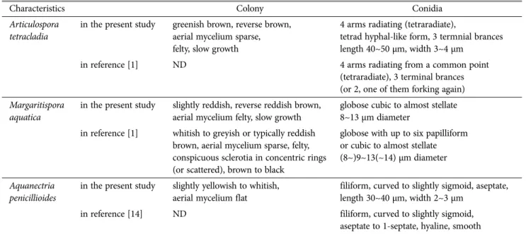
Table 1. Morphological characteristics of three aquatic fungi isolated in this study
Characteristics Colony Conidia
Articulospora tetracladia
in the present study greenish brown, reverse brown, aerial mycelium sparse, felty, slow growth
4 arms radiating (tetraradiate),
tetrad hyphal-like form, 3 termnial brances length 40~50 μm, width 3~4 μm
in reference [1] ND 4 arms radiating from a common point
(tetraradiate), 3 terminal brances (or 2, one of them forking again) Margaritispora
aquatica
in the present study slightly reddish, reverse reddish brown, aerial mycelium felty, slow growth
globose cubic to almost stellate 8~13 μm diameter
in reference [1] whitish to greyish or typically reddish brown, aerial mycelium sparse, felty, conspicuous sclerotia in concentric rings (or scattered), brown to black
globose with up to six papilliform or cubic to almost stellate (8~)9~13(~14) μm diameter
Aquanectria penicillioides
in the present study slightly yellowish to whitish, aerial mycelium flat
filiform, curved to slightly sigmoid, aseptate, length 30~40 μm, width 2~3 μm
in reference [14] ND filiform, curved to slightly sigmoid,
aseptate to 1-septate, hyaline, smooth ND, no description.
Fig. 2. Colony shape and conidia of three aquatic fungi. A, Articulospora tetracladia NNIBRFG329; B, Margaritispora aquatica NNIBRFG339; C, Aquanectria penicillioides NNIBRFG19 (scale bars = 20 μm).
Fig. 3. Effect of light for sporulation rates of three aquatic fungi. A, Articulospora tetracladia NNIBRFG329; B, Margaritispora aquatica NNIBRFG339; C, Aquanectria penicillioides NNIBRFG19.
기는 지름이 8~13 μm이었다(Table 1; Fig. 2B). 광조건을 계속 유지한 3일 이후부터 포자를 생성하였으며, 암조건에 서는 9일이 지나도 포자를 생성하지 않았다 (Fig. 3B).
Aquanectria penicillioides (Ingold) L. Lombard & P.W.
Crous, Stud. Mycol. 80:207 (2015)
Aquanectria penicillioides는 Flagellospora penicillioides로 Ingold [13]에 의해 최초로 보고되었으나, 이후 Lombard 등[14]에 의해서 Aq. penicillioides로 재명명되었다. NNIB RFG19 균주는 강원도 삼척시 소한천의 담수침전식물체 시 료에서 분리되었다. 이 균주의 ITS rDNA 부분 염기서열을 NCBI에 등록된 염기서열과 비교하여 분석한 결과, Aq.
penicillioides (GenBank Accession number KM231743)와 99.5%(547/550 bp)의 상동성을 보였다(Fig. 1).
NNIBRFG19 균주를 MEA 배지에 배양했을 때 15oC에서 7일 동안 40 mm 정도로 생장하였으며, 균사체는 가운데 부 분이 연노란색을 띄고 가장자리로 갈수록 흰색을 보였다.
Aq. penicillioides는 약간 S자 모양으로 구부러진 길고 가는 형태의 투명한 포자를 가지고 있으며, 크기는 길이는 30~
40 μm, 너비는 2~3 μm이었다(Table 1; Fig. 2C). NNIBRF G19 균주는 광조건과 암조건 모두에서 3일이 지난 후부터 포자를 생성하였다(Fig. 3C).
적 요
본 연구에서는 강원 삼척 및 경북 영주 지역의 하천에서 채집해 온 담수 침전식물체 및 담수 포말에서 균을 분리하 여 수생균류 3종을 발굴하였다. 분리된 균을 형태학적 특징 관찰과 internal transcribed spacers rDNA 유전자 분석을 통해 동정한 결과, NNIBRFG329, NNIBRFG339, NNIBR FG19 균주는 각각 Articulospora tetracladia, Margaritispora aquatica, Aquanectria penicillioides이었다. 이들 3종의 균류 는 불완전균강에 속하는 담수 서식 균류로 국내 미기록종 으로 보고하는 바이다.
Acknowledgements
This study was supported by a grant of the Nakdong- gang National Institute of Biological Resources (Project:
The survey and discovery of freshwater bioresources; NN IBR, 2016) of the Republic of Korea.
References
1. Ingold CT. Aquatic hyphomycetes of decaying alder leaves.
Trans Br Mycol Soc 1942;25:339-417.
2. Ferreira V, Encalada AC, Graça MA. Effects of litter diversity on decomposition and biological colonization of submerged litter in temperate and tropical streams. Freshw Sci 2012;31:
945-62.
3. Ferreira V, Goncalves AL, Pratas J, Canhoto C. Uranium adsorption by Articulospora tetracladia: can aquatic hypho- mycetes be natural bioremediators of uranium contaminated streams? In: Mendez-Vilas A, editor. Microorganisms in ind- ustry and environment: From scientific and industrial res- earch to consumer products. London: World Scientific; 2011.
p. 265-9.
4. Baschien C, Tsui CK, Gulis V, Szewzyk U, Marvanová L. The molecular phylogeny of aquatic hyphomycetes with affinity to the Leotiomycetes. Fungal Biol 2013;117:660-72.
5. Chauvet E, Suberkropp K. Temperature and sporulation of aquatic hyphomycetes. Appl Environ Microbiol 1998;64:1522- 5.
6. Glass NL, Donaldson GC. Development of primer sets designed for use with the PCR to amplify conserved genes from fila- mentous ascomycetes. Appl Environ Microbiol 1995;61:1323- 30.
7. Larkin MA, Blackshields G, Brown NP, Chenna R, McGettigan PA, McWilliam H, Valentin F, Wallace IM, Wilm A, Lopez R, et al. Clustal W and Clustal X version 2.0. Bioinformatics 2007;
23:2947-8.
8. Hall TA. BioEdit: a user-friendly biological sequence alignment editor and analysis program for Windows 95/98/NT. Nucleic Acids Symp Ser 1999;41:95-8.
9. Tamura K, Stecher G, Peterson D, Filipski A, Kumar S. MEGA 6: Molecular Evolutionary Genetics Analysis version 6.0. Mol Biol Evol 2013;30:2725-9.
10. Abdullah SK, Descals E, Webster J. Teleomorphs of three aqua- tic hyphomycetes. Trans Br Mycol Soc 1981;77:475-83.
11. Baral HO, Krieglsteiner GJ. Bausteine zu einer Askomyzeten- Flora der Bundesrepublik Deutschland: In Süddeutschland ge- fundene Inoperculate Discomyzeten: mit taxonomischen, öko- logischen, chorologischen Hinweisen. Verbreitung und Ökolo- gie ausgewählter Nichtblätterpilze in der Bundesrepublik Deu- tschland (Mitteleuropa). Tübingen: Dt. Ges. für Mykologie;
1985.
12. Fungal Barcoding Database. Identification; Pairwise sequence alignment [Internet]. Utrecht, The Netherlands: CBS-KNAW Fungal Biodiversity Centre; 2011 [cited 2016 Nov 4]. Available from: http://www.fungalbarcoding.org/BioloMICSSequences.
aspx?file=all.
13. Ingold CT. Some new aquatic hyphomycetes. Trans Br Mycol Soc 1944;27:35-47.
14. Lombard L, van der Merwe NA, Groenewald JZ, Crous PW.
Generic concepts in Nectriaceae. Stud Mycol 2015;80:189-245.