1
분당서울대학교병원 구강악안면외과 강동우, 김영균
턱관절 장애로 인한 청각장애의 치료: 증례보고
Treatment of hearing loss due to temporomandibular joint disorders: Case Report Department of Oral and Maxillofacial Surgery, Section of Dentistry, Seoul National University Bundang Hospital
Dong-Woo Kang. D.D.S., Young-Kyun Kim. D.D.S., PhD
Introduction : Patients with temporomandibular disorder may have various ear-related symptoms. If an excessive load is applied to the ear area due to the skeletal shape of the mandibular condyle or malposition of the disc, an auditory problems may occur.
Case report : The patient was referred to our clinic due to the suspicion of temporomandibular disorder from the local otorhinolaryngology clinic. A few days ago, his right ear could not be heard. MRI showed that the left TMJ disc was anterior displacement with reduction, the right TMJ disc was anteromedial displacement without reduction. Also Right mandibular condyle showed sclerotic bone change, subchondral cyst and was compressing the frontal wall of the ear on MRI view. Right TMJ arthroplasty was done under the diagnosis of right TMJ osteoarthritis and osteochondroma. Postoperative intermaxillary fixation was done with SAS screw and elastics for 2 weeks. One month after the operation, hearing and TMJ discomfort were recovered without any complications.
Conclusions As seen in this case, hearing loss due to benign tumor-like lesions of the temporomandibular joint should be treated surgically to restore the TMJ function and hearing.
Key words : Temporomandibular joint disorder, Hearing loss, Deafness
Corresponding Author
Young-Kyun Kim D.D.S., M.S.D., Ph.D.
Department of Oral and Maxillofacial Surgery, Section of Dentistry, Seoul National University Bundang Hospital E-mail: [email protected]
ABSTRACT
Ⅰ. 서론
턱관절 장애 환자들에게 이명, 귀 통증, 어지럼증, 청력소실, 현훈, 귀 충만감 등 귀와 관련된 다양한 증상들이 발생 할 수 있다1). 과두의 골격적인 해부학적 형태 이상이나 관절원판의 위치변화 등으로 인해 귀 부위에 과도하게 압박이 가해지는 경우 청각 이상이 발생할 수 있다. 하악 과두에서 발생하는 발생하는 증식성 병소들은 골종(osteomas), 연골종(chondromas), 골아세포종(osteoblastomas), 골연골종(osteochondromas), 연골모세포종(chondroblastomas), 거대세포종양 (giant cell tumor), 외골종(exostosis), 과형성 (hyperplasia) 등이 있는데, 임상 및 방사선학적으로 이들을 확실하게 구분하기는 쉽지 않다2). 과두의 증식성 병소로 인해 흔하게 나타나는 임상 증상들은 안면 비대칭, 부정교합, 개구 제한 및 귀 관련 증상 등이 있다. 임상적 특징, 방사선학적 소견, 조직병리학적 소견을 종합하여 최종 진단이 이루어지는데 Scintigraphy가 해당 부위의 집중적인 uptake 소견을 확인하기 위해 유용하게 사용될 수 있다3,4).
턱관절에 발생하는 증식성 병소들에 대한 궁극적인 치료 목표는 병소의 제거를 통해 해부학적으로 정상에 가까운 상태로 회복시킴으로써 안모 개선, 정상 교합 수복, 턱관절 기능 개선, 재발 방지의 목표를 달성하는 것이다. 증식성 병소를 완벽히 제거하지 못할 경우 재발을 초래할 가능성이 있지만, 병소를 완전히 제거하는 것은 해부학적으로 한계가 있다.
병소의 크기가 너무 커서 forcep으로 잡지 못하는 경우, 병소에 screw를 식립하여 screw에 steel wire 를 묶어 견인하여 제거하거나 교근이 부착된 관골궁 (zygomatic arch)을 일시적으로 골절시켜 제거 할 수 있다5).
저자 등은 하악 과두에 발생한 종양성 병소로 인해, 간헐적으로 우측 귀가 들리지 않는 증상, 부정교합, 우측 턱관절 염발음(crepitus), 턱관절 통증을 호소하는 환자를 외과적 치료를 통해 청력 회복 및 턱관절 기능을 회복한 증례를 문헌고찰과 함께 보고하고자 한다.
Ⅱ
.증례보고
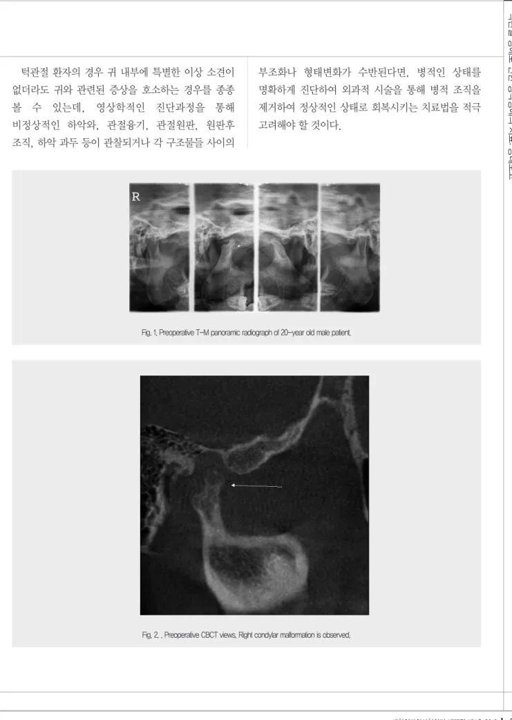
특별한 기저질환 없이 건강한 20세 남자 환자가 개인 이비인후과 의원에서 의뢰되었다. 환자는 우측 귀가 잘 들리지 않는 증상을 주소로 이비인후과 진찰을 받았지만 특별한 이상 소견이 발견되지 않았고 턱관절 증상이 의심되어 치과 구강악안면외과로 의뢰되었다. 초진 시 부정교합과 우측 턱관절 염발음 (crepitus)이 관찰되었고 개구 시 혹은 저작 시 양측 턱관절 통증과 턱 불편감 등을 호소하였다. T-M panorama, TMJ CBCT 및 MRI 촬영 결과 좌측 관절원판은 정복성 전방 변위 상태였고 우측 관절원판은 비정복성 전내측 변위 상태였으며 우측 과두는 연골 하방의 낭종성 변화와 경화성 골 변화를 보이며 귀 전벽을 압박하는 듯한 양상을 보였다.(Fig.
1,2,3) 우측 턱관절 골 관절염(osteoarthritis) 혹은 골 연골종(osteochondroma)으로 잠정 진단하고 전신마취 하에 턱관절 과두 성형술(Arthroplasty of TMJ) 및 종양성 병소 제거술을 시행하였다. 전이개 접근법으로 피부판 절개 및 박리를 시행하여 상관절강을 노출시킨 후 우측 턱관절 과두 후방부위 융기를 제거하였으며 제거된 시편은 조직검사를 의뢰하였다.(Fig. 4) 또한 우측 턱관절 과두의 외측으로 형성된 골극(osteophyte)을 제거한 후 관절융기절제술(eminectomy)을 시행하였다(Fig.
5,6). 전방으로 전위된 관절 원판은 후방으로 재위치
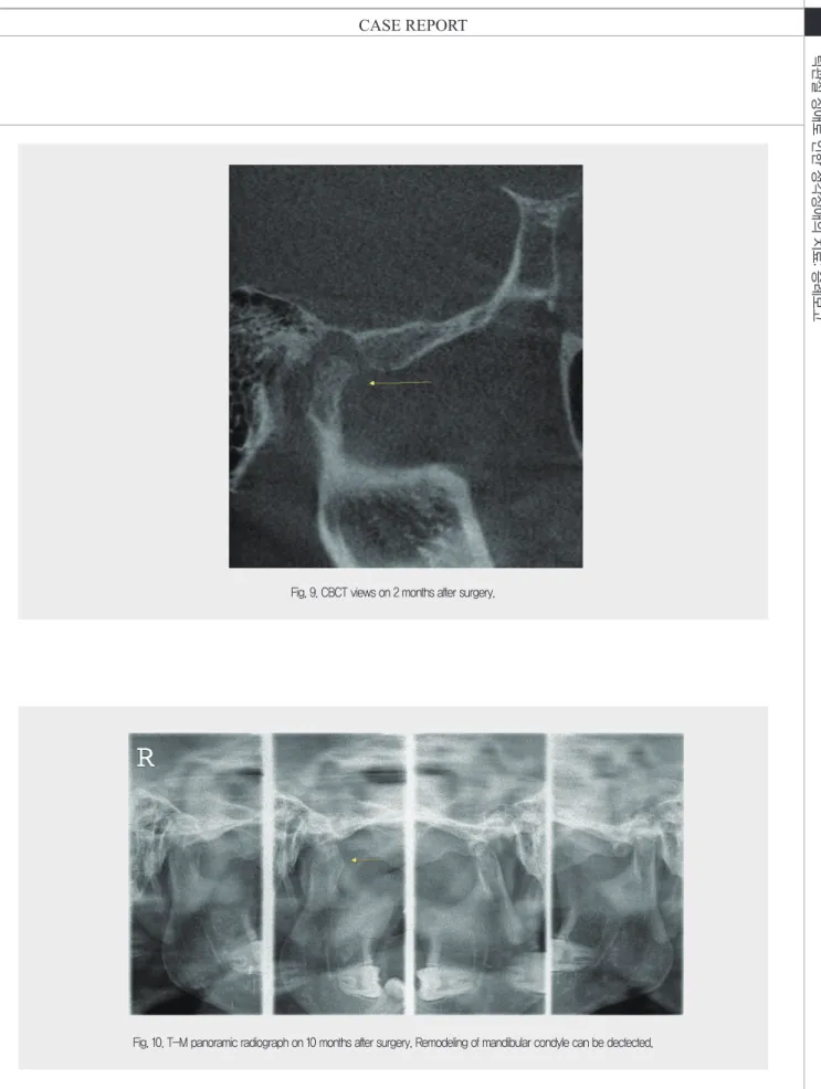
시킨 후에 봉합하여 고정하였다. 시킨 후에 봉합하여 고정하였다.(Fig. 7) 상관절강에 silastic drain을 삽입하고, Hyaluronic acid (Guardix-sol; Hanmi Pharm., Seoul, Korea)를 주입한 후에 층별 봉합을 시행하였다. 수술 직후 T-M panorama 영상에서 과두 성형술 및 종양성 병소가 제거된 상태를 확인하였다.(Fig. 8) 교합의 안정을 위해 약 2주간 악간 고정을 시행하였다. 조직검사 결과는 골종 (osteoma)으로 확인되었다. 수술 1개월 후 특별한 합병증 없이 청력, 부정교합 및 턱관절 불편감이 회복되었다. 수술 2개월 후의 TMJ CBCT에서도 큰 골성 변화 없이 잘 유지되고 있는 과두의 모양을 확인 하였다(Fig. 9). 술 후 10개월 뒤 최종 촬영한 T-M panorama 영상에서 잘 회복되어 재형성된 과두 및 피질골 형성을 확인 할 수 있었으며, 특이 합병증 없이 치료를 종결하였다(Fig. 10).
Ⅲ. 고찰
하악 과두의 증식, 관절원판의 후방 변위, 턱관절 내부의 염증이나 혈종으로 인해 외이도(external auditory meatus)에 압력이 가해지면 부분적인 청력 저하 증상이 생길 수 있다. Goyal과 Sidhu 등은 청각 이상을 유발하는 하악 과두의 광범위한 골 연골종 (osteochondroma)에 대해 보고한 바 있다6). Koole 등은 하악 과두의 광범위한 골 연골종으로 인해 청력 소실과 외이도의 폐쇄에 대해 보고한 바 있다7). 이전 연구들에 따르면, 골 연골종의 병인론은 명확히 밝혀지지 않았지만, 외상이나 감염이 기여요인으로 알려져 있다. Seki 등은 종양의 과증식으로 유발되는 측두골에서의 골경화성 변화가 중이염이나 외이염을 유발하여 완전한 청력 소실을 유발 할 수 있다고 보고하였다8).
턱관절 부위의 증식성 병소로 인해 턱관절이나 귀
혹은 턱얼굴 부위에 이상 증상들이 발생할 경우 외과적 처치가 필요하다. 하악 과두 절제술 (condylectomy), 불규칙한 하악 과두의 표면을 다듬는 하악 과두 성형술(condyloplasty) 등을 고려해 볼 수 있다. 그러나 증식성 병소를 제거하기 위해 하악 과두를 완전히 절제하는 것은 불가능하기 때문에 병소에 수반된 과두를 최대한 제거하고 남은 하악 과두나 과두의 경부를 재형성한 후 관절원판을 재위치 시키는 보존적인 하악 과두의 절제법이 추천된다9). 한편 관절융기 절제술은 습관적인 턱관절 탈구나 관절 내 압력을 줄이기 위해 관절융기를 제거하거나 높이를 낮추어 과두 걸림을 해소하고 관절강을 확대시켜 관절 내부의 압력을 줄여 조직이 눌려서 발생하는 통증, 염증을 감소시켜준다10). 관절원판 성형술 및 복위술은 관절을 재위치 시키거나 적절히 성형하여 외이도에 가해지는 압력을 줄일 수 있다11).
본 증례에서도 수술적 치료로, 우측 턱관절 외측의 골극(osteophyte) 제거를 포함한 과두 성형술 및 종양성 병소 제거, 관절융기절제술(eminectomy)을 시행하였다. 후방으로 전위된 관절 원판을 재위치하여 봉합하였다. 술 후에는 관절원판 및 교합의 안정을 위해 2주가량 악간 고정을 시행하였다. 술 후 일시적으로 청각이상이 보였으나 회복하였고, 턱관절의 기능 또한 회복하면서 특이 합병증은 발생하지 않았다. CT, MRI 등으로 진단하여 수술계획이 수립되더라도, 수술 전후에 이비인후과와 협진하여 청력 검사, 이경 검사법(otoscopy) 등을 시행하여 외이도나 고막의 상태를 확인하는 것이 필요하다. 본 증례에서는 수술 후 2주가량 청력이 회복되었다가 일시적인 청력 소실이 재발되어 이비인후과 진료를 꾸준히 보았는데, 특별한 처치 없이 1개월 후에 점차 회복되면서 자연치유 되었다.
수술 후 발생한 출혈, 부종, 염증성 물질, 귀지 (cerumen) 등에 의해 일시적으로 발생한 부분적 청력 소실로 추정된다.
턱관절 환자의 경우 귀 내부에 특별한 이상 소견이 없더라도 귀와 관련된 증상을 호소하는 경우를 종종 볼 수 있는데, 영상학적인 진단과정을 통해 비정상적인 하악와, 관절융기, 관절원판, 원판후 조직, 하악 과두 등이 관찰되거나 각 구조물들 사이의
부조화나 형태변화가 수반된다면, 병적인 상태를 명확하게 진단하여 외과적 시술을 통해 병적 조직을 제거하여 정상적인 상태로 회복시키는 치료법을 적극 고려해야 할 것이다.
Fig. 2. . Preoperative CBCT views. Right condylar malformation is observed.
Fig. 1. Preoperative T-M panoramic radiograph of 20-year old male patient.
Fig. 3. Preoperative MRI views. Subcondral cyst and sclerotic change at right mandible condyle were suspected.
Fig. 4. Intraoperative clinical photos on right TMJ. Right mandibular condyle posterior protuberance was dectected
Fig. 5 . Intraoperative clinical photos on right TMJ. Lateral osteophyte and posterior protuberance of right mandibular was resected.
Fig. 6. Intraoperative clinical photos on right TMJ. Lateral osteophyte and posterior protuberance of right mandibular condyle were removed.
Fig. 7. Intraoperative clinical photos on right TMJ. Displacement of right TMJ disc was repositioned and sutured.
Fig. 8. Postoperative T-M panoramic radiograph. Rt. TMJ arthroplasty was done.
Fig. 9. CBCT views on 2 months after surgery.
Fig. 10. T-M panoramic radiograph on 10 months after surgery. Remodeling of mandibular condyle can be dectected.
1. Fricton JR, Kroening R, Haley D, Siegert R.
Myofascial pain syndrome of the head and neck: a review of clinical characteristics of 164 patients. Oral Surg Oral Med Oral Pathol 1985;60:615-623
2. Thoma KH: Tumor of the mandibular joint. J Oral Surg 1964;22:158
3. Holmlund AB, Gynther GW, Reinholt FP. Surgical treatment of osteochondroma of the mandibular condyle in the adult. A 5-year follow-up. Int J Oral Maxillofac Surg 2004;33:549-553
4. Vezeau PJ, Fridrich KL, Vincent SD. Osteochondroma of the mandibular condyle: literature review and report of two atypical cases. J Oral Maxillofac Surg 1995;53:954-963
5. Chen MJ, Yang C, Qiu YT, He DM, Zhou Q,, Huang D, et al. Local resection of the mass to treat the osteochondroma of the mandibular condyle:
indications and different methods with 38-case series. Head Neck 2014;36:273–9
6. Goyal M, Sidhu SS: A massive osteochodroma of the mandibular condyle. Br J Oral Maxillofac Surg 1992;30:66
7. Koole R, Steeks MH, Witkamp TD, et al: Osteochondroma of the mandibular condyle: A case report. Int J Oral Maxillofac Surg 1996;25:203
8. Seki H, Fukuda M, Takahashi T, Iino M. Condylar osteochondroma with complete hearing loss: report of a case. J Oral Maxillofac Surg 2003;61:131-3 9. de Melo WM, Pereira-Santos D, Sonoda CK, de
Moura WL, de Paulo-Cravinhos JC. conservative condylectomy for management of osteochondroma of the mandibular condyle. J Craniofac Surg 2013;24:e209-e211
10. Kim HG, Choi HS, Huh JK, Park KH. Surgical treatment of recurrent TMJ dislocation by eminectomy with discoplasty. J. Kor. Oral Maxillofac.
Surg. 2002;28:141-146
11. Shim CH, Kim YK, An CM. Hearing difficulty according to traumatic disk displacement: A case report. J Korean Assoc Maxillofac Plast Reconstr Surg 2002;24:172-175
참 고 문 헌