The Whole Extract of Enterococcus faecalis Has Suppressive Effect on the Allergic Responses in Asthmatic Mouse Model
Jeong Hyun Chang
1, EunJu Yang
1, Sun Nyoung Yu
2and Soon-Cheol Ahn
2*
1
Department of Clinical Laboratory Science, Daegu Haany University, Gyeongsan 38610, Korea
2
Department of Microbiology and Immunology, Pusan National University School of Medicine, Yangsan 50612, Korea Received August 24, 2017 /Revised October 23, 2017 /Accepted October 25, 2017
Probiotics are usually defined as intestinal bacteria that provide healthy benefit to the host and may offer new therapeutic materials for the treatment of inflammatory diseases. Lactobacillus, Bifidobacterium and Enterococcus are known as typical probiotics. But, these bacteria have mostly a weak viability and thus decreased probiotics-mediated effects in the intestinal tract. Asthma is an inflammatory airway disease, which is characterized by the releases of inflammatory mediators including cytokine and IgE.
They are mainly associated with the recruitment, activation and disregulation of specific inflammatory cells, especially mast cells, monocytes, T cells, eosinophils and neutrophils in asthma. We performed these studies as in vitro and in vivo test the human inflammatory cell lines and ovalbumin (OVA)-induced asthma mouse model. And then the inhibitory effects of Enterococcus faecalis whole ex- tract on inflammatory responses were examined. For our examinations, the E. faecalis whole extract (Ef extract) was acquired from whole bacteria of E. faecalis using freeze/thawing after ultrasonication method. As results, OVA-mediated THP-1 cell viability was decreased by the treatment of Ef extract.
In the asthmatic mouse model, Ef extract inhibited the infiltration of inflammatory cells into the in- flammatory sites and blood. This whole extract may have anti-asthmatic effects associated with the regulation of IL-5 and IgE expression. It may also be a promising candidate in anti-allergic medicine for the treatment of asthma.
Key words : Asthma, Enterococcus faecalis, IgE, IL-5, inflammatory cell
*Corresponding author
*Tel : +82-51-510-8092, Fax : +82-55-382-8090
*E-mail : [email protected]
This is an Open-Access article distributed under the terms of the Creative Commons Attribution Non-Commercial License (http://creativecommons.org/licenses/by-nc/3.0) which permits unrestricted non-commercial use, distribution, and reproduction in any medium, provided the original work is properly cited.
Journal of Life Science 2017 Vol. 27. No. 10. 1168~1175 DOI : https://doi.org/10.5352/JLS.2017.27.10.1168
Introduction
Asthma is an inflammatory disease of the lungs charac- terized by increased infiltration of leukocytes, especially eo- sinophils, into the airways and reduced respiratory function.
The inflammation leads to bronchoconstriction, increased airway hyperresponsiveness, and mucus production [5]. In asthma, mast cells migrate into the inflammatory sites and are then activated by IgE, which is produced by activated plasma cells [18]. The activated mast cells secrete various pro-inflammatory mediators such as histamine, cytokines and prostaglandin, which induce eosinophilia and mucus production in lung tissue [1]. Eosinophils are infiltrated into the airways and act as primary effector cells by the release of specific granule proteins and reactive oxygen species [6].
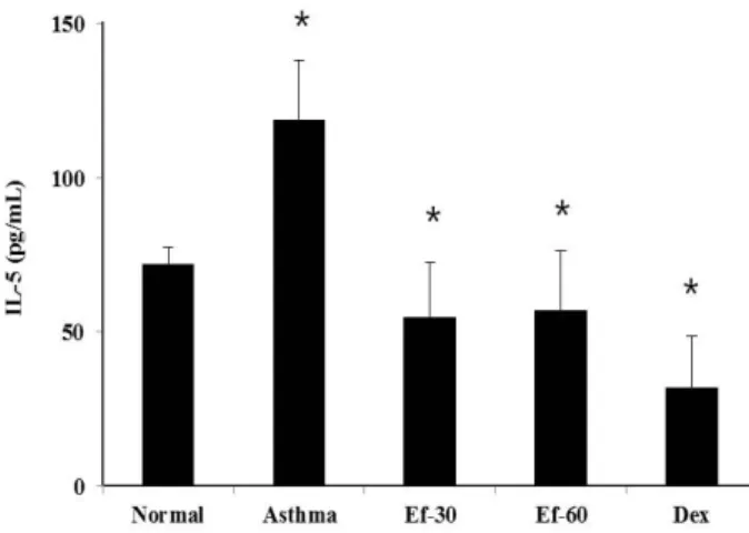
T helper (Th) 2 cytokines induce elevation of IgE in serum and BAL fluid [7]. In Th2 cytokines, IL-4 plays important roles in immunoglobulin E (IgE) switching in B cells, mucus hypersecretion, and eosinophil infiltration into the lung tis- sues [10]. IL-5 promotes the proliferation and activation of eosinophils and increases eosinophil infiltration into the air- ways [16]. In addition to IL-5, the modulations of other cyto- kines are closely linked with the development of asthma.
The prevalence of asthma is rapidly increasing around the world, especially in young children, and it has become a significant cause of morbidity and mortality in developed countries [2]. However, because the absence of proper treat- ment, we have been needed the development of therapeutic agents without adverse effect.
Normal flora is a microorganism that resides on a number
of tissues and organs. In various normal flora, probiotics are
usually defined as intestinal bacteria that provide healthy
benefit to the host and may offer new therapeutic materials
for the treatment of inflammatory diseases. Lactobacillus,
known as common probiotics, reduced 50% of prevalence
in early atopic dermatitis in children [8], and suppressed
proliferation and inflammatory response of CD4+ T cells
[22]. But, because these bacteria have almost a weak viability in the intestinal tract, Lactobacillus-mediated probiotic effects lead to be decreased. Other probiotics, Enterococcus may also be founded in vaginal tract and can contribute to the acidic environment. However, this bacteria can easily acquire an antibiotic resistance ability and then transmit resistance fac- tor to other harmful bacteria [19]. For these reasons, WHO has avoided that Enterococcus is being used as a probiotics.
Nevertheless, in other study, it has been reported that Enter- ococcus has suppressive effect on superantigen toxic syn- drome toxin-1 induced IL-8 from vaginal inflammation [3].
In the present study, to investigate the inhibitory effects of Enterococcus faecalis whole extract on asthmatic responses, its cytotoxic effects on the asthma-related cell was de- termined in vitro. The effects of E. faecalis whole extract on leukocyte infiltration, IgE level and IL-5 level were de- termined using in vivo Ovalbumin-specific asthma mouse model.
Materials and Methods
Preparation of Enterococcus faecalis whole extract Enterococcus faecalis (ATCC® 19433™) was purchased from American Type Culture Collection (ATCC, Manassas, VA, USA). E. faecalis was grown overnight in brain-heart in- fusion broth (BD, Franklin lakes, NJ, USA) at 37℃. After 24 hr, cultured bacteria were concentrated by centrifugation at 20,000× g for 10 min. Cell pellets were washed three times with PBS and suspended in 1 ml of PBS. To obtain bacteria whole extract, E. faecalis suspension was disrupted ultrasoni- cally at 20 kHz for 10 cycles of 30 sec (at 4°C). And then bacterial debris was heated at 75℃ for 30 sec, and lyophi- lized. The E. faecalis (Ef) whole extract was resuspended in PBS at a concentration of 10 mg/ml.
Cell viability
Cell viability of THP-1 cells was determined using an MTT assay kit (Roche, Penzberg, Germany). In brief, 100 μl of these cells (1×10
6cells/ml) were seeded in 96-well plate and incubated for 24 hr after treatment with various concen- trations (0, 30 and 60 μg/ml) of Ef extract and 3 μg/ml of dexamethasone (Dex), respectively. 10 μl of MTT solution (0.5 mg/ ml) was added, and the cells were incubated at 37℃ for 4 hr. 100 μl of solubilization solution was then add- ed to each well. After 24 hr incubation, the optical densities of 96-well culture plate were measured using a spectropho-
tometer (Bio-Tek Instruments, Winooski, VT, USA) at 540 nm. The optical density of untreated control cells was taken as 100% viability.
Induction of asthma in BALB/c mice
Six-week-old female BALB/c mice were obtained from DaehanBiolink Co. LTD (Seoul, Korea) and kept in an air-conditioned room at 22±1℃ and a humidity of 55±10%.
The mice were divided into five groups (n=4) and the aller- gic lung inflammation of the mice in four groups was in- duced by ovalbumin (OVA) (Grade III) (Sigma-Aldrich, MO, USA) using the established protocol in a previous study [23]. Asthmatic mice groups were induced four times OVA challenges with PBS or Ef extract. Briefly, mice were sensi- tized via intra-peritoneal injection with 75 μg of OVA ad- sorbed to 2 mg of aluminum hydroxide (Alum; Sigma- Aldrich) in 200 μl of 0.9% sterile saline on day 1, 2, 8 and 9. For challenge, 50 μg of OVA was intra-nasally ad- ministered to the mice on day 15, 16, 22.
The asthma-induced mice were treated 10 times with oral injection of 30 mg/kg and 60 mg/kg of Ef extract and 3 mg/kg of Dex during 20 days, respectively. The normal group was sensitized and challenged with PBS without OVA and Ef extract treatment.
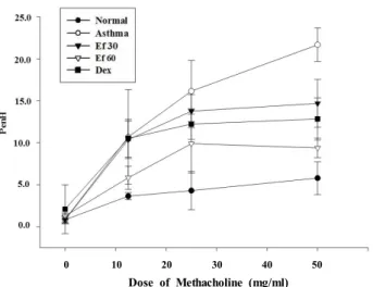
Measurment of airway hyperresponsiveness And day 23 and airway hyperresponsiveness (AHR) was measured on day 24 and the mice were sacrificed on day 25. After last OVA challenges, airway responsiveness was monitored a change using whole-body plethysmography for animals (Allmedicus, Anyang, Korea) in response to aero- solized methacholine (Sigma-Aldrich). The enhanced pause (PenH) was evaluated at baseline (PBS) and after treatment with increasing doses of aerosolized methacholine (0-50 mg/ml) for measuring bronchoconstriction. The mice were permitted to acclimate for 3 min, exposed to nebulized PBS for 10 min, and then subsequently treated in methacholine using an ultrasonic nebulizer (Omron, Koyto, Japan). After each nebulization, the average of PenH values were meas- ured during each 150 sec periods. All procedure for the han- dling and care of animals were approved by animal ethics committee of Pusan national university (ED-PNU2016-1056).
Collection of blood and serum
At day 25 after the first ovalbumin (OVA) sensitization,
the mice were sacrificed. And the blood was collected by
A B
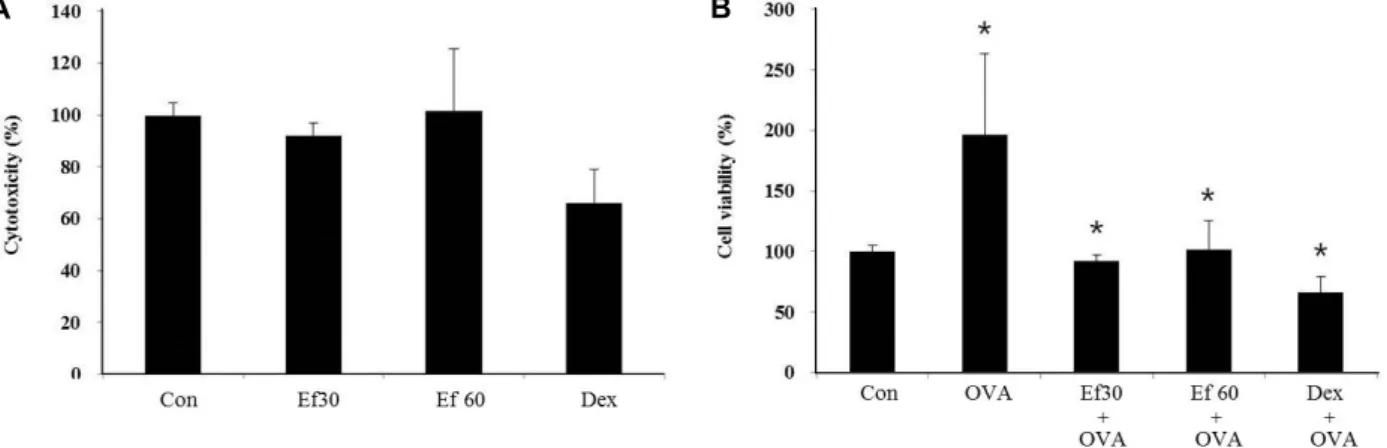
Fig. 1. The effect of Ef extract on the viability of THP-1 cells. THP-1 cells were seeded into 96-well plate at 5×10
4cells/100 μl per well. (A) These cells were treated with Ef extract at the concentration of 30 μg/ml (Ef 30), 60 μg/ml (Ef 60), or with 1 μg/ml of dexamethasone (Dex) for 24 hr. (B) THP-1 cells were pre-treated with the Ef extract at the concentration of 30 μg/ml (Ef-30), 60 μg/ml (Ef 60), or with 1 μg/ml of dexamethasone (Dex) for 1 hr. And then the cells were treated with 100 μg/ml of OVA for 24 hr. The cell viability was measured by MTT assay as described in Materials and Methods section. The data were expressed as the relative ratio to the absorbance of the untreated cells (Con), which was set at 100%.
All data were expressed as the means ± S.D. of three independent experiments. *p<0.05 indicates statistically significant differences between the control group .
heart puncture. After blood sampling, the blood smear films were prepared for leukocyte differential counting and stained with Wright staining solution. The cells in blood smear film were divided into neutrophils, eosinophils, baso- phils, monocyte and lymphocyte according to general leuko- cyte morphology and staining features. The percentage of each cell was determined by counting 300 cells per slide.
Also, blood were concentrated by centrifugation at 2,000×
g for 10 min and the supernatant (serum) was stored at -70℃
until analysis of IL-5 and immunoglobulin E (IgE).
Collection of bronchoalveolar lavage (BAL) fluid After the mice were sacrificed, BAL fluid was collected by lavage of the lung via the trachea with 1 ml of PBS. After three lavages, the total cells were separated from the super- natant of BAL fluid. The BAL fluid were used for the deter- mination of total cells and differential counting. Total cell number was counted using a Neubauer hemocytometer.
And the cells were cytospined and stained with Wright stain solution for differential counting. The cells in BAL fluid were divided into neutrophils, eosinophils, lymphocyte, and al- veolar macrophage according to general leukocyte morphol- ogy and staining features. The percentage of each cell was determined by counting 300 cells per cytospined slide.
Determination of cytokine concentrations
In the serum, the concentrations of IL-5 and the total IgE
were measured using OptEIA Set mouse IL-5 and IgE (BD, Franklin lakes, NJ, USA) according to the manufacturer’s instructions. All assays were performed in triplicate. The concentration of each protein was calculated from the stand- ard curve of indicated protein.
Statistical analysis
All data were expressed as the means ± S.E.M. Data were analyzed by one-way ANOVA or Student’s t test using the SPSS statistical software package, Version 10.0 (SPSS Inc., Chicago, IL, USA). A p value less than 0.05 was considered significant.
Results
Ef extract has the inhibitory effect on cellular inflammation induced by OVA
Prior to examining the effect of Ef extract on asthma
mouse model, it was investigated whether Ef extract has a
cytotoxic effect. For this examination, we used the THP-1
cell, the human monocytic cell line. Monocytes act on the
pathogenesis of asthma. The survival rate of THP-1 cell was
not changed after treatment with Ef extract for 24 hr in a
dose-dependent manner (Fig. 1A). These results indicate that
Ef extract used in this study have no cytotoxic effect on the
cells. Also Ef extract considerably decreases the elevated rate
of survival by OVA in THP-1 cells (Fig. 1B). Dex was used
0 10 20 30 40 50