龍眼肉 물추출물이 대식세포의 염증반응과 Cytokine에 미치는 영향
가천대학교 한의과대학 한방부인과학교실 김미림, 임은미
ABSTRACT
Effects of Longanae Arillus Water Extract on Inflammatory Response and Cytokines in Mouse Macrophage Cells
Mi-Rim Kim, Eun-Mee Lim
Dept. of Gynecology, College of Oriental Medicine, Ga-Chon University Objecives: The purpose of this study was to investigate the effects of Longanae Arillus water extract (LA) on the production of inflammatory mediators in RAW 264.7 cell. LA is used for forgetfulness, insomnia, palpitation symptoms in korean medicine.
Methods: In order to evaluate cytotoxicity of LA, cell viability was measured.
To investigate anti-inflammatory effects of LA in the lipopolysacharide (LPS)-induced RAW 264.7 cell, the concentration of nitric oxide (NO) and cytokines were measured.
And when p-value is below 0.05, it is judged to have the significant difference statistically.
Results:
1. LA showed no cytotoxicity.
2. LA inhibited significantly the production on NO at the concentration of 50 and 200 μg/ml.
3. LA inhibited significantly the production on interleukin (IL)-1β at the concentration of 25, 50, 100 and 200 μg/ml.
4. LA inhibited significantly the production on tumor necrosis factor (TNF)-α at the concentration of 50, 100 and 200 μg/ml.
5. LA inhibited significantly the production on lipopolysaccharide-induced chemokine (LIX) at the concentration of 25, 100 and 200 μg/ml.
6. LA inhibited significantly the production on regulated on activation normal T cell expressed and secreted (RANTES) at the concentration of 200 μg/ml.
Conclusions: These results suggest that LA has anti-inflammatory effect.
Key Words: Longanae Arillus, Anti-inflammation, Macrophage, NO, Cytokine
1)
Corresponding author(Eun-Mee Lim) : Dept. of Gynecology, Gil Oriental Medical, Gachon University, 1200-1, Guwol 1-dong, Namdong-gu, Incheon, Republic of Korea
Tel : 070-7120-5010 E-mail : [email protected]
Ⅰ. 서 론
염증반응이란 손상이나 괴사가 일어난 조직과 미생물 등을 제거하는 생명체의 생존에 필수적인 기능으로 최근에는 염 증반응이 신경계 퇴행의 진행 초기부터 관여할 수 있으며 병리현상의 완급을 조 절하고 신경세포의 생존과 사멸 조절에 적극적으로 관여한다고 알려졌다 1-3) .
이러한 염증 질환의 치료를 위하여 현 재 다양한 항염증제제 개발에 대한 연구 가 이루어지고 있고 이와 더불어 한약재 로부터 유발되는 생리활성물질이나 단일 추출물 및 한약복합화합물이 주목을 받 고 있다 4-7) .
용안육(龍眼肉, Longanae Arillus)은 용 안나무(龍眼樹, Euphoria lingan Steud, Dimocarpus longan Lour.)의 假種皮로, 性味가 甘溫質潤하여 心, 脾 二經에 들어 가 補益하고 營血을 滋養하므로서 양호한 安神作用을 가지고 있어 心脾血虛의 병증 을 치료하는 효과가 있다고 알려져 있다 8) . 용안육 추출물의 항산화 기능 및 면역 조절 기능 9) , 항염증 효능 10,11) 등이 이미 보 고된 바 있으나 염증 기전에 관계된 cytokine 에 대한 연구는 미비한 것으로 조사되었 기에 마우스 RAW 264.7 cell을 이용하여 용안육의 염증 기전에 미치는 영향과 한 의학적으로 益神志하는 효능과의 관련성 을 찾아보고 이러한 용안육의 효능이 향 후 뇌신경질환에 응용될 수 있는지를 알 아보고자 하였다.
이에 저자는 용안육을 열수추출하여 RAW 264.7 cell의 cell viability와 lipopolysaccharide (LPS)로 유발된 염증반응에서 nitric oxide (NO) 생성, cytokine 생성을 측정하여 유
의한 항염 활성 결과를 얻었기에 보고하 는 바이다.
Ⅱ. 재료 및 방법
1. 재 료 1) 약 재
실험에 사용된 용안육은 한국 대구의 옴니허브 주식회사로부터 2012년 10월에 구입(NO; 20121013)하였으며, 모든 약재 는 초음파 세척기를 이용하여 불순물을 제거하고 사용하였다.
2) 세포주
실험에 사용된 세포는 mouse macrophage RAW 264.7 cell line으로 한국 세포주 은 행(KCLB, Korea)에서 구입하였다.
3) 시약 및 기기 (1) 시 약
본 실험을 위해서 FBS(Sigma, USA), ethyl alcohol(Samchun Chemical, Korea), penicillin(Sigma, USA), streptomycin(Sigma, USA), DMEM(Sigma, USA), methyl alcohol (Samchun Chemical, Korea), DMSO(Sigma, USA), 1×PBS(Sigma, USA), EDTA(Sigma, USA), trypsin-EDTA(Sigma, USA), MTT assay kit(Sigma, USA), NO assay kit(Sigma, USA), Bio-Plex cytokine assay kit(Panomics, USA) 등이 사용되었다.
(2) 기 기
본 실험에 사용된 기기는 centrifuge(Hanil,
Korea), CO 2 incubator(NUAIRE, USA),
rotary vacuum evaporator(Eyela, Japan),
air compressor(Tamiya, Japan), homogenizer
(O-mni, USA), research microscope(Olympus,
Japan), fume hood(Hanil, Korea), clean
bench(Jeio thec, Korea), ultrasonic cleaner
(Branson, USA), deep freezer(Ilshin Lab Co, Korea), microplate reader(Bio-Rad, USA), thermo aluminum bath(Fine PCR, USA), vortex mixer(Vision Scientific Co, Korea), water bath(In-Tron biotech., Korea), ice-maker(Vision Scientific Co, Korea), Bio-Plex 200(Bio-Rad, USA) 등이다.
2. 방 법
1) 龍眼肉 열수추출물 제조
龍眼肉 30 g 중량을 정확하게 측정한 뒤, 환류추출기에 1차 증류수 2,000 ml와 함께 넣고 탕액이 끓는 시점으로부터 2 시 간 동안 가열하여 추출한 액을 filter paper 로 감압 여과한 후 rotary vacuum evaporator 를 이용하여 농축시켰다. 이 농축액을 동 결건조기를 이용하여 건조시켜 얻은 분말 을 시료로 사용하였다. 건조분말은 16.79 g을 얻었으며, 수율은 55.6%였다.
2) 세포 배양
RAW 264.7 cell은 37℃, 5% CO 2 조건에서 10% FBS, penicillin(100 U/ml), streptomycin (100 μg/ml)이 첨가된 DMEM 배지에서 배양되었다. 세포를 75 cm 2 flask에서 충 분히 증식시키고 배양 3일 간격으로 배양 세포 표면을 PBS 용액으로 씻어준 후 50 ml flask 당 1 ml의 0.25% trypsin-EDTA 용액을 넣고 실온에서 1분간 처리한 다 음 trypsin용액을 버리고 37℃에서 5분간 보관하여 세포를 탈착하여 계대 배양하 였다. 탈착된 세포는 10% FBS가 첨가된 DMEM 배양액 10 ml에 부유시킨 다음 새로운 배양용기(50 ml culture flask)에 옮겨 1:2의 split ratio로 37℃, 5%의 CO 2
incubator에서 배양하였다.
3) 세포 생존율 측정
용안육 물추출물이 RAW 264.7 cell의
세포 생존율에 미치는 효과를 알아보기 위하여 MTT assay를 다음과 같이 실시하 였다. 96 well plate에 1×10 5 cells/well의 cell을 100 μl씩 넣고 37℃, 5% CO 2 incubator 에서 24 시간동안 배양한 후 배지를 버리 고 배양세포 표면을 1×PBS 용액으로 씻 어주었다. 같은 양의 배지와 PBS에 녹인 다양한 농도의 용안육 물추출물을 각 well 에 처리하고 24시간 동안 배양하였다. 배 양이 끝난 후 PBS에 녹인 1 mg/ml MTT 를 100 μl씩 각 well에 처리하여 알루미늄 호일로 차광시킨 후 2시간 동안 같은 조 건에서 배양하였다. 배양액을 모두 제거 한 후 DMSO를 100 μl 처리하고 37℃에 서 2시간 방치한 뒤 microplate reader를 이용하여 490 nm에서 흡광도를 측정하고 cell viability를 비교하였다.
4) NO 생성 측정
NO의 기질인 L-arginine은 L-citrulline 과 NO로 변하는데, 이는 빠르게 안정된 NO 2 , nitrite, nitrate로 변한다. 그리스 시 약(griess reagent: 0.5%의 sulfanilamide, 2.5%의 phosphoric acid 및 0.5%의 naphthylethyleneamine)은 nitrite와 화학 반응하여 보라색의 아조염을 형성하고 이 것은 NO의 농도와 일치하기 때문에, 아조 염의 농도로부터 nitrite의 농도를 추정하 기 위해 microplate reader를 이용하여 540 nm에서 흡광도를 측정하여 NO의 생성 정 도를 비교하였다. 이를 위해 다음과 같이 실험하였다. LPS를 단독 혹은 다양한 농 도의 용안육 물추출물과 함께 배지에 담아 각 well에 처리하고 24시간 동안 37℃, 5%
CO 2 incubator에서 배양한 후 세포 배양
상등액 100 μl을 채취하여 여기에 그리스
시약 100 μl을 혼합하여 15분 동안 반응
시킨 후 microplate reader를 이용하여 540
nm에서 흡광도를 측정하고 NO의 생성 을 비교하였다.
5) Cytokine 생성 측정
Cytokine과 관련된 용안육 물추출물의 영향을 알아보기 위해 Park 12) 과 Politch 등
13) 의 선행보고에 따라 다음과 같이 실험을 시행하였다. 96 well plate에 1×10 5 cells/ml 의 cell을 100 μl씩 넣고 37℃, 5% CO 2
incubator에서 24시간동안 배양한 후 배지를 버리고 배양세포 표면을 1×PBS 용액으로 씻어준 뒤 각 well에 LPS를 단독 혹은 다 양한 농도의 용안육 물추출물과 함께 배지에 담아 처리하고 24시간 동안 배양하였다. 배 양이 끝나면 상등액을 채취하여 filter plate 에 미리 준비되어 있던 antibody-conjugated capture beads와 결합시켰다. 결합된 capture beads가 담긴 filter plate의 각 well을 150 μl의 wash buffer로 세척하였다. 세척이 끝난 뒤 각 well에 detection antibody를 추가한 후 30분간 배양하였다. 배양이 끝나면 wash buffer 로 3회 세척한 뒤 각 well에 streptavidin-PE 를 분주하고 상온에서 300~500 rpm의 조 건으로 30분간 진동배양 한다. 배양이 끝 나면 wash buffer로 3회 세척한 뒤 각 well 에 120 μl의 reading buffer를 분주하고 상 온에서 300~500 rpm의 조건으로 5분간 진 동배양 한 후 Bio-Plex array reader를 이 용, 측정하고자 하는 cytokine의 양을 조사, 비교하였다.
3. 통계처리
본 실험에서 얻은 결과에 대해서는 평 균치±표준오차(mean±SEM)로 나타내었 으며, 대조군과 각 실험군과의 평균값의 차이는 Student’s t-test와 Moses test로 분석하여 p-value값이 0.05 미만일 때 통 계적으로 유의한 차이가 있는 것으로 판
정하였다.
Ⅲ. 결 과
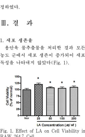
1. 세포 생존율
용안육 물추출물을 처리한 결과 모든 농도 군에서 세포 생존이 증가되어 세포 독성을 나타내지 않았다(Fig. 1).
Fig. 1. Effect of LA on Cell Viability in RAW 264.7 Cell.
LA : Longanae Arillus water extract Nor : Normal, Treated with media only Cells were incubated with LA at the concentration of 25, 50, 100, 200 μg/ml for 24 hr.
Results are represented as mean±SEM of three independent experiments.
* : Represents p<0.05 compared to the normal
2. 용안육 물추출물의 NO 생성에 대한 영향
용안육 물추출물은 50, 200 μg/ml 농도
에서 LPS에 의해 증가된 NO 생성을 유의
하게 억제하였다(Fig. 2).
Fig. 2. Effect of LA on NO Production of RAW 264.7 Cell Treated with LPS.
LA : Longanae Arillus water extract Nor : Normal, Treated with media only Con : Control, Treated with LPS (1 μg/ml) only LPS-induced cells were incubated with LA at the concentration of 25, 50, 100, 200 μg/ml for 24 hr.
Results are represented as mean±SEM of three independent experiments.
* : Represents p<0.05 compared to the normal
†: Represents p<0.05 compared to the control
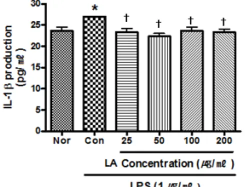
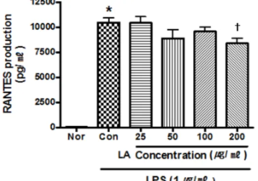
3. 용안육 물추출물의 cytokine 생성에 대한 영향
1) IL-1β 생성에 대한 영향
용안육 물추출물은 25 μg/ml 이상의 모 든 농도에서 LPS에 의해 증가된 IL-1β 생성을 유의하게 억제하였다(Fig. 3).
2) TNF-α 생성에 대한 영향
용안육 물추출물은 50 μg/ml 이상의 농도에서 LPS에 의해 증가된 TNF-α 생 성을 유의하게 억제하였다(Fig. 4).
3) LIX 생성에 대한 영향
용안육 물추출물은 25, 100, 200 μg/ml 의 농도에서 LPS에 의해 증가된 LIX 생 성을 유의하게 억제하였다(Fig. 5).
4) RANTES 생성에 대한 영향 용안육 물추출물은 200 μg/ml의 농도 에서 LPS에 의해 증가된 RANTES 생성 을 유의하게 억제하였다(Fig. 6).
Fig. 3. Effect of LA on IL-1β Production of RAW 264.7 Cell Treated with LPS.
LA : Longanae Arillus water extract Nor : Normal, Treated with media only Con : Control, Treated with LPS (1 μg/ml) only LPS-induced cells were incubated with LA at the concentration of 25, 50, 100, 200 μg/ml for 24 hr.
Results are represented as mean±SEM of three independent experiments.
* : Represents p<0.05 compared to the normal
†: Represents p<0.05 compared to the control
Fig. 4. Effect of LA on TNF-α Production of RAW 264.7 Cell Treated with LPS.
LA : Longanae Arillus water extract Nor : Normal, Treated with media only Con : Control, Treated with LPS (1 μg/ml) only LPS-induced cells were incubated with LA at the concentration of 25, 50, 100, 200 μg/ml for 24 hr.
Results are represented as mean±SEM of three independent experiments.
* : Represents p<0.05 compared to the normal
†: Represents p<0.05 compared to the control
Fig. 5. Effect of LA on LIX Production of RAW 264.7 Cell Treated with LPS
LA : Longanae Arillus water extract Nor : Normal, Treated with media only Con : Control, Treated with LPS (1 μg/ml) only LPS-induced cells were incubated with LA at the concentration of 25, 50, 100, 200 μg/ml for 24 hr.Results are represented as mean±SEM of three independent experiments.
* : Represents p<0.05 compared to the normal
†: Represents p<0.05 compared to the control
Fig. 6. Effect of LA on RANTES Production of RAW 264.7 Cell Treated with LPS.
LA : Longanae Arillus water extract Nor : Normal, Treated with media only Con : Control, Treated with LPS (1 μg/ml) only LPS-induced cells were incubated with LA at the concentration of 25, 50, 100, 200 μg/ml for 24 hr.
Results are represented as mean±SEM of three independent experiments.
* : Represents p<0.05 compared to the normal
†: Represents p<0.05 compared to the control