Biochemical Characterization of Recombinant Equine Chorionic Gonadotropin (rec-eCG), Using CHO Cells and PathHunter Parental Cells Expressing Equine Luteinizing Hormone/Chorionic Gonadotropin Receptors (eLH/CGR)
So-Yun Lee
1, Munkhzaya Byambaragchaa
1, Jeong-Soo Kim
1, Hun-Ki Seong
1, Myung-Hwa Kang
2and Kwan-Sik Min
1,3*
1Animal Biotechnology, Graduate School of Future Convergence Technology, Institute of Genetic Engineering, Hankyong National University, Ansung 17579, Korea
2Department of Food Science and Nutrition, Hoseo University, Asan 31499, Korea
3Department of Animal Resource Science, Hankyong National University, Ansung 17579, Korea Received March 14, 2017 /Revised July 14, 2017 /Accepted July 20, 2017
Equine chorionic gonadotropin (eCG) consists of highly glycosylated α- and β-subunits and is a unique member of the gonadotropin family, because it elicits the response characteristics of follicle stimulat- ing hormone (FSH) and luteinizing hormone (LH) in species other than the horse. To directly assess the biological function of rec-eCGβ/α, we constructed mammalian expressing vectors of equine lutei- nizing hormone/chorionic gonadotropin receptors (eLH/CGR). The activity of rec-eCGβ/α in vitro as- sayed in transient transfected CHO-K1 cells and in stably transfected PathHunter Parental cells with eLH/CGR was investigated. rec-eCGβ/α was efficiently secreted in the CHO-K1 suspension cell me- dia, and the quantity detected was about 200 mIU/ml from 1 to 7 days after transfection. In the west- ern blot analysis, the rec-eCGβ/α protein was broadly identified to be about 40~45 kDa molecular weight. The cAMP stimulation in CHO-K1 cells expressing eLH/CGR was determined to evaluate the activity of rec-eCGβ/α. The cAMP concentration increased in direct proportion to the concentration of the rec-eCGβ/α. The EC
50value in the transient transfected CHO-K1 cells was 8.1±6.5 ng. The sta- ble cell lines of eLH/CGR were established in the PathHunter Parental cells expressing β-arrestin. We found that rec-eCGβ/α had full LH activity in the PathHunter Parental cells expressing eLH/CGR.
The EC
50value in transient and stable cells was 5.0±4.7 ng/ml and 4.5±5.2 ng/ml, respectively. These results suggest that rec-eCGβ/α has a biological activity in a cell expressing eLH/CGR. These stable cells expressed in PathHunter Parental cells could be useful for elucidating the functional mechanisms of deglycosylated rec-eCGβ/α mutants.
Key words : cAMP, CHO cells, eLH/CGR, PathHunter Parental cells, rec-eCGβ/α
*Corresponding author
*Tel : +82-31-670-5421, Fax : +82-31-670-5417
*E-mail : [email protected]
This is an Open-Access article distributed under the terms of the Creative Commons Attribution Non-Commercial License (http://creativecommons.org/licenses/by-nc/3.0) which permits unrestricted non-commercial use, distribution, and reproduction in any medium, provided the original work is properly cited.
Journal of Life Science 2017 Vol. 27. No. 8. 864~872 DOI : https://doi.org/10.5352/JLS.2017.27.8.864
Introduction
Equine chorionic gonadotropin (eCG) is a member of the glycoprotein hormone family which includes luteinizing hormone (LH), follicle-stimulating hormone (FSH), and thy- roid-stimulating hormone (TSH) [20, 25]. eCG is a unique member of the gonadotropin family since it appears to be a single molecule that possesses both LH- and FSH-like ac- tivities in other species than the horse [1, 5, 6, 13, 18].
eCG in mares is secreted by the endometrial cups, which
binucleate trophoblast that detach from the chorionic girdle of the conceptus, between 37 and120 during pregnancy [3, 8, 11]. eCG appearance coincides with revival of progester- one secretion by the primary corpus luteum (CL) [8]. The formation of the endometrial cups and secretion of eCG ini- tiated subsequently stimulates progesterone synthesis by the primary CL [7]. The primary CL also synthesizes estrone sul- fate [9]. High eCG concentrations in mares impregnated by horses protect the CL of pregnancy against the luteolytic ef- fects of PGF
2α. Low eCG concentrations carrying mule fe- tuses afford them less protection against the luteolytic effect of PGF
2α, and thus these results may be a cause of the in- creased foetal mortality that occurs between days 60 and 90 of pregnancy in these mares [11].
Along with LH receptors (LHR), FSH receptors (FSHR)
and LH/CG receptor (LH/CGR) belong to a subfamily of
glycoprotein hormone receptors that include G-protein-asso-
ciated seven transmembrane-domain receptors [2, 11].
According, eCG displays dual LH and FSH activities in non-equid species [29]. eCG treatment in dairy cattle results in fewer atretic follicle, the recruitment of smaller follicles (<5 mm) with an increase growth rate, and sustains the growth of medium (6-8 mm) and large (>9 mm) follicles [10, 23]. eCG administration has been related to an increase in the ovulation rate [15, 23] and especially in early post-par- tum cows [26] and in cows suffering long anoestrus or are under seasonal heat stress [15]. A dairy cows received single dose of 600 IU eCG intramuscularly (i.m.) between 9 and 15 postpartum to increase reproductive performance could not be recommended under the given circumstance [12]. In
vitro, eCG stimulated luteal cells progesterone productionas well as INSR and GLUT protein expression [28]. In sheep, the administered eCG (20 ug/ml) in oocytes was sig- nificantly higher FSHR and GnRHR protein levels compared with those matured in the absence of eCG (without eCG) [30]. Thus eCG enhances maturation and decreases apoptosis of oocytes undergoing IVM, and heightens FSHR, LHR, and GnRHR expression [30].
In the recombinant eCG studies, the 102-104 sequence in eCG β-subunit appears to be of utmost importance for their binding to FSHR [5]. The 104-109 region of the eCG β-sub- unit is essential for the secretion of a fully folded eCGβ/α and for its FSH activity but not for its LH activity [13]. The heterodimeric rec-eCGβ/α exhibits the same thermal stabil- ity as natural pituitary LH and its advantages over the eCGβ /α include higher in vitro bioactivity, and reduced potential risk of immunogenicity [18]. We also reported that rec-eCG produced from CHO cells displays dual LH- and FSH- activ- ities in the rat Leydig cells and rat granulose cells [19, 20]
and in the rat LH/CGR and rat FSHR cells [16, 24, 25].
In the present study, we constructed the mammalian ex- pressing vector of eLH/CGR and investigated the biological activity of rec-eCGβ/α using cells expressing eLH/CGR in the CHO cells and PathHunter Parental cell lines expressing β-arrestin.
Materials and Methods
Materials
The oligonucleotides used in this study were synthesized by Genotech (Daejon, Korea). The following reagents and materials were also used: restriction enzymes and a DNA ligation kit (Takara, Tokyo, Japan); CHO cells (Japanese
Cancer Research Resources Bank, Tokyo, Japan); Ham’s F-12 medium, Opti-MEM I, serum-free CHO-S-SFM II, Geneticine and Lipofectamine 2000 (Gibco BRL, Grand Island, NY, USA); fetal bovine serum (Hyclone Laboratories, Logan, UT, USA); Pro-Prep
TMprotein-extraction solution (Intron Bio- technology, Seoul, Korea); Lumi-Light Western blot kit (Roche, Basel, Switzerland); pcDNA3 mammalian expressing vector, CHO-K1 suspension cells, FreeStyle MAX reagent, and FreeStyle CHO expression medium, pCMV-ARMS1-PK2 expression vector, anti-myc antibody, antibiotics and Assay Complete medium (Invitrogen Corporation, San Diego, CA, USA), PathHunter CHO-K1 β-arrestin Parental cell line (DiscoveRx, San Diego, CA, USA), Disposable spinner flask (Corning Incorporated, NY, USA). PMSG ELISA kit (DRG International Inc., Mountainside, NJ). cAMP Dynamic 2 im- munosaasy kit (Cisbio Bioassay, France). All other reagents used were from Sigma-Aldrich Corp (St. Louis, MO, USA).
Mammalian expression vector construction of rec- eCGβ/α
To obtain the mammalian expressing vector, the cDNA encoding the full-length eCG β-subunit was fused with the mature protein part of the α-subunit using the method of overlapping PCR mutagenesis as previously report [20, 24, 25]. The same method was used to add myc-tag (10 amino acids; ) between the first amino acid and the second amino acid of the mature protein in eCG β-subunit as previously report [24]. These PCR fragments were digested with EcoRI and SalI enzymes and then ligated into the EcoRI and XhoI sites of the eukaryotic expression vector pcDNA3 (designated as pcDNA3-eCGβ/α). The direction was confirmed through restriction mapping. Finally, this vector was sequenced com- pletely to confirm the Kozak site, myc-tag and PCR errors.
Production of rec-eCGβ/α in CHO suspension cell
The expression of rec-eCGβ/α as described previously
[22] was transfected into CHO-S cells by using the FreeSytle
MAX reagent transfection method according to the suppli-
er’s instructions. Briefly, CHO-S cells were cultured with
FreeStyle CHO expression medium at 1×10
7cells/ 30 ml for
3 days. One day prior to transfection, the cells were passage
at 5-6×10
5cells/ml with CHO expression medium of 125 ml
in disposable spinner flask (125 ml). On the day of trans-
fection, the cell density is about 1.2-1.5×10
6cells/ml. Next,
DNA (160 μg) was mixed gently in 1.2 ml of the OptiPRO
serum free medium (SFM), and FreeStyle MAX reagent (160
μl) for transfection was mixed gently in 1.2 ml of the OptiPRO serum free medium. Both of the mixed medium was incubated for 5 minutes at RT. After then, the complex (2.4 ml) added to each cell suspension flask. For rec-protein assay, the culture medium was collected each 2 ml on days 1, 3, 5, 6 and 7. Finally, the culture media were collected on day 7 after transfection and centrifuged at 15,000 rpm at 4℃ for 10 min to remove cell debris. Supernatants were collected and frozen at -80℃. And then the samples were concentrated by frozen-dry method and mixed by PBS. A rec-protein was analyzed by Western blot or ELISA.
Analysis of rec-eCGβ/α protein and Western blot analysis
The rec-hormones were quantified PMSG enzyme-linked immunosorbant assay (ELISA) using anti-PMSG monoclonal antibody and enzyme conjugate with couple to horseradish peroxidase and TMB substrate according to the supplier’s protocol [25]. The culture media (100 μl) analyzed the sam- ples collected on days 1 3, 5, 6 and 7. After the frozen-dry, the concentrated sample was also measured by mixed 10 to 20 times. After add 50 μl of stop solution to each well in final step, the plate reader determined the absorbance at 450 nm within 30 minutes.
For Western blot analysis, the concentrated sample (10 μg) was subjected to reducing 12% sodium dodecyl sulfate poly- acrylamide gel electrophoresis (SDS-PAGE) by the method of Laemmi [17]. After SDS-PAGE, the proteins were trans- ferred onto a polyvinylidene difluoride (PVDF) membrane (0.2 um) using a Bio-Rad Mini Trans-Blot electrophoresis cell. The membranes were washed with 1x Tris-Buffered Saline and Tween 20 (TBS-T) and incubated with primary antibody (anti-myc antibody) diluted 1:5,000. Next, the membranes were reacted with the secondary antibody (goat anti-mouse IgG-HRP) diluted with 1:3,000. Subsequently, membranes were incubated for 1 min with 2-ml of the Lumi-Light substrate solution and, after the substrate sol- ution was removed, the membranes were placed on the Saran wrap, covered with a second piece of the Saran wrap, and exposed on X-ray films for 1-10 min.
Construction of eLH/CGR expression vector eLH/CGR cDNA was cloned using cDNA of testis and ovary as previously reported [24]. The PCR fragments were ligated into the pcDNA3 mammalian expressing vector by
EcoR1 and Xho1 enzyme sites (designated as pcDNA3-eLH/CGRwt). For the Parental cells, the digested eLH/CGR cDNA by Nhe1 and Sac1 enzymes were cloned the same en- zyme sites of pCMV-ARMS1-PK2 expression vector (desig- nated as pCMV-ARMS1-PK2-eLH/CGRwt). There is no stop codon in the C-terminal region of Sac1 enzyme site. The di- rection was confirmed through restriction mapping.
Transient transfection of CHO-K1 cell and stable transfection of PathHunter CHO-K1 EA-Parental cells
Transfections of CHO cells were done using the liposome tranfection method as previously described [25]. CHO cells were cultured in growth medium [Ham’s F-12 media con- taining penicillin (50 U/ml), streptomycin (50 μg/ml) and glutamine (2 mM) and 10% fetal bovine serum]. CHO cells grow at 80-90% in 6 well plates and the plasmid DNAs were tranfected using lipofectamine reagent. After combined the diluted DNA with lipofectamine reagent, the mixed tube was incubate about 20 mins. CHO cells were washed by Opti-MEM and DNA-lipofectamine complex was added to each well. After 5 hr, CHO growth medium included 20%
FBS added to each well. The transfected culture medium was changed by new CHO growth medium at 24 hr after transfection. And then the cells were adjusted for cAMP analysis at the 48-72 hr after transfection.
PathHunter CHO-K1 EA-Parental cells, which engineered to stably express the enzyme acceptor-tagged b-arrestin fu- sion protein, were transiently and stably transfection as sup- plier protocol. PathHunter CHO-K1 EA-Parental cells were cultured in AssayComplete
TMCHO-K1 culture medium [AssayComplete CHO-K1 medium with 10% fetal bovine se- rum and antibiotics mixed (penicillin, streptomycin and glu- tamine)]. For the stably cell lines, transfected cells were seed- ed 500 and 1,000 cells in 100 mm culture dish at 24-48 hr after transfection. The cells cultured in AssayComplete me- dium containing G418 to isolate the cells expressing eLH/
CGR for 2-3 weeks. Approximately 20 clones were recovered and cultured in a 24 well plate. The grown cells were trans- fer to 6 well plate and 25 cm
2culture flask. Finally, 5 cell clone lines were isolated and stocked according to the meth- od previously reported [20].
cAMP assay via homogenous time-resolved foster resonance energy transfer (HTRF)
Measurement of AMP accumulation in CHO cells and
PathHunter CHO-K1 EA-Parental cells was performed using
cAMP Dynamics 2 competitive immunoassay kits (Cisbio
A B
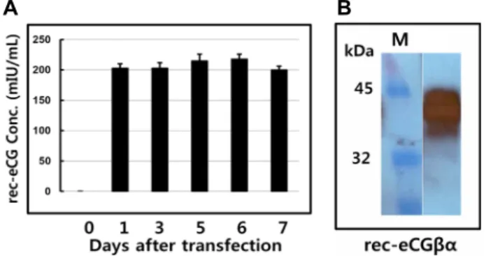
Fig. 1. Quantification of rec-eCGβ/α for transient transfection in CHO suspension cells. A) ELISA results of rec-eCGβ/
α. The media were collected and centrifuged on days 1, 3, 5, 6 and 7 after transfection. And then expression quantity of rec-eCGβ/α was analyzed by ELISA as transcribed in Materials and Methods. Values are ex- pressed as mean ± SEM for at least three independent experiments. B) Western blot of rec-eCGβ/α. Sample of rec-eCGβ/α was electrophoresed on 1 12.5% SDS-PAGE.
First antibody was used anti-myc antibody. The second antibody used goat anti-mouse IgG-HRP. The band of rec-eCGβ/α was detected. M: Marker.
Bioassays) as described previously [4]. The cAMPs assay uses a cryptate-conjugated anti-cAMP monoclonal antibody and d2-labeled cAMP. The tranfected cells of eLH/CHRs were divided 10,000 cells per well into 384 well. And cell dilution buffer was added MIX in order to prevent cAMP degradation. The standard samples prepared to cover an average range of 0.17~712 nM (final concentration of cAMP per well). After the cells (10,000) were seeded into 384 well and 5 μl compounds medium buffer were added to each well. The plate was seal and incubated for cell stimulating at RT for 30 min. And then cAMP-d2 (5 μl) and anti cAMP-cryptate (5 μl) were added to each wells. After the plate was seal and incubated at RT for 1 hr. The plate was read on a compatible HTRF reader. Results are calculated from the 665 nm/620 nm ratio and expressed in Delta F
% (cAMP inhibition).
Delta F% = (Standard or sample ratio-sample negative)
×100 / ratio negative
The cAMP concentration for Delta F% value were calcu- lated by GraphPad Prism.
Data analysis
Dose-response curves were fitted with a nonlinear re- gression, variable slope equation using GraFit 5.0 (Erithacus Software Limited, Surrey, UK) and GraphPad Prism 6.0 (GraphPad Software, Inc, La Jolla, CA, USA). Curves fitted in a single experiment were normalized to the background signaling measured for mock-transfected cells (0%). Each sum curve was calculated from at least three independent experiments.
Results
Production of rec-eCGβ/α in CHO-S cells and western blot
First, we examined the production quantity of rec-eCGβ/
α in CHO-S cells during culture days. The level of rec-eCGβ /α produced was shown in Fig. 1A. The secreted quantity in media was about 200 mIU/ml from 1 to 7 days after transfection. rec-eCGβ/α expression did not detected on day of tranfection. The expression quantity was detected as 201 mIU/ml on day 1 after transfection. The expression was al- most the same pattern from 3 to 7 day during cell culture.
These patterns were similar to that of the transient ex- pression in attached CHO-K1 cells. Next, we examined ex- pression of rec-eCGβ/α protein in the western blot analysis.
The size of the rec-eCGβ/α protein was identified to be about 40~45 kDa. The band was detected broadly as shown in Fig. 1B. Thus, we suggested that oligosaccharides of about 10~15 kDa were added to the rec-eCG produced in CHO-S cells system.
Biological activity in CHO cells expressing transient eLH/CGR
The effects of the rec-eCGβ/α on cAMP stimulation in CHO cell lines expressing eLH/CGR genes were determined to evaluate the activity of rec-eCG. Receptor cells were in- cubated with dose-dependent concentrations (0.008~1,500 ng/ml) of rec-eCG. As shown in Fig. 2B, Delta F% in rec-eCG β/α was gradually decreased by the dose-response depend- ent as standard curve (0.17~712 nM) (Fig. 2A). Here, the cAMP production presented by Delta F% was inhibited by activation of the transfected eLH/CGR (IC
503.10 ng; Fig. 2B).
Next, these data were calculated by cAMP concentration (nM) as shown Fig. 2C. The cAMP concentration increased in direct proportion to the concentration of the rec-eCGβ/α.
The EC
50value in the transient transfected CHO cells of the eLH/CGR was 8.1±6.5 ng (Table 1).
Biological activity in stably cell expressing eLH/
CGR in PathHunter Parental cells
First, eLH/CGR plasmids were transfected into Path-
Table 1. Bioactivity of the rec-eCGb/a between CHO cells and PathHunter Parental cells expressing eLH/CGR
Cells type and transfection method cAMP responses
Basal (nM/104 cells) EC50 (ng) Rmax (nM/104 cells) CHO cells (transient)
Path-Hunter PA CHO cells (transient) Path-Hunter PA CHO cells (stably)
1.5±0.2 0.6±0.1 0.5±0.1
8.1±6.5 5.0±4.7 4.5±5.2
84.4±4.2 66.6±5.2 74.5±3.9
Values are the means ± SEM of triplicate experiments. The EC50 values used to determine the potencies were determined from the concentration-response curves for the in vitro bioassays.
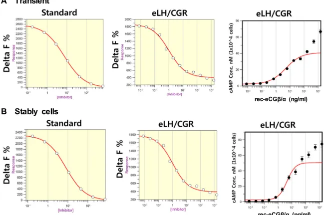
A Standard B eLH/CGR C eLH/CGR
rec-eCGβ/α (ng/ml) Fig. 2. Dose dependent inhibition and increase in cAMP accumulation induced by a rec-eCGβ/α in transient CHO-K1 cells express-
ing eLH/CGR. CHO-K1 cells were transfected with eLH/CGR. The transfected culture media were changed by new CHO growth medium at 24 hr after transfection. And then the cells were adjusted for cAMP analysis at the 48-72 hr after transfection (see Materials and Methods for details). A) Standard curve, B) Delta F% value was shown by inhibition (Grafit), C) cAMP nM (1×10⁴ cells) value by calculated by GraphPad Prism.
Hunter-EA CHO Parental cells expressing β-arresting.
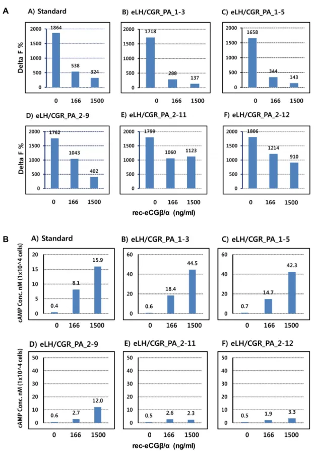
Stably clones were selected by G418 treatment. 5 clones were subjected into the cAMP analysis by rec-eCGβ/α (0, 166 and 1,500 ng/ml). As shown in Fig. 3A, the Delta F% value was the most highly inhibited in two cell clones (eLH/CGR_
PA_1-3 and 1-5) than other three clones (eLH/CGR_PA_2-9, 2-11 and 2-12). We also calculated the data as cAMP concen- tration nM as shown in Fig. 3B.
Next, the cAMP stimulation was analyzed between tran- sient cell and stably cell (eLH/CGR_PA_1-3) selected by G418 as shown Fig. 4. Delta F% IC
50values in the transient and stably cells (1-3) were 1.0482 ng and 1.0461 ng, re- spectively. There is no difference between transient and sta- bly cells. The EC
50value calculated by cAMP stimulation in both of these cells was 5.0±4.7 and 4.5±5.2 ng, respectively (Table 1). This data suggest for the first time in cells express- ing the eLH/CGR. Our data also suggest that rec-eCG dis- plays having a biological activity in cells expressing eLH/
CGR. These clone cell could be useful in the biological activ- ity in the deglycosylated rec-eCG mutants.
Discussion
The presented study indicates that rec-eCGβ/α has full activity in the CHO cells and PathHunter Parental cells ex- pressing eLH/CGR. Our results showed that rec-eCGβ/α was efficiently secreted into the medium in the CHO-S cells on day 1 after transfection. The bands of western blot were detected broadly about 40~45 kDa. We also established the stably cells of eLH/CGR in the PathHunter-EA Parental cells expressing β-arrestin which was function as adaptor pro- teins specifically targeting GPCRs for dynamin-dependent endocytosis via clathrin-coated vesicles. To our knowledge, these results suggest that rec-eCGβ/α has activity in the cells expressing eLH/CGR.
In the secretion pattern of rec-eCGβ/α, we also reported on activity in attached CHO cells [16, 20, 24, 25]. Our results are consistent with those of earlier studies in demonstrating that rec-eCGβ/α in CHO-S cells could be translated and effi- ciently secreted as biologically active tethered form [20].
Thus, the non-covalent heterodimeric structure is not critical
for the glycoprotein hormone family function [24]. On the
other hands, the deletion mutant of C-terminal region (Δ87)
A
rec-eCGβ/α (ng/ml)
B
rec-eCGβ/α (ng/ml)
Fig. 3. Selection of stably cells expressing eLH/CGR in PathHunter CHO-K1 EA Parental cells. A: Delta F% value was shown by inhibition. B: cAMP stimulation concentration. PathHunter CHO-K1 Parental cells were cultured in AssayCompleteTM CHO-K1 culture medium. After transfection, the cells cultured in AssayCompleteTM medium containing G418 to isolate the cells expressing eLH/CGR for 2-3 weeks. Finally, 5 cell clone lines were isolated and cAMP accumulation analyzed by rec-eCGβ/α concentrations (0, 166, and 1,500 ng/ml). Each point represents the average of three independent experiments.
A Transient
rec-eCGβ/α (ng/ml)
B Stably cells
rec-eCGβ/α (ng/ml)
Fig. 4. Dose dependent inhibition and increase in cAMP accumulation induced by a rec-eCGβ/α in transient and stably cells expressing eLH/CGR. PathHunter CHO-K1 EA Parental cells were transfected with eLH/CGR. The transient method described in Fig.2 legend. In the stably cells, cells (eLH/CGR_PA_1-3) were divided 10,000 cells per well into 384 well. The standard samples prepared to cover an average range of 0.17-712 nM. The plate was incubated for 30 min at RT after rec-eCGβ/α adding (0 to 1,500 ng/ml). cAMP d2 and anti cAMP-cryptate were added and incubated at RT for 1 hr. Inhibition of cAMP accumu- lation was shown by Delta F%. And the data of cAMP concentration was calculated by GraphPad Prism. A) Standard curve, B) Delta F% value was shown by inhibition (Grafit), C) cAMP nM (1×10⁴ cells) value by calculated by GraphPad Prism.
in the eCG β-subunit did not secreted rec-eCG protein in the medium and. However, mRNA of the Δ87 mutant was transcribed by RT-PCR and northern blot analyses [24]. In the Sf9 system, rec-eCGβ/α was produced 1.5±0.1 ug/ml [18]. The deletion of the 104-149 sequence was the most dras- tic since no correctly folded hormone was detected in the culture media after 48 hr of expression in the COS-7 cells [13].
In the Western blot results, the band was detected about 40~45 kDa. These results are consistent to the previous stud- ies suggested as approximate size of 43~45 kDa in the at- tached CHO cells [16, 24, 25]. Our results are consistent that the secreted eCGβ/α single- chain was detected as a doublet of ~46 and 44 kDa in the COS-7 cells [13] and rec eCGβ/α expressed in Sf9 insect cells appear at ~45 kDa and hetero- dimeric eCG with an upper band at 45 kDa and a lower one at ~38-40 kDa [18]. rec-eCGβ/α produced from milk of transgenic rabbits was also heterogeneous and identified by
three bands of 35, 90 and 100 kDa under non-reducing con- ditions [14]. But the main band of rec-eCGβ/α in the trans- genic rabbit was supposed to 35 kDa. Thus, the carbohydrate chains in rec-eCGβ/α were decreased about 10 kDa in the milk of transgenic rabbit. However, rec-eCGβ/α in mamma- lian cultured cells (CHO-K1, COS7 and CHO-K1 suspension cells) was consistent with the molecular weight observed in these cells.
In the presented study, our results indicate that rec-eCGβ
/α displays having a biological activity by a dose-dependent
manner in the cells expressing eLH/CGR. In the previous
studies, we reported that the heterodimeric rec-eCGα/β
showed similar LH- and FSH-like activities to native eCG
in the in vitro bioassay using primary rat Leydig cells and
granulosa cells, respectively [19] and in the tethered-eCGβ/α
[20]. We also suggest that rec-eCG has LH- and FSH activ-
ities in nonequid species in vitro using cells expressing
ratLH/CGR and ratFSHR [16, 24, 25]. However, no reports
have been published about the functions of rec-eCG in the cells expressing eLH/CGR. Thus, the functional significance of rec-eCG protein in the equine must be elucidated. To the best of our knowledge, this is the first study that reported on the biological activity in cells expressing the eLH/CGR.
FSH-like activities of two gonadotropins, rec-eFSHβ/α and rec-eCGβ/α, are evoked through the distinct molecular mechanisms regarding the biological role of oligosaccharide at Asn
56of the α-subunit [27]. Our results also agree with the finding that the dual activities of rec-eCG displayed in the progesterone stimulation using MLTC-1 cell line from mouse Leydig tumor and using Y1 cell line from a mouse adrenal cortex tumor stably expressing the hFSH receptor [5, 13]. The other groups reported that LH bioactivity of rec-eCG was determined by their steroidogenic activity in MLTC-1 cells expressing an endogenous mouse LH receptor [18].
In conclusion, the eLH/CGR cells expressed in CHO-K1 and PathHunter Parental cells provides us with a valuable model with which to research the function of deglycosylated rec-eCG mutants on the receptor binding and activation in equid. Our results suggest that rec-eCG produced from CHO-K1 suspension cells could be utilize to reproductive physiology in equine. Thus, further investigation of func- tional significance of rec-eCGs for equids has to be carried out. Further studies are required to elucidate the functional mechanisms that regulate the roles in the ovary and testis of equine.
Acknowledgement
The authors thank Dr. HW Seong (Institute of Animal Science) for his helpful discussions.
References
1. Apparailly, F. and Combarnous, Y. 1994. Role of sialic acid residues in the in vitro superactivity of human choriogona- dotropin (hCG) in rat Leydig cells. Biochim. Biophys. Acta 1224, 559-569.
2. Ascoli, M., Fanelli, F. and Segaloff, D. L. 2002.The lutropin/
choriogonadotropin receptor, a 2002 perspective. Endocr.
Rev. 23, 141-174.
3. Boeta, M. and Zarco, L. 2012. Luteogenic and luteotropic effects of eCG during pregnancy in the mare. Anim. Reprod.
Sci. 130, 57-62.
4. Brule, C., Perzo, N., Joubert, J. E., Sainsily, X., Leduc, R., Castel, H. and Prezeau, L. 2014. Biased signaling regulates the pleiotropic effects of the urotensin II receptor to modu-
late its cellular behaviors. FASEB J. 28, 5148-5162.
5. Chopineau, M., Martinat, N., Galet, C., Guillou, F. and Combarnous, Y. 2001. β-subunit 102~104 residues are cru- cial to confer FSH activity to equine LH/CG but are not sufficient to confer FSH activity to human CG. J. Endocrinol.
169, 55-63.
6. Combarnous, Y., Guillo, F. and Martinat, N. 1984. Compar- ison of in vitro follicle-stimulating hormone (FSH) activity of equine gonadotropins (luteinizing hormone, FSH, and chorionic goandotropin) in male and female rats. Endocrinol- ogy 115, 1821-1827.
7. Condon, W. A., Ganjam, W. K. and Kenney, R. M. 1979.
Catecholamines and equine luteal progesterones. J. Reprod.
Fertil. Suppl. 27, 199-203.
8. Conley, A. J. 2016. Review of the reproductive endocrinol- ogy of the pregnant and parturient mare. Theriogenology 86, 355-365.
9. Daels, P. F., Shideler, S., Lasley, B. L. and Hughes, J. P. 1990.
Source of oestrogen in early pregnancy in the mare. J.
Reprod. Fertil. 90, 55-61.
10. Drancourt, M. A., Thatcher, W. W., Terqui, M. and Andrieu, D. 1991. Dynamics of ovarian follicular development in cat- tle during the estrous cycle, early pregnancy and response to PMSG. Domest. Anim. Endocrinol. 8, 209-221.
11. Flores-Flores, G., Velazquez-Canton, E., Boeta, M. and Zarco, L. 2014. Luteoprotective role of equine chorionic gonado- tropin (eCG) during pregnancy in the mare. Reprod. Dom.
Anim. 49, 420-426.
12. Freick, M., Passarge, O. and Weber, J. 2017. Lack of effects of an equine chorionic gonadotropin (eCG) administration between days 9 and 15 postpartum on reproductive per- formance in a Holstein dairy herd. Reprod. Domest. Anim.
52, 429-436.
13. Galet, C., Guillou, F., Foulon-Gauze, F., Combarnous, Y. and Chopineau, M. 2009. The b104-109 sequence is essential for the secretion of correctly folded single-chain ba horse LH/CG and for its activity. J. Endocrinol. 203, 167-174.
14. Galet, C., Menck, Le., Bourhis, C., Chopineau, M., Le Griec, G., Perrin, A., Magallon, T., Attal, J., Viglietta, C., Houde- bine, L. M. and Guillou, F. 2000. Expression of a single
βα
chain protein of equine LH/CG in milk of transgenic rabbits and its biological activity. Mol. Cell. Endocrinol. 174, 31-40.15. Garcia-Ispierto, I., Lopez-Helguera, I., Martino, A. and Lopez- Gatius, F. 2012. Reproductive performance of anoestrous high-producing dairy cows improved by adding equine chorionic gonadotrophin to a progesterone-based oestrous synchronizing protocol. Reprod. Domest. Anim. 47, 752-758.
16. Jeoung, Y. H., Yoon, J. T. and Min, K. S. 2010. Biological functions of the COOH-terminal amino acids of the a-sub- unit of tethered equine chorionic gonadotropin. Reprod. Dev.
Sci. 34, 47-53.
17. Laemmli, U. K. 1970. Cleavage of structural proteins during the assembly of the head of bacteriophage T4. Nature 227, 680-685.
18. Legardinier, S., Poirier, J. C., Klett, D., Combarnous, Y. and Cahoreau, C. 2008. Stability and biological activities of heter-
초록:말의 LH/CGR를 발현하는 CHO 세포와 PathHunter Parental 세포에서 유전자 재조합 eCGβ /α의 생화학적 특성
이소연
1․뱜바락차 뭉흐자야
1․김정수
1․성훈기
1․강명화
2․민관식
1,3*
(1한경대학교 미래융합기술대학원 동물생명공학전공, 2호서대학교 식품영양학과, 3한경대학교 동물생명환경과학과)
eCG는 다른 포유동물에서 FSH와 LH의 활성을 나타내기 때문에 성선자극 호르몬 family에서 아주 특이적이고 많은 당쇄가 수식되어진 알파와 베타의 비공유결합으로 구성되어 있다. 유전자 재조합 eCGβ/α의 생물학적 기능 을 규명하기 위하여 말의 LH/CGR의 포유동물발현용 벡터를 구축하였다. 재조합 eCGβ/α의 활성분석은 말의 LH/CGR가 일시적으로 발현되는 CHO-K1 세포와 지속적으로 발현되는 PathHunter Parental 세포를 이용하여 분석하였다. 유전자 재조합 eCGβ/α는 CHO-K1 부유세포의 상층으로 효율적으로 분비되었으며, 분비량은 trans- fection 후 1일에서 7일까지 약 200 mIU/ml이었다. Western blot 분석결과는 재조합 eCGβ/α의 분자량은 약 40-45 kDa으로 검출되었다. eLH/CGR가 발현되는 CHO-K1 세포에서의 cAMP분비량으로 재조합 eCGβ/α의 활 성을 분석하였다. 그 결과 cAMP농도는 재조합 eCGβ/α의 농도의존적으로 증가하였다. eLH/CGR가 일시적으로
발현하는 CHO-K1 세포에서 EC
50값은 8.1±6.5 ng이었다. 또한 일시적 및 지속적으로 eLH/CGR가 발현하는
PathHunter Parental 세포에서도 재조합 eCGβ/α의 LH 활성 분석결과 높은 활성을 나타내는 것으로 확인되었으 며, 이들의 EC
50값은 각각 5.0±4.7 ng/ml, 4.5±5.2 ng/ml으로 나타났다. 따라서 이러한 결과에 의하면 재조합 eCGβ/α는 말의 LH/CGR가 발현하는 세포에서 생물학적 활성을 나타난다는 것을 확인하였으며, PathHunter Parental 세포에서 지속적으로 발현되는 세포의 확보는 당쇄제거에 의한 재조합 eCG의 돌연변이등에 관한 기능 적인 메커니즘을 밝히는데 유용할 것으로 사료된다.
odimeric and single-chain equine LH/chorionic goando- tropin variants. J. Mol. Endocrinol. 40, 185-198.
19. Min, K. S., Hattori, N., Aikawa, J. I., Shiota, K. and Ogawa, T. 1996. Site-directed mutagenesis of recombinant equine chorionic gonadotropin/luteinizing hormone: differential role of oligosaccharides in luteinizing hormone- and follicle- stimulating hormone-like activities. Endocr. J. 43, 585-593.
20. Min, K. S., Hiyama, T., Seong, H. H., Hattori, N., Tanaka, S. and Shiota, K. 2004. Biological activities of tethered equine chorionic gonadotropin (eCG) and its deglycosylated mutants.
J. Reprod. Dev. 50, 297-304.
21. Min, K. S., Liu, X., Fabritz, J., Jaquette, J., Abell, A. N. and Ascoli, M. 1998. Mutations that induce constitutive activa- tion and mutations that impair signal transduction modu- late the basal and/or agonist-stimulated internalization of the lutropin/choriogonadotropin receptor. J. Bio. Chem. 273, 34911-34919.
22. Nanjidsuren, T. and Min, K. S. 2014. The transcription factor Ap-1 regulates monkey 20α-hydroxysteroid dehydrogenase promoter activity in CHO cells. BMC Biotechnol. 14, 71.
23. Pacala, N., Corin, N., Bencsik, I., Dronca, D., Cean, A., Bole- man, A., Caraba, V. and Papp, S. 2010. Stimulation of the reproductive function at cyclic cows by ovsynch and PRID/
ECG. Anim. Sci. Biotech. 43, 317-320.
24. Park, J. J., JarGal, N., Yoon, J. T. and Min, K. S. 2009.
Function of the tethered rec-eCG in rat and equine receptors. Reprod. Dev. Biol. 33, 229-236.
25. Park, J. J., JarGal, N., Yoon, J. T. and Min, K. S. 2010. β- subunit 94-96 residues of tethered recombinant equine cho- rionic gonadogropin are important sites luteinizing hor- mone and follicle stimulating hormone like activities.
Reprod. Dev. Biol. 34, 33-40.
26. Rostami, B., Niasari-Naslaji, A., Vojgani, M., Nikjou, D., Amanlou, H. and Gerami, A. 2011. Effect of eCG on early resumption of ovarian activity in postpartum dairy cows.
Anim. Reprod. Sci. 128, 100-106.
27. Saneyoshi, T., Min, K. S., Ma, X., Nambo, Y., Hiyama, T., Tanaka, S. and Shiota, K. 2001. Equine follicle-stimulating hormone: molecular cloning of
β
-subunit and biological role of the asparagine-linked oligosaccharide at asparagines56 ofα
-subunit. Biol. Reprod. 65, 1686-1690.28. Sousa, M., Mendes, G. P., Campos, D. B., Baruselli, P. S.
and Papa, P. C. 2016. Equine chorionic gonadotropin modu- lates the expression of genes related to the structure and function of the bovine corpus luteum. Plos One 11, e0164089.
29. Stewart, F. and Allen, W. R. 1981. Biological functions and receptor binding activities of equine chorionic goando- tropins. J. Reprod. Ferti. 62, 527-536.
30. Wei, S. C., Gong, Z. D., Zhao, H. W., Liang, H, Q., Lai, L. J. and Deng, Y. Y. 2016. Equine chorionic gonadotropin influence on sheep oocyte in vitro maturation, apoptosis, and follicle-stimulating hormone receptor and luteinizing hormone receptor expression. Genet. Mol. Res. 15, doi:
10.4238/gmr15049162.