소복축어탕의 마우스 경구 단회투여 독성 평가
대구한의대학교 한의과대학 부인과교실 김동철
ABSTRACT
Single Oral Dose Toxicity Test of Sobokchuko-tang, a Polyherbal Formula in ICR Mice
Dong-Chul Kim
Dept. of Oriental Obstetrics & Gynecology, College of Oriental Medicine, Daegu Haany University
Objectives: This study was to evaluate the single dose toxicity of Sobokchuko-tang (SBC) in male and female mice.
Methods: Aqueous extract of SBC (yield=6.60%) was administered to female and male mice as an oral dose of 2,000, 1,000 and 500 mg/kg (body weight) according to the recommendation of Korea Food and Drug Administration (KFDA) Guidelines.
Animals were monitored for the mortality and changes in body weight, clinical signs and gross observation during 14 days after dosing, upon necropsy; organ weight and histopathology of 14 principle organs were also examined.
Results: we could not find any SBC treatment related mortality and clinical signs, changes in the body and organ weights, gross findings and changes in histopathology of principle organs, except for pharmacological immunomodulatory effects related findings including significant increases of submandibular lymph node weights, hypertrophy and hyperplasia of lymphoid cells in the submandibular lymph nodes restrictly detected in 2,000 mg/kg treated female and male mice with some sporadic accidental findings.
Conclusions: The results obtained in this study suggest that the 50% lethal dose and approximate lethal dose of SBC aqueous extracts in both female and male mice were considered as over 2,000 mg/kg, the limited highest dosage recommended by KFDA Guidelines, and can be safety used in clinics.
Key Words: Sobokchuko-tang, Single Oral Dose Toxicity, Mice, Histopathology
3)
Corresponding author(Dong-Chul Kim) : Daegu Haany Univ. Pohang Korean Hospital of Daegu Haany University, 907-8, Daejam-dong, Nam-gu, Pohang-si, Gyeongsangbuk-do, Korea
Tel : 054-271-8002 Fax : 054-281-7464 E-mail : [email protected]
Ⅰ. 서 론
최근 중국산 한약재의 수입과 대량 생 산에 따른 농약 등의 오염에 의한 독성 문제가 심각한 사회문제로 대두됨에 따 라 오랫동안 사용되어 온 한약 역시 독 성으로부터 완전히 벗어나지 못하게 되 었다. 따라서 최근 한약 자체에 대한 독 성에 대한 문제가 폭넓게 제기되어 왔으 나 1,2) , 한약이 장기복용 약물이며 또한 생약 복합물이기 때문에 실험의 진행이 매우 어렵고 동물의 생체 내에서 약물의 동태를 파악하기 어려워 잔류 가능성이 있는 잠재적인 독성 평가는 거의 이루어 지지 않고 있다. 하지만 오랫동안의 시 행착오와 경험을 바탕으로 확립된 처방 전은 처방 자체로 하나의 의약품과 같은 역할을 하게 되었다. 이에 따라 양방의 학에서 말하는 의약품의 독성평가에 준 하여 한의학의 처방을 적용하여 안전성 평가가 이루어져야 할 것으로 생각된다 3) .
少腹逐瘀湯은 淸代 醫家인 王淸任의 著書 ≪醫林改錯≫ 4) 에 최초로 기록되어 소복에 허한성 어혈로 인한 동통, 종양, 출혈, 월경통 등을 치료하고 5) , 임상에서는 만성골반염, 불임, 자궁내막증식증, 자궁 근종, 자궁암 등의 질환에 응용되고 있 다 6-8) . 현재까지 소복축어탕의 자궁내막 증 9) 및 월경불순 10) 에 대한 예방 및 치료 가능성이 실험동물 및 적출 자궁의 평활 근 이완 효과를 통해 비교적 잘 밝혀져 있으며, 흉부 대동맥 평활근 확장효과, Fibronectin 합성 억제에 의한 사구체 신 염에 대한 치료 효과 11) 및 항암 효과 12) 역시 잘 알려져 있으나, 독성학적 측면에
단회 투여 독성에 대한 보고조차 찾아볼 수 없다. 따라서 본 연구에서는 부인과 질환 치료에 널리 사용되어 온 소복축어 탕의 일반적인 독성 시험 중 현재 한국 식품의약품안전청의 독성 시험 기준에 명시되어 있는 마우스 경구 단회투여 독성 시험을 실시하여, 장기투여 독성 시험과 생식, 발생 독성 시험을 위시한 특수 독 성시험에 대한 기초 자료를 제공하고, 객관적인 안전성을 확보하고자 하였다.
Ⅱ. 재료 및 방법
1. 실험동물 및 사양관리
본 동물실험은 대구한의대학교 동물실 험윤리위원회(IACUC)의 승인(DHU2012 -018)을 받아 수행하였다. 암수 각 20마리 의 ICR 마우스(6-wk old upon receipt, SLC, Japan)를 11일간의 순화과정을 거 쳐 실험에 사용하였으며, 순화과정 및 실 험 전 기간 동안 온도(20~25℃)와 습도 (30~35%)가 조절된 사육실에서 마우스 용 polycarbonate 사육상자에 5마리씩 수용하여 사육하였고, 명암 주기(light : dark cycle)는 12시간 주기로 조절하였으 며, 사료(Samyang, Korea)와 음수는 자 유롭게 공급하였다. 모든 실험동물은 투 여일 및 최종 부검일 18시간 전 절식을 실시하였으며(이 기간에도 음수는 자유 롭게 공급하였다), picric acid로 개체를 식별하였다.
2. 소복축어탕 추출물
본 실험에 사용된 약재는 약업사(대원
약업사, 대구, 한국)에서 매입한 것을 현
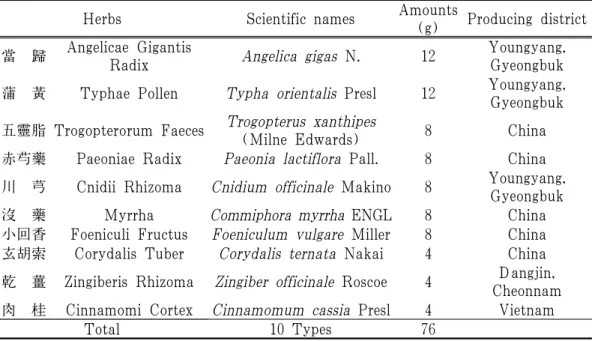
사용하였으며, 본 실험에 사용된 소복축 어탕의 1첩 분량의 조성은 아래와 같다 (Table 1). 선정된 약제 1첩 분량(총량 76 g)을 취하여 정제수 1,000 ml 로 60℃에 서 3시간 동안 3번 가열 추출한 후, 흡인 여과한 여과액을 rotary vacuum evaporator (Rotavapor R144; Buchi Labortechnik AG, Switzerland)로 감압・농축하여, 점조 성의 추출물을 얻은 다음 programmable
freeze dryer(Freezone 1; Labconco Corp., MO, USA)를 사용하여 동결 건조시켜, 총 5.02 g(수율 6.60%)의 연갈색의 물 추출 물을 얻어 실험에 사용하였다. 준비한 소 복축어탕 물 추출물은 -20℃로 냉동 보관 후 실험에 사용하였으며, 본 실험에서 사용한 용매는 증류수에 100 mg/ml 의 농도까지 비교적 잘 용해되었다.
Herbs Scientific names Amounts
(g) Producing district 當 歸 Angelicae Gigantis
Radix Angelica gigas N. 12 Youngyang, Gyeongbuk 蒲 黃 Typhae Pollen Typha orientalis Presl 12 Youngyang, Gyeongbuk 五靈脂 Trogopterorum Faeces Trogopterus xanthipes
(Milne Edwards) 8 China 赤芍藥 Paeoniae Radix Paeonia lactiflora Pall. 8 China 川 芎 Cnidii Rhizoma Cnidium officinale Makino 8 Youngyang,
Gyeongbuk 沒 藥 Myrrha Commiphora myrrha ENGL 8 China 小回香 Foeniculi Fructus Foeniculum vulgare Miller 8 China 玄胡索 Corydalis Tuber Corydalis ternata Nakai 4 China 乾 薑 Zingiberis Rhizoma Zingiber officinale Roscoe 4 Dangjin,
Cheonnam 肉 桂 Cinnamomi Cortex Cinnamomum cassia Presl 4 Vietnam
Total 10 Types 76
Table 1. Composition of Sobokchuko-tang Used in This Study
3. 소복축어탕 추출물의 투여
실험동물은 군당 5마리씩, 암수 매체 대조군, 암수 2,000, 1,000 및 500 mg/kg 투여군의 8군으로 구분하여, 실험을 실 시하였다. 현재까지 소복축어탕의 독성 에 대한 보고를 찾아볼 수 없어, 한국식 품의약품안전청 고시 제 2009-116호 13) 에 의거하여, 설치류 최고 한계투여용량인 2,000 mg/kg을 최고 용량으로 설정하였 으며, 공비 2로 1,000 및 500 mg/kg을 중
간 및 저용량 투여군으로 설정하였다.
또한 암수 각각에 대한 매체 대조군을
추가하였다. 모든 투여군에서는 소복축
어탕 추출물을 멸균 증류수에 용해시켜
20 ml/kg의 용량으로 존데(zonde)가 부
착된 1 ml 주사기를 이용하여, 강제 경
구투여하였다. 식이와 음수에 따른 약물
의 흡수 변화를 최소화하기 위해, 투여
후 대략 3시간 동안 사료와 음수 공급을
제한하였다.
4. 임상증상 및 체중의 관찰
모든 실험동물의 임상증상을 투여 전 후에 각각 functional observational battery test 14) 를 기초하여 동물의 행동, 자극에 대한 반응성, 각성도 및 경계성, 자세 및 보행 이상 등에 관한 일반증상을 관찰, 기록하였으며, 투여일 이후에도 하루에 최소한 2번씩 모든 실험동물의 임상증상 을 관찰, 기록하였다. 또한 모든 실험동 물의 체중을 투여전 1일, 투여직전, 투여 후 1, 2, 7, 13 및 14일(최종 희생일)에 각각 측정하였다.
5. 부 검
투여 14일 후 모든 실험동물은 이산화 탄소 마취 하에 부검을 실시하고, 주요 장기를 위주로 이상 육안소견을 각각 관 찰, 기록하였다.
6. 장기중량 측정
모든 실험동물은 육안부검 소견을 관 찰 기록한 후 하기의 장기에 대한 절대 중량을 각각 측정하였으며, 체중의 변화 에 수반된 이차적 변화를 최소화하기 위 해 체중에 대한 각각의 장기 절대중량의 비율인 상대 중량을 산출하였다.
폐, 심장, 가슴샘, 좌측 신장, 좌측 부 신, 비장, 좌측 고환, 간, 췌장 비장엽, 좌 측 부고환, 좌측 악하임파절, 좌측 난소, 뇌 및 자궁.
7. 조직병리
상기의 조직을 10% 중성포르말린에 18시간 이상 고정시킨 다음, 탈수를 거 쳐 파라핀 포매 후 4 μm의 절편을 제작
하였다. 이후 Hematoxylin&eosin(H&E) 염색을 실시하고, 광학현미경 하에서 이 상 유무를 관찰, 기록하였다.
8. 통계처리
모든 수치는 평균±표준편차로 표시하 였으며, 다중비교검증을 이용하여 통계 처리를 실시하였고, 분산동질성을 Levene test를 실시하여 검증하였다 15) . 등분산일 경우, one way ANOVA test를 실시한 다음 Scheffe test 로 사후 검증을 실시하 여 군간의 유의성을 측정하였다. 비등분 산일 경우에는 비모수 검증인 Kruskal- Wallis H test 를 실시하여 유의성이 인정 된 경우에는, Mann-Whitney U(MW) test 를 실시하여 군간의 유의성을 검증하였다 16) . 반수치사량 및 95% 신뢰한계(confidence limits)를 Probit 방법으로 측정하였으며, 임상 증상, 육안부검 및 조직병리학적 소견은 각각 그 정도에 따라 0(normal), 1+(slight), 2+(moderate) 및 3+(severe) 로 구분하였다 17-20) . 통계처리 및 Probit 방법은 SPSS for Windows(Release 14.0K, SPSS Inc., USA)를 이용하여 평가하였 으며, p-value가 0.05 이하인 경우 통계적 유의성을 인정하였다.
Ⅲ. 결 과
1. 사망례
소복축어탕 추출물 투여와 관련 있는
사망례는 실험 전 기간동안 관찰되지 않
아, 모든 실험동물(5/5; 100%)을 최종부
검을 실시하였다(Table 2).
Groups
Male vehicle control
SBT treated male rats (mg/kg)
Female vehicle control
SBT treated female rats (mg/kg) 2,000 1,000 500 2,000 1,000 500 Mortality 0/5 0/5 0/5 0/5 0/5 0/5 0/5 0/5 Gross findings
Lung focal congestion 1/5 1/5 0/5 1/5 1/5 1/5 0/5 1/5 Thymus atrophy 1/5 1/5 1/5 1/5 0/5 0/5 0/5 0/5 Spleen atrophy 1/5 0/5 1/5 1/5 2/5 1/5 0/5 0/5 Lymph node hypertrophy* 1/5 3/5 1/5 2/5 1/5 3/5 1/5 1/5
Uterus edema 2/5 2/5 1/5 2/5
Histopathological findings
Lung focal congestion 1/5 1/5 0/5 1/5 1/5 1/5 0/5 1/5 Kidney fTV † 1/5 0/5 0/5 0/5 0/5 0/5 0/5 0/5 Spleen wDE ‡ 1/5 0/5 0/5 1/5 1/5 1/5 0/5 0/5 Spleen rHP § 1/5 1/5 0/5 1/5 1/5 1/5 1/5 1/5 Liver IF-FN ∥ 0/5 0/5 0/5 1/5 1/5 0/5 0/5 1/5 Lymph node HP* ¶ 1/5 3/5 2/5 2/5 1/5 3/5 1/5 1/5
Values were observed animals/total observed animals.SBC=Sobokchuko-tang aqueous extracts (yield=6.60%)
*Submandibular lymph node
†Focal tubular vacuolation
‡White pulp lymphoid cell decreases
§Red pulp lymphoid cell hyperplasia
∥Focal inflammatory cell infiltration and focal necrosis
¶Diffused hyperplasia of lymphoid cells
Table 2. Mortality, Clinical Signs, Gross and Histopathological Findings of Animals Exposed with SBT in the Single Dose Toxicity Study
2. 임상증상
소복축어탕 추출물 투여와 관련된 임 상증상은 14일간의 실험 전 기간 동안 인정되지 않았다.
3. 체중의 변화
각각의 동일 성별의 매체 대조군에 비 해, 소복축어탕 추출물 투여와 관련있는 체중의 변화는 인정되지 않았다(Fig. 1).
4. 장기 중량의 변화
본 실험 결과, 소복축어탕 2,000 mg/kg
암수 투여군에 국한되어, 각각의 동일한
성별의 매체 대조군에 비해 유의성 있는
(p<0.05) 악하임파절 중량의 증가가 인
정된 이외에, 소복축어탕 추출물 투여와
관련된 의미있는 장기중량의 변화는 인
정되지 않았다(Table 3).
Fig. 1. Changes on the Body Weights during 14 Days of Observation in Female (A) and Male (B) Mice.
No meaningful changes on the body weights were detected in all SBC extracts treated male and female groups as compared with equal genders of vehicle control, throughout whole 14 days experimental periods, respectively.
Values are expressed as mean±SD of five mice.
SBC=Sobokchuko-tang aqueous extracts (yield=6.60%)
‡: all rats were overnight fasted; before means 1 day before administration. 0 means the day of administration
Groups Male vehicle control
SBT treated male rats
(mg/kg) Female vehicle control
SBT treated female rats (mg/kg)
2,000 1,000 500 2,000 1,000 500 Organ weights (% of body weight)
Lung 0.56±0.03 0.55±0.06 0.61±0.05 0.57±0.03 0.67±0.05 0.68±0.06 0.67±0.05 0.65±0.06 Heart 0.50±0.04 0.51±0.03 0.52±0.01 0.51±0.02 0.52±0.04 0.53±0.05 0.50±0.04 0.50±0.06 Thymus 0.17±0.01 0.18±0.04 0.16±0.02 0.16±0.03 0.22±0.02 0.25±0.02 0.21±0.05 0.23±0.05 Kidney (left) 0.91±0.18 0.80±0.15 0.82±0.07 0.81±0.08 0.61±0.06 0.62±0.05 0.64±0.05 0.66±0.03 Adrenal gland (left) 0.01±0.01 0.02±0.01 0.01±0.01 0.01±0.01 0.02±0.01 0.01±0.01 0.02±0.01 0.02±0.01 Spleen 0.33±0.05 0.32±0.07 0.32±0.03 0.33±0.03 0.38±0.07 0.44±0.06 0.40±0.04 0.38±0.06 Testis/Ovary (left) 0.32±0.03 0.35±0.04 0.35±0.04 0.37±0.06 0.08±0.01 0.09±0.04 0.07±0.02 0.07±0.01 Liver 4.18±0.23 4.37±0.30 4.50±0.32 4.38±0.07 4.39±0.32 4.49±0.32 4.52±0.23 4.31±0.33 Pancreas 0.55±0.07 0.50±0.06 0.51±0.03 0.50±0.04 0.57±0.04 0.57±0.04 0.57±0.06 0.55±0.07 Brain 1.51±0.08 1.47±0.12 1.55±0.12 1.43±0.14 1.96±0.08 1.94±0.15 1.93±0.13 1.91±0.14 Epididymis (left)
/Uterus 0.12±0.01 0.12±0.01 0.14±0.02 0.13±0.01 0.65±0.29 0.65±0.25 0.58±0.30 0.54±0.07 Lymph node (left) † 0.01±0.01 0.03±0.01* 0.02±0.01 0.01±0.01 0.01±0.01 0.04±0.02* 0.02±0.02 0.01±0.01
Values are expressed as mean±S.D. of five mice.SBC=Sobokchuko-tang aqueous extracts (yield=6.60%)
*p<0.05 as compared with equal genders of vehicle control by LSD test
†Submandibular lymph node
Table 3. Relative Organ Weights of Animals Exposed with SBT in the Single Dose
Toxicity Study
5. 부검소견
암수 2,000 mg/kg 투여군에서 각각의 동일 성별 매체 대조군에 비해 경미한 (1+) 악하임파절 종대 소견의 출현빈도 증가가 인정되었으며, 폐 충출혈, 가슴샘 및 비장 위축 및 자궁부종 소견이 암수 매체 대조군을 포함한 모든 실험군에 걸쳐 산발적으로 관찰된 이외에 의미 있는 육안 부검소견은 인정되지 않았다(Table 2).
6. 조직병리학적 관찰
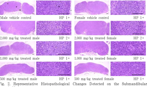
경미하거나 중등도(2+)의 악하임파절 미만성 임파구 증생(Fig. 2) 소견이 암수
소복축어탕 추출물 2,000 mg/kg 투여군 에서 각각 3례(3/5; 60%) 인정되어, 각 각 1례(1/5; 20%)의 악하임파절 미만성 임파구 증생 소견이 인정된 암수 매체 대 조군에 비해, 현저한 출현 빈도의 증가를 나타내었으며, 경미한 국소 폐 충출혈(Fig.
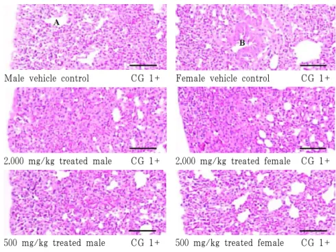
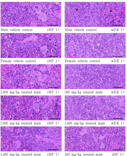
3), 신장 국소 세뇨관 상피 공포화(Fig. 4) 비장 백색 수질 임파구 감소 또는 적색 수질 임파구 증생(Fig. 5), 간의 국소 괴사 -염증세포 침윤(Fig. 6) 소견이 암수 매체 대조군을 포함한 모든 실험군에 걸쳐 산발 적으로 관찰된 이외에 의미 있는 조직병리 학적 변화는 인정되지 않았다(Table 2).
Male vehicle control HP 1+ Female vehicle control HP 1+
2,000 mg/kg treated male HP 2+ 2,000 mg/kg treated female HP 2+
1,000 mg/kg treated male HP 1+ 1,000 mg/kg treated female HP 1+
500 mg/kg treated male HP 1+ 500 mg/kg treated female HP 1+
Fig. 2. Representative Histopathological Changes Detected on the Submandibular Lymph Node.
Note that marked increase trends of the frequency of slight (1+) to moderate (2+) hyperplasia of lymphoid cells (HP) at histopathological on the submandibular lymph node were detected in 2,000 mg/kg treated female and male mice as compared with equal genders of slight and lower frequencies of diffused lymphoid cell hyperplasia were demonstrated. These findings are considered as effects of SBC related changes rather than toxicological signs.
SBC=Sobokchuko-tang aqueous extracts (yield=6.60%)
C : cortex, P : paracortex, M : medullary sinus, SF : secondary lymphatic follicle All Hematoxylin & Eosin stain, Scale bars=80 μm.
Male vehicle control CG 1+ Female vehicle control CG 1+
2,000 mg/kg treated male CG 1+ 2,000 mg/kg treated female CG 1+
500 mg/kg treated male CG 1+ 500 mg/kg treated female CG 1+
Fig. 3. Representative Histopathological Changes Detected on the Lung.
Note that slight (1+) lung focal congestional spots. thickening of alveolar lung inflammatory cell infiltration with/without focal hemorrhages were randomly detected throughout most of all experimental groups including both genders of vehicle controls as sporadic finings not SBC treatment related toxicological signs.
SBC=Sobokchuko-tang aqueous extracts (yield=6.60%) A=alveolar sac-respiratory bronchiole, B=bronchus All Hematoxylin & Eosin stain, Scale bars=80 μm.
Male vehicle control fTV 1+
Fig. 4. Representative Histopathological Changes Detected on the Kidney.
Note that slight (1+) focal tubular vacuolation (fTV) was restrictly detected in one (1/5;
20%) male vehicle control as sporadic accidental finings.
SBC=Sobokchuko-tang aqueous extracts (yield=6.60%) G=glomerulus
All Hematoxylin & Eosin stain, Scale bars=80 μm.
Male vehicle control rHP 1+ Male vehicle control wDE 1+
Female vehicle control rHP 1+ Female vehicle control wDE 1+
2,000 mg/kg treated male rHP 1+ 500 mg/kg treated male wDE 1+
2,000 mg/kg treated male rHP 1+ 2,000 mg/kg treated male wDE 1+
1,000 mg/kg treated male rHP 1+ 500 mg/kg treated male rHP 1+
Fig. 5. Representative Histopathological Changes Detected on the Spleen.
Note that slight (1+) hyperplasia of lymphoid cells and megakaryocyte in the red pulp (rHP) or decreases of white pulp lymphoid cells (wDE) were sporadically observed throughout most of all experimental groups including both female and male vehicle control mice as accidental finings not PS treatment related toxicological signs, respectively.
Male vehicle control rHP 1+ Male vehicle control wDE 1+
Female vehicle control rHP 1+ Female vehicle control wDE 1+
2,000 mg/kg treated male rHP 1+ 500 mg/kg treated male wDE 1+
2,000 mg/kg treated female rHP 1+ 2,000 mg/kg treated female wDE 1+
1,000 mg/kg treated female rHP 1+ 500 mg/kg treated female rHP 1+
SBC=Sobokchuko-tang aqueous extracts (yield=6.60%) M=megakaryocyte, W=white pulp, R=red pulp All Hematoxylin & Eosin stain, Scale bars=80 μm.