Retrospective Study on the Survival Rate of Preformed Metal Crowns in Permanent First Molars
Nayoung Oh
1, Soonhyeun Nam
2, Jaesik Lee
2, Hyunjung Kim
21
Department of Pediatric Dentistry, Kyungpook National University Dental Hospital
2
Department of Pediatric Dentistry, College of Dentistry, Kyungpook National University
This study analyzed the longevity of preformed metal crowns (PMCs) in first permanent molars and evaluated factors influencing their survival during a long-term follow-up period. In all, 115 first permanent molars treated with PMCs between June 2008 and June 2018 were retrospectively analyzed. The overall combined success rate for the study group was 84.3%. The 5-year survival rate was 82.8%. Multivariate Cox regression analyses identified distal cavities and mandibular PMC placement as risk factors for restoration failure. Careful placement of PMCs at the final try-in stage augments the longevity of the crown.
Key words : Permanent first molar, Preformed metal crown, Stainless steel crown, Survival rate Abstract
Corresponding author : Hyunjung Kim
Department of Pediatric Dentistry, School of Dentistry, Kyungpook National University, 2177, Dalgubeol-daero, Jung-gu, Daegu, 41940, Republic of Korea Tel: +82-53-600-7201 / Fax: +82-53-426-6608 / E-mail: [email protected]
Received September 24, 2019 / Revised November 20, 2019 / Accepted November 4, 2019
Ⅰ. Introduction
Among the permanent teeth, the first permanent molars play the greatest role in occlusion, function, and development of the dentition[1,2]. Proper crown coverage is essential when a permanent first molar is extensively damaged by severe car- ies or hereditary anomalies, such as molar incisor hypominer- alization (MIH), dentinogenesis imperfecta (DI), or amelogen- esis imperfecta (AI)[3]. This task is compounded by the fact that young permanent teeth are partially erupted and will con- tinue to erupt and alter their position in the mouth, which will change the margins of any existing restoration[4]. Moreover, moisture-control problems, difficulty with coordination due to young age, and short crown height make treatment difficult.
In such cases, preformed metal crowns (PMCs, also known as stainless steel crowns) may be useful.
PMCs have long been used to cover molars with defective
enamel and they are still recommended as a treatment option
for MIH of the posterior teeth[5,6]. In many cases of severely
damaged permanent first molar crowns in children, PMCs
are a successful interim restorative option until a permanent
restoration, usually of the full coverage type, can be placed
later[4]. No other type of restoration offers the convenience,
low cost, durability, and reliability of such crowns when interim
full coronal coverage is required[7]. They prevent further tooth
loss, control sensitivity, and establish correct interproximal and
proper occlusal contacts. PMCs have a very long history of
use, although their use in the primary dentition is by far the
most common. Several studies have reported survival times in
excess of 5 years and success rates of 92 - 94% in the primary
dentition[8]. Although the use of PMCs in permanent first mo-
lars is widespread, only limited data on their long-term prog-
nosis are available[9]. Therefore, this study was carried out to evaluate the longevity of PMCs in permanent first molars and identified clinical preoperative variables that may correlate with treatment outcomes.
Ⅱ. Materials and Methods
The protocol was approved by the Institutional Review Board of Kyungpook National University Dental Hospital (IRB No.
KNUDH-2019-05-02-00).
1. Subjects
This study retrospectively analyzed 158 children (212 teeth) treated with PMCs in permanent first molars between June 2008 and June 2018 at the Department of Pediatric Dentistry, College of Dentistry, Kyungpook National University. Children were excluded if they were older than 15 years old, lacked pre- operative and postoperative radiographs, were lost to follow- up (minimum 1 year), or had a molar root incisor malformation with a questionable prognosis. Preoperative and postoperative radiographs were required for all teeth subjected to review, as was a record of at least one follow-up appointment that included a clinical and radiographic re-evaluation of the PMC during the cumulative 10-year period of study.
Overall, 115 teeth of 82 patients were selected for inclusion.
Assessments of preoperative factors, prognosis of the PMC, and reasons for failure were examined by analyzing the pa- tients’ electronic dental records and radiographs.
2. Methods
1) Determination of survival and failure.
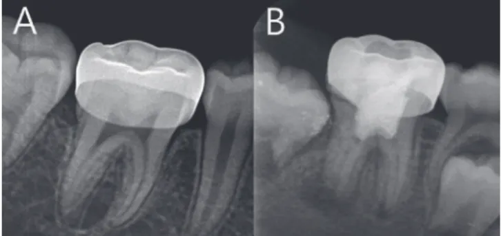
The crown’s viability as an interim restoration was assessed at its follow-up appointment within the study period. Deter- mination of survival and failure were defined as follows. For survival, the PMC not only survived in function until the com- pletion of the study period, having served its purpose until a cast crown could be placed, but was also devoid of any major clinical or radiographic issues (Fig. 1A). The date of the last re- call visit of the patient was recorded as the censoring date for PMC-treated teeth that survived. The time lapse between PMC placement and the last recall visit was calculated for these teeth. To be categorized as failure, the crown had to either be lost (debonded, intentionally sectioned and replaced, or ex-
tracted) or exhibit major clinical or radiographic issues (Fig. 1B, e.g., a short crown with open margins, impaction of adjacent teeth under the margins of the crown, perforation due to wear, or the presence of a periapical pathology). Failed PMCs were no longer suitable as an interim restoration and needed to be retreated.
2) Classification of preoperative parameters.
The following data were collected from the patients’ records:
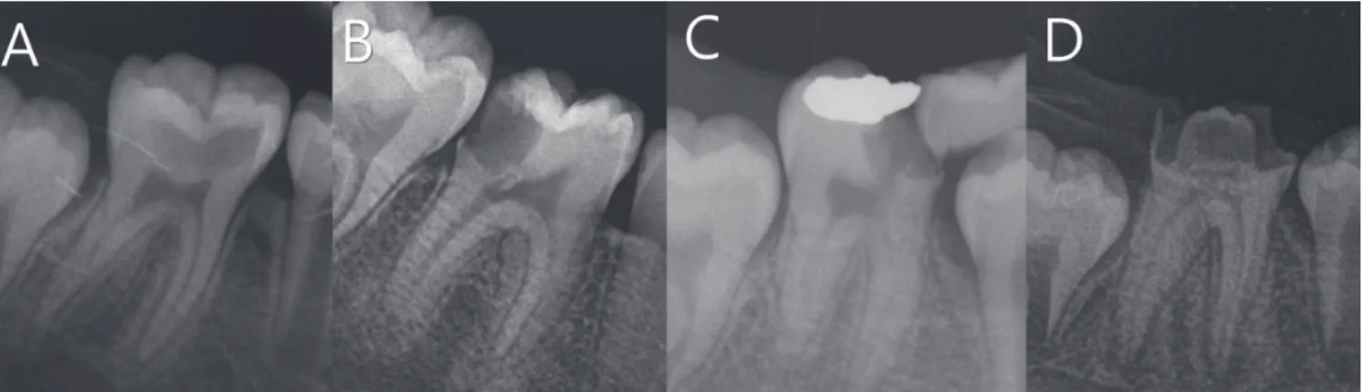
age at time of restoration, sex, dental arch (maxilla versus mandible), operator experience, number of proximal contacts, precipitating factors, and pulp involvement. The subjects were divided into three age groups: 6 - 8, 9 - 11, and 12 - 14 years old. The number of proximal contacts was classified as either 2 (both walls intact), 1 (distal cavity or mesial cavity), or 0 (bro- ken-down wall) based on the remaining tooth structure (Fig.
2)[10]. The precipitating factors for crown placement included caries, AI or DI, MIH, and post-endodontic therapy. Pulp in- volvement was classified as no pulp treatment, indirect pulp capping, direct pulp capping and pulpotomy, or pulpectomy.
3) Statistical analysis
Data were analyzed using SPSS software (ver. 23.0, SPSS Inc., Chicago, IL, USA). The 10-year survival rate of PMCs was analyzed using the Kaplan-Meier method and log-rank test for differences between groups. Multivariate Cox regression analy- ses were performed to analyze the influence of variables that showed significant differences.
Fig. 1. (A) A successful preformed metal crown restoration.
(B) A failed preformed metal crown showing defective res-
toration with open margins and periapical pathology.
Ⅲ. Results
1. Survival rate and reasons for failure
The minimum time of follow-up was 12 months and the maximum was 118 months (average 44.3 months). The patients ranged in age from 6 to 14 years old (average 9.27 years old).
Success was documented in 97 of the 115 PMCs. The total number of failed PMCs was 18. Failures included defective res- toration (n = 10), debonding (n = 4), periapical pathology (n = 3), and perforation from wear (n = 1). The overall success rate for the study group was 84.3% (Table 1).
The mean survival time was 98 months (range: 88.4-107 months, SD = 4.9 months). The survival rate was 97.4% at 1 year, 84.8% at 3 years, and 82.8% at 5 years (Fig. 3).
2. Analysis of risk factors
Prognostic variables for univariate survival analyses included sex, age, operator experience, pulp involvement, number of proximal contacts, dental arch, and precipitating factors (Table 2). The 115 PMCs were similarly distributed between males and females. The mean age of the patients at PMC placement was 9.17 years old. Log-rank tests revealed that pulp involve- ment, number of proximal contacts, dental arch, and precipi- tating need were correlated with the survival rate of the PMCs.
Regarding pulp involvement, the success rate decreased in the following order: pulpectomy, no pulp treatment, indirect pulp capping, and pulpotomy. Notably, the success rate of pulp- otomy was 66.7%.
Table 1. Reasons for failure of preformed metal crowns in perma- nent first molars
Reasons for Failure Number of Failures (%) Defective restoration 10 (55.6)
Debonding 4 (22.2)
Periapical pathology 3 (16.7) Perforation due to wear 1 (5.6)
Total 18 (100.0)
Fig. 2. Number of proximal contacts. (A) 2, Both walls intact. (B) 1, Distal cavity. (C) 1, Mesial cavity. (D) 0, Broken down wall.
Fig. 3. Kaplan-Meier survival analyses of preformed metal
crowns.
Based on precipitating the need for crown placement, the success rates of PMCs were 61.9% for caries, 84.3% for end- odontically treated teeth, 86.0% for MIH, and 91.7% for AI and DI.
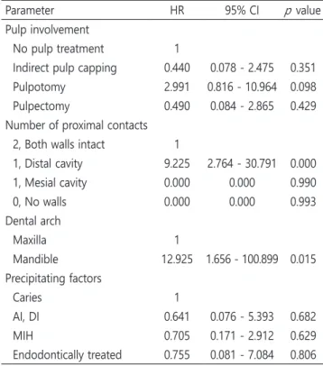
To analyze the effects of preoperative parameters on the PMC survival rate further, hazard ratios (HRs) were calculated for the variables that showed statistical significance on the Kaplan-Meier test. According to the Cox multivariate hazard
model, the number of proximal contacts and dental arch were the factors that significantly affected the survival of the PMCs (Table 3). Teeth with distal cavities were 9.225 times more likely to fail (HR = 9.225). There were no failures among 6 cases of mesial cavity and 4 cases of broken-down wall. Thus, the hazard ratios for a mesial cavity and broken-down wall were 0, compared to cases with both walls intact. The number of sample cases was small and the results were not statistically significant ( p = 0.990 and 0.993, respectively).
In addition, lower permanent first molars were 12.925 times more likely to fail compared to upper first permanent molars.
Precipitating need and pulp involvement showed no signifi- cance in failure rates.
Ⅳ. Discussion
The longevity of dental restorations depends on many dif- ferent factors related to the restorative material, the patient, and to the dentist[11]. Although limited data are available on Table 3. Multivariate Cox proportional hazard regression model for determining the survival rate of preformed metal crowns
Parameter HR 95% CI p value
Pulp involvement
No pulp treatment 1
Indirect pulp capping 0.440 0.078 - 2.475 0.351 Pulpotomy 2.991 0.816 - 10.964 0.098 Pulpectomy 0.490 0.084 - 2.865 0.429 Number of proximal contacts
2, Both walls intact 1
1, Distal cavity 9.225 2.764 - 30.791 0.000 1, Mesial cavity 0.000 0.000 0.990
0, No walls 0.000 0.000 0.993
Dental arch
Maxilla 1
Mandible 12.925 1.656 - 100.899 0.015 Precipitating factors
Caries 1
AI, DI 0.641 0.076 - 5.393 0.682
MIH 0.705 0.171 - 2.912 0.629
Endodontically treated 0.755 0.081 - 7.084 0.806 p value from multivariate Cox-regression analyses
HR = Hazard ratio, CI = Confidence interval
AI = Amelogenesis imperfecta, DI = Dentinogenesis imperfecta, MIH = Molar incisor hypomineralization
Table 2. Preoperative parameters influencing the overall survival rate and the results of univariate analyses using the Kaplan-Meier method
Parameters
Preformed Metal Crowns No. p
Performed
No.
Survival (%) Sex
Male 49 41 (83.7)
0.732
Female 66 56 (84.8)
Age
6 - 8 39 32 (82.1)
0.668
9 - 11 64 54 (84.4)
12 - 14 12 11 (91.7)
Operator
Professor 28 26 (92.9)
0.086
Resident 87 71 (81.6)
Pulp involvement
No pulp treatment 70 62 (88.6)
0.003 Indirect pulp capping 7 5 (71.4)
Pulpotomy 18 12 (66.7)
Pulpectomy 20 18 (90.0)
Number of proximal contacts
2, Both walls intact 80 74 (92.5)
0.000 1, Distal cavity 25 13 (52.0)
1, Mesial cavity 6 6 (100)
0, Broken down wall 4 4 (100) Dental arch
Maxilla 44 43 (97.7)
0.003
Mandible 71 54 (76.1)
Precipitating factors
Caries 21 13 (61.9)
0.012
AI,DI 24 22 (91.7)
MIH 50 43 (86.0)
Endodontically treated 20 19 (84.3) p value from Kaplan-Meier test
AI = Amelogenesis imperfecta, DI = Dentinogenesis imperfecta
MIH = Molar incisor hypomineralization
the longevity of PMCs, they are considered long-lasting by many dentists. This is confirmed by the 82.8% 5-year survival rate for PMCs on permanent molars in the present study. Simi- larly, other studies have shown promising results for PMCs.
Chen et al .[12] found that most PMCs were judged as clinically acceptable (22/23, 96%) from an evaluation based on modi- fied US Public Health Services criteria with at least 52 months of clinical service. Discepolo and Sultan[4] reported an 88%
overall success rate during an average service period of 45.18 months. Moreover, significant success was noted in patients less than 9 years of age in their study. In contrast, greater suc- cess was noted for older subjects in the present study. Teeth emerge, and coordination improves, with age, which may pro- mote proper tooth isolation and adaptation. However, there was no statistical correlation between age and success rate.
In this study, a commonly observed problem was defective restorations. This finding is in agreement with the report by Chen et al .[12], who studied permanent molars with AI. They found that 9 out of 27 PMCs were placed with faulty marginal limits, most likely due to the nature of a prefabricated crown and the operator’s inability to identify the size required cor- rectly. Poor marginal sealing may allow microleakage along the interface between the tooth and crown[13]. This lack of ad- aptation would allow for bacterial invasion, and thus failure of any present endodontic treatment. Debonding failure can also occur if the crown is not seated properly or does not adapt well to the margins of the tooth, leading to cement wash- out[4,13]. Thus, defective restorations, debonding, and pulp pathology are all closely related.
Defective restorations, such as a short crown with open margins, an overall poor fit, or lodging, were predominantly related to initial seating. In view of the potential longevity of these crowns, a periapical or bitewing radiograph is recom- mended before cementation to check the marginal fit mesially and distally, as it is often not possible to check these areas adequately using an explorer[14].
The number of proximal contacts was significantly associ- ated with survival, in that teeth with distal cavities failed at 9.225 times the rate of teeth with two proximal contacts. The significant decrease in the survival of teeth with distal cavities may be the result of increased difficulty of the preparation procedure due to compromised access, restricted visibility, and moisture-control problems[12]. It is often inevitable to place a PMC margin subgingivally in partially erupted permanent mo- lars. Increased subgingival crown height makes isolation and
margin placement difficult compared to mesial cavities[12,14].
This problem is particularly serious if the permanent second molar is close to emergence.
With regard to dental arch, mandibular molars showed a higher failure rate than did maxillary molars within the con- fines of this study. This may be related to the fact that the rate of distal cavities in the maxilla was 13.6%, whereas it was 26.8% in the mandible. Jeong et al .[15] reported that the re- pair rate of composite resin restorations in the permanent first molars of children under 12 years old was 1.4 times higher in the mandible than in the maxilla. This may be because the posterior operculum lasts longer in the mandible than in the maxilla, which makes it more difficult to isolate clinically[15,16].
In relation to pulp involvement, pulpotomy showed the low- est success rate, Presumably, the lower survival rate of PMCs with pulpotomy occurred because the failure of pulp treat- ment caused the failure of the PMCs. Moreover, bacterial con- tamination can occur through the marginal gap of the coronal restoration; thus jeopardizing the long-term success of the pulpotomy procedure[17]. However, this was not found to be significant according to the Cox multivariate hazard model.
Based on precipitating the need for crown placement, AI and DI had the highest success rate. This was probably be- cause AI and DI teeth are often covered with PMCs for pre- ventive purposes before post eruptive breakdown occurs. This might have led to the higher success rate of PMCs.
This study was performed retrospectively based on medical
records, and thus it was difficult to establish a cause of failure
when pulp pathology was involved, and failure may in fact
have been multifactorial in origin. Roberts et al .[18] assessed
PMC failure as ‘true’ and ‘false’ failures. Crown loss following
cement failure or perforation of the occlusal surface as a result
of wear was considered true failure, and failure related to end-
odontic treatment was considered false failure. False failure of
PMCs may occur when a pulpotomy is not carried out when
it should have been, or when a pulp treatment is performed
but fails due to operator error. An example of true failure of a
restoration resulting from pulp pathology would be when end-
odontic therapy is carried out but failure occurs as a result of
restoration leakage leading to a recurrence of pulp inflamma-
tion[19]. In addition, coronal leakage is often associated with
endodontic failure and a well-constructed coronal restoration
has a greater effect on endodontic success than the quality
of the endodontic obturation[20-22]. Therefore, it is not clear
whether pulp pathology is due mainly to a defective restora-
tion or a failed pulp treatment. However, in this study, all pulp- related complications were classified as failures, therefore overestimation of PMC failure might have occurred.
The record review method for identifying PMC failure relies exclusively on data from patients’ electronic dental records and radiographs. Only events documented in patients’ electronic dental records are included in the analysis; therefore, available information regarding events in the records may be insuffi- cient for failure assessment. When conducting routine clinical examinations, perforation due to wear and loosening of PMCs due to cement loss may be overlooked, thus leading to under- estimation of PMC failure.
The retrospective nature of this study made it impossible to identify a true causative relationship between preoperative pa- rameters and survival rate. Therefore, the effects of preopera- tive parameters should be further evaluated in future prospec- tive randomized controlled trials.
Nonetheless, this study is significant in that it evaluated se- quelae of PMCs over a long-term follow-up period and deter- mined factors influencing the outcome of PMCs in permanent first molars. The results imply that PMCs can be used in per- manent first molars as interim restorations until a permanent restoration can be placed. Interim PMCs do not replace the need for permanent restorations in the future, but the interval allows for the practitioner to determine the optimal timing for replacement. This study identified the factors that influence the success rate of PMCs and may be helpful in establishing treatment guidelines for extensively damaged permanent first molars.
Ⅴ. Conclusion
PMCs can function as a viable treatment option for the in- terim restoration needs of extensively compromised permanent first molars, with an 89% overall success rate and an 82.8%
5-year survival rate. Distal cavities and mandibular placement are risk factors for failure. To increase their longevity, dentists should understand these failure factors and ensure careful placement of PMCs.
Authors' Information
Nayoung Oh https://orcid.org/0000-0002-8981-5868 Soonhyeun Nam https://orcid.org/0000-0002-8309-7658 Jaesik Lee https://orcid.org/0000-0001-5514-4595
Hyunjung Kim https://orcid.org/0000-0001-6568-9687
References
1. Jeong HK, Yang YM, Kim JG, et al . : A pattern of the forma- tion and eruption of first permanent molars. J Korean Acad Pediatr Dent , 37:317-327, 2010.
2. Celebi AA, Lee SH, Kau CH : Size discrepancies in molars and first key to optimal occlusion. Eur J Dent , 11:250-252, 2017.
3. Korean Academy of Pediatric Dentistry : Textbook of Pedi- atric Dentistry, 5th ed. Yenang, Seoul, 385, 2014.
4. Discepolo K, Sultan M : Investigation of adult stainless steel crown longevity as an interim restoration in pediatric pa- tient. Int J Paediatr Dent , 27:247-254, 2017.
5. Lygidakis NA : Treatment modalities in children with teeth affected by molar-incisor enamel hypomineralisation : a systematic review. Eur Arch Paediatr Dent, 11:65-74, 2010.
6. Fayle SA : Molar incisor hypomineralization: restroative management. Eur J Paediatr Dent, 4:121-126, 2003.
7. Croll TP : Preformed posterior stainless steel crowns: an up- date. Compend Contin Educ Dent , 20:89-92, 1999.
8. Roberts JF, Sherriff M : The fate and survival of amalgam and preformed crown molar restorations placed in special- ist paediatric dental prectice. Br Dent J , 169:237-244, 1990.
9. Ha N, Kim YJ, Kim HJ, et al . : A prognostic assessment of first permanent molars showing molar-incisor hypominer- alization based on restorative materials and defect class. J Korean Acad Pediatr Dent , 44:263-271, 2017.
10. Aquilino SA, Caplan DJ : Relationship between crown placement and the survival of endodontically treated teeth.
J Prosthet Dent , 87:256-263, 2002.
11. Hickel R, Manhart J : Longevity of restorations in posterior teeth and reasons for failure. J Adhes Dent , 3:45-64, 2001.
12. Chen CF, Hu JC, Estrella MR, et al . : Assessment of restor- ative treatment of patients with amelogenesis imperfecta.
Pediatr Dent , 35:337-342, 2013.
13. Memarapour M, Mesbahi M, Rezvani G, et al . : Microleakge of adhesive and non-adhesive luting cements for stainless steel crowns. Pediatr Dent , 33:501-504, 2011.
14. Randall RC : Preformed metal crowns for primary and per- manent molar teeth: review of the literature. Pediatr Dent, 24:489-500, 2002.
15. Jeong YY, Lee HS, Choi SC, et al . : Repair rate of composite
resin restorations in permanent first molar in children un-
der 12 Years Old. J Korean Acad Pediatr Dent, 45:370-377, 2018.
16. Kim IY, Kim JM, Jeong TS, Kim S : 5 Years evaluation of composite restoration on permanent first molar in children.
J Korean Acad Pediatr Dent, 35:110-117, 2008.
17. Zanini M, Hennequin M, Cousson PY : A review of criteria for the evaluation of pulpotomy outcomes in mature per- manent teeth. J Endod , 42:1167-1174, 2016.
18. Roberts JF, Attari N, Sherriff M : The survival of resin modi- fied glass ionomer and stainless steel crown restorations in primary molars placed in a specialist paediatric dental practice. Br Dent J , 198:427-431, 2005.
19. Randall RC, Vrijhoef MM, Wilson NH : Efficacy of preformed metal crowns vs. amalgam restorations in primary molars:
a systematic review. J Am Dent Assoc , 131:337-343, 2000.
20. Cheung GS : Endodontic failures-changing the approach.
Int Dent J , 46:131-138, 1996.
21. Ray HA, Trope M : Periapical status of endodontically treated teeth in relation to the technical quality of the root filling and the coronal restoration. Int Endod J , 28:12-18, 1995.
22. Whitworth JM, Walls AW, Wassell RW : Crowns and extra-
coronal restorations: endodontic considerations: the pulp,
the root-treated tooth and the crown. Br Dent J , 192:315-
320, 323-327, 2002.
국문초록
제1대구치 기성금속관 생존율에 관한 후향적 연구
오나영
1전공의 ㆍ남순현
2교수 ㆍ이제식
2교수 ㆍ김현정
2교수
1
경북대학교치과병원 소아치과
2