천련자 메탄올 추출물이 Bcl-2 발현 억제를 통해 유방암 세포의 자멸사에 미치는 영향
대구한의대학교 한의과대학 한방부인과학교실 윤우경, 김동철
ABSTRACT
Toosendan Fructus Induces Apoptotic Cell Death in MCF-7 Cell, Via the Inhibition of Bcl-2 Expression
Woo-Kyeong Yoon, Dong-Chul Kim
Dept. of Gynecology, College of Oriental Medicine, Daegu Haany University Purpose: The research is to investigate the effect of TFE on apoptosis of human-derived breast cancer cells, to find out the relationship with apoptosis.
Methods: Human-derived breast adenocarcinoma cell line, MCF-7 cells were treated by TFE with various concentration. The inducement effect of TFE on cell apoptosis was observed with MTT assay and the relationship between the treatment and apoptosis was investigated with FACS analysis, TUNEL assay and DNA laddering assay and the change in the protein levels of PARP and caspase-3 activities were also observed. The release of cytochrome-c was observed to find out the pathway of apoptosis induced by TFE.
Results: The cell apoptosis was significantly induced in MCF-7 cells treated with TFE in concentration-dependent and time-dependent manner. It was verified by FACS anaylsis, TUNEL assay, DNA laddering assay that cell-death was caused not by necrosis but by apoptosis. The activity of PARP and caspase were increased concentration-dependently. The release of cytocrome-c was decreased in proportion to the concentration of the fruit extract. It therefore demonstrated that mitochondria were involved in apoptosis induced by TFE. The appearance of Bcl-2 protein was decreased concentration-dependently.
Conclusion: The treatment by TFE induced apoptosis of human breast adenocarcinoma cell line, MCF-7. It seems likely that cell-death was caused by apoptosis and mitochondria were involved in it. The mechanism of protein change causing apoptosis seems related to the inhibition of Bcl-2 protein, the promotion of inversion from cytochrome-c into cytosol, the activation of caspase and the promotion of PARP cleavage.
Key Words: Toosendan Fructus, apoptosis, caspase-3, Bcl-2, cytochrome-c, human breast adenocarcinoma
2)
교신저자(김동철) : 경북 포항시 남구 대잠동 907-8 대구한의대 부속 포항 한방병원 전화 : 054-271-8002 이메일 : [email protected]
Ⅰ. 緖 論
유방암은 세계적으로 가장 빈번한 여 성의 사망원인 질환 중의 하나이며, 매 년 약 100만명 이상의 환자가 발생한다
1) . 우리나라에서도 유방암은 여성암 중 자궁암, 위암 다음으로 많은 비중을 차 지하고 있다 1) . 특히, 유방암은 2002년 새 로 발생한 암환자 중에서 증가율이 가장 높아, 1995년과 비교하면 166%의 급격한 증가세를 보이고 있으며, 또한 사망자수 에 있어서도 급격한 증가세를 보이고 있 다 2) .
유방암과 유사한 한의학적 질환으로는 石癰 3) , 乳巖 4) , 奶巖 5) , 番花奶 6) , 㛋巖 7) 등 이 있다. 증상으로는 乳房腫塊. 不痛, 不 痒, 不赤하고, 或 內熱, 夜熱, 五心煩熱, 肢體倦瘦, 月經不調 등이 나타나기도 하 며 4,8) , 病因으로 肝鬱氣滯 4) , 憂怒抑鬱 9) , 憂鬱傷肝 10) , 思慮傷肝 10) 등의 七情所傷과, 이것이 오래되어 氣血損傷 4) , 衝任失調 4) 되어 장부기능이 실조된 것으로 볼 수 있다. 치료로는 초기에 肝氣鬱結로 인한 때는 疏氣行血, 疏肝解鬱 하는 치법을 이용하였고 10) , 氣血虧損할 때는 大補氣 血하는 치법을 이용하였다 4) .
유방암에 관해 국내 연구 발표된 단일 약재로는, 鬼箭羽 11) , 黃芩 12) , 半枝蓮 13) , 槲寄生 14) , 三稜 15) , 탕약으로는 抗癌丹 16) , 加味雙和湯 17) , 淸肝解鬱湯 18) , 歸朮破癥湯
19) , 活絡交靈丹 20) , 益氣養榮湯 21) , 橘葉散 變方 22) 등이 있으며, 蜂毒 藥鍼液 23) 을 이 용한 연구도 보고되고 있다.
川楝子(Toosendan Fructus)는 苦寒有 小毒하며, 肝胃小腸經으로 入하고, 胸脇 痛, 脘腹脹痛, 疝痛, 蟲積腹痛을 치료한 다 24) . 현재까지 천련자에 대한 연구로는,
주로 천련자의 毒性에 관한 연구 25) 및 천련자가 肝膽에 미치는 영향에 대한 약 리학적 연구 26,27) 및 전립선에 미치는 영 향 28) 등이 보고된 바 있으나 천련자에 대한 항암 효과와 기전 연구는 부족한 편이다.
이에 본 연구자는 천련자를 인간유래 breast adenocarcinoma cell line인 MCF-7 cell의 apoptosis에 미치는 영향을 알아보 고자 본 연구를 시행하여 유의한 결과를 얻었기에 보고하는 바이다.
Ⅱ. 材料 및 方法
1. 재 료
DMEM(Dulbecoo's Modified Eagle's Medium)과 fetal calf serum은 BioWhittaker (Walkersville, MD, USA)와 Life Technologies (Gaithersburg, MD, USA)로부터 구입 하여 사용하였다.
3- (4,5 -dime th ylth iaz ol-2- yl)- 2, 5-diphenyl-tetrazolium bromide(MTT)와 기타 다른 reagent는 Sigma Chemical (St. Louis, MO, USA)에서 구입하여 사 용하였다.
2. 천련자 추출물의 제조
천련자 추출물(Toosendan Fructus Extract,
TFE)은 대원약업사 (Daegu, Korea)에
서 구입한 천련자(Toosendan Fructus)
200g을 1 L의 메탄올에 72시간 추출한
후에 여과농축하여 동결건조하였다. 천
련자의 최종 수율은 7.34%이었으며
Sigma-aldrich (St. Louis, MO, USA)로
부터 구입한 DMSO(Dimethyl Sulfoxide)
에 녹여 0.2 μm filter (Millipore Corporation,
Bedford, MA, USA)로 여과하여 사용하 였다.
3. 세포배양
인간 유래 breast adenocarcinoma cell line인 MCF-7 cell은 한국세포주은행 (Korea Cell Line Bank, Seoul, Korea) 으로 부터 구입하였으며, 10% fetal calf serum, 50 units/㎖ penicillin과 50 ㎎/㎖
streptomycin이 포함된 DMEM 배지에 서 37℃의 온도와 5%의 CO 2 가 유지되 는 환경에서 배양하였다. 1×10 6 cells 개 의 MCF-7 cells을 10 cm 2 plastic dish에 24시간 배양하여 (80% 이상의 confluence 를 유지), 24시간 배지를 고갈한 뒤, MCF-7 cells에 지정된 시간동안 TFE를 농도별로 처치하였다.
4. MTT 세포생존율 측정
96 well plate의 well당 5×10 4 개의 MCF-7 cells을 배양하여 confluency가 80%이상이 된 경우, 24시간 배지를 고갈 한 다음, TFE를 농도별(0.03-0.30 ㎎/
㎖), 시간별(24, 48 h)로 처치하여, TFE 의 세포생존율을 측정하였다. 세포배양 후 생존한 세포를 0.5 ㎎/㎖의 MTT로 처치한 후, 4시간 incubation하였다. 그 후 배지를 제거하고 생성된 formazan crystals에 200 μl의 DMSO를 가하여 용 해하였다. 흡광도는 Titertek Multiskan Automatic ELISA microplate reader (Model MCC/340, Huntsville, AL, USA) 를 사용하여 540 nm에서 측정하였다. 세 포생존율은 어떠한 처치도 가하지 않은 control cells과의 비율로 나타내었다.
[즉, viability (% control) = 100 x (absorbance of treated sample)/(absorbance
of control)]
5. Flow cytometric analysis
붉은색 형광염색약인 propidium iodide (PI)와 초록색 형광염색약인 FITC annexin V (Molecular Probes, Eugene, OR, USA)를 사용하여 apoptotic cell을 분석하였다. 1×10 6 개의 cells를 10 cm 2 plastic plate에 깔고 70-80%의 confluence 를 유지하였다. 먼저 cells에 24시간 배지 고갈을 하고, 다음 24시간 동안 TFE를 농도별로 처치하였다. 부착된 cells를 trypsin으로 처치하여 회수하였고 부유 cells과 trypsin처치 cells는 모두 70%
ethanol로 resuspension하여 -20℃에서 overnight하여 고정시켰다. 고정된 cell은 1×10 6 개/㎖의 농도로 PBS에 suspension 시킨 후, FITC annexin V와 PI를 처리 하여 얼음위에서 20분간 반응시켜 flow cytometer (Particle Analysis System, Partec GmbH, Münster, Germany)로 분 석하였다.
6. TUNEL assay
MCF-7 cell을 4 well chamber slide에 well당 1×10 5 개의 농도가 되게 배양하고 TFE를 농도별로 24시간 처리하였다.
Cell은 PBS로 2회 washing한 후에 4%
paraformaldehyde로 고정하여 Tunel (Terminal deoxynucleotidyl transferase -medited dUTP -biotin nick end labeling) assay에 사용하였다. Tunel assay는 In Situ cell death detection kit-POD (Roche, Mannheim, Germany)를 사용하여 kit에 포함된 protocol에 맞춰 분석하였다. 즉, paraformaldehyde로 고정된 cell을 0.3%
H 2 O 2 를 사용하여 blocking시키고 0.1%
triton X-100/0.1% sodium citrate로 permeabilisation 상태로 만든 후에 TdT- enzyme이 처리된 용액으로 labeling하였 다. PBS로 2회 washing한 후에 converter -peroxidase(POD)로 37℃에서 30분간 처리하고 DAB로 발색하여 light microscope 로 관찰하였다.
7. DNA laddering assay
Cell은 회수하여 PBS로 2회 washing 한 후, 5×10 6 개의 cell을 PBS 200 μl로 suspension시켰다. DNA laddering assay 는 DNA purification kits (Nucleogen, Seoul, Korea)를 사용하여 agarose gel electrophoresis를 실시하여 분석하였다.
즉, 5×10 6 개/200 μl PBS로 준비된 cell에 proteinase K(100 μg/㎖)와 RNase A(20 μg/㎖)를 첨가하고 kit에서 제공되는 lysis buffer를 사용하여 56℃에서 10분간 반응시켰다. 400 μl의 순수 ethanol을 가 하여 vortexing한 후, kit에서 제공되는 protocol에 따라 filtering과 washing 과정 을 반복하여 genomic DNA를 회수하였 다. DNA 전기영동은 1.8% agarose gel 을 사용하여 50 V에서 1시간 동안 전개 시켰으며 ethilium bromide(0.5 μg/㎖)로 염색하여 band를 확인하였다.
8. Poly-ADP Ribose Polymerase(PARP) cleavage
세포핵추출은 Kim 등 29) 의 방법에 따 라 시행하였다. 즉, cell을 ice-cold PBS 로 2회 세척한 후 PBS를 가하여 수거한 다음 microtubes에 보관하였다. 그 후 cells을 2,000×g에서 5분간 원심분리하고, 10 mM의 HEPES (pH 7.9), 10 mM의 KCl, 0.1 mM의 EDTA, 0.5%의 Nonidet
P-40, 1 mM의 DTT와 0.5 mM의 phenylmethylsulfonyl fluoride를 함유한 저장액의 buffer를 가하여 세포를 rupture 시켰다. Cells lysates를 얼음위에 10분간 방치한 다음 7,200×g로 5분간 4℃에서 원심분리 하였다. Crude nuclei를 함유한 pellets에 20 mM의 HEPES (pH 7.9), 400 mM의 NaCl, 1 mM의 EDTA, 10 mM의 DTT와 1 mM의 PMSF를 함유 한 추출 buffer를 50 μl를 가하여 현탁시 킨 후, 얼음에 30분간 방치하였다. 그 후 15,000×g에서 10분간 원심분리한 후, nuclear fractions을 함유한 상층액을 얻었다. Nuclear fractions 50 μg을 7.5% SDS-polyacrylamide gels을 사용하여 분해시키고, nitrocellulose membranes에 이전하였다. 이 membranes 을 5%의 BSA를 함유한 PBS tween으로 4℃에서 overnight하여 blocking하였다.
그 후 anti-PARP antibody (1:2000)로 실온에서 배양하였다. 발색은 immunoblot analysis에서 기술한 방법으로 하였다.
9. Caspase-3 activity
Caspase-3 activation은 procaspase-3 로부터 proteolytic processing에 의해 cleaved된 p17, p12절편을 만들어 낸다.
따라서 cleaved caspase-3 sandwich
ELISA kit(Cell signaling, MA, USA)를
사용하여 caspase-3의 activity를 측정하
였다. Kit에 제공되는 cell lysis buffer를
이용하여 cell을 분쇄하여 15,000×g에서
10분간 원심분리하여 cell lysates (상등
액)를 준비하였다. Cell lysates로 부터
cleaved caspase-3는 제품에 첨부된
protocol에 맞게 ELISA법으로 분석하였
다.
10. Cell lysates의 준비
Cells은 10 mM의 Tris (pH 7.4), 100 mM 의 NaCl, 30 mM의 sodium pyrophosphate, 1 mM의 EGTA, 0.5%의 Triton X-100, 10%의 glycerol, 1 mM의 phenylmethylsulfonyl fluoride와 100 μM의 sodium orthovanadate 를 함유한 buffer로 녹였다. Cell lysates 는 매 5분마다 vortexing하면서 30분간 얼음에 방치한 후, 15,000×g에서 15 분간 원심분리하여 찌꺼기를 제거하고 상등액 을 취하여 total cell lysates로 사용하였 다.
11. Immunoblot analysis
SDS-PAGE전기영동과 immunoblot analyses는 Kim 등의 방법 28) 에 따라 시 행하였다. 단백질을 12% gel에 전기영동 으로 분리하고, 이것을 nitrocellulose paper로 이전하였다. Nitrocellulose paper에 Bad, Bcl-2, , Bcl- XL, Bax, Actin (Zymed Laboratory, San Francisco, CA, USA), caspase-9, caspase-3 (Pharmingen, San Diego, CA, USA)등의 antibody를 가하 여 배양하였다. 면역반응성 단백질은 ECL chemiluminescence detection kit (Amersham Biosciences, Buckinghamshire, UK)를 사용하여 발색하였다.
12. Cytosolic fraction에서 cytochrome-c analysis
MCF-7 cell은 PBS로 2회 washing 한 후, lysis buffer (250 mM sucrose, 20 mM Hepes, 10 mM KCl, 1.5 mM MgCl 2 , 1 mM EDTA, 1 mM EGTA, 1 mM dithiothreitol, protease inhibitors)에 resuspension시켰다. 얼음 위에서 20분간 incubation 한 후, Dounce homogenizer
(Wheaton, Millville, NJ)를 사용하여 세 포를 homogenization (15 strokes)시켰 다. Homogenates는 1,000×g에서 10분간 원심분리한 후, 상등액만을 취하여 15,000×g 에서 20분간 더 원심분리하였고 회수된 cytosolic fraction (상등액)을 cytochrome-c 분석에 사용하였다. Cytosolic fractions 20 μg을 12.5% SDS-polyacrylamide gels 을 사용하여 전개시키고, nitrocellulose membranes에 이전하였다. 이 membranes 을 5%의 BSA를 함유한 PBS tween으로 4℃에서 overnight하여 blocking하였다.
그 후 anti-cytochrome-c antibody (1:2000) 로 실온에서 배양하였다. 발색은 immunoblot analysis에서 기술한 방법으로 하였다.
13. Statistical analysis
실험결과는 mean ± S.D.로 나타내었 으며, student t-test 통계처리방법으로 유의성을 검정하였다. 유의수준은 p<0.05 로 하였다. 통계 프로그램은 spss 11.0을 사용하였다.
Ⅲ. 結 果
1. 천련자가 MCF-7 cell의 세포생존율 에 미치는 영향
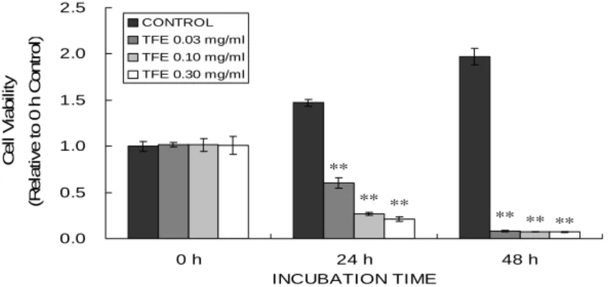
다른 농도에서의 천련자추출물(TFE) 이 MCF-7 cell의 생존율에 미치는 영향 을 분석하기 위하여, MCF-7 cell에 24시 간 배지고갈을 한 후, TFE를 0.03-3.0 ㎎/㎖
의 농도로 24-48시간 처치하였다. 그 결
과 대조군은 0 h에서 1.0000±0.0524, 24 h
에서 1.4720±0.0376, 48 h에서 1.9725±0.0873
으로 시간의 경과에 따른 세포의 증식을
나타내었다. 그러나 TFE 0.03 ㎎/㎖을
처리한 경우는 24 h에서 0.6039±0.0575, 48 h에서 0.0835±0.0063으로 유의하게 감 소하였다(P<0.01). TFE 0.10 ㎎/㎖을 처 리한 경우에는 24 h에서 0.2674±0.0154, 48 h에서 0.0729±0.0016으로 유의하게 감
소했으며(P<0.01), TFE 0.30 ㎎/㎖을 처 리한 경우에는 24 h에서 0.2086±0.0247, 48 h에서 0.0710±0.0029로 유의하게 감소 하였다(P<0.01).
**
** **
**
**
**
0.0 0.5 1.0 1.5 2.0 2.5
0 h 24 h 48 h
INCUBATION TIME C e ll V ia b il it y (R el at iv e t o 0 h C ont ro l)
CONTROL TFE 0.03 mg/ml TFE 0.10 mg/ml TFE 0.30 mg/ml
Fig. 1. The effects of TFE on the changes of cell viability.
MCF-7 cells were exposed to TFE (0.03-0.30 ㎎/㎖) for 24-48 h. Cell viability was assessed by MTT assay. Data represent the mean ± S.D. with eight separate experiments. significance was compared with untreated control at the same treated time, **P < 0.01, (TFE; Toosendan
Fructus Extract)
2. 천련자가 MCF-7 cell의 cell membrane 변화에 미치는 영향
TFE가 0.03-0.30 ㎎/㎖의 농도에서 24, 48 h에서 유도한 세포사가 세포자멸 사에 의한 것인지를 알아보기 위해서, 붉은색 형광염색약인 PI와 초록색 형광 염색약인 FITC annexin V를 사용하여 apoptotic cell을 분석하였다. FACS analysis결과 TFE의 농도가 증가할수록 세포외막으로 돌출된 PS와 결합한 FITC annexin V의 증가로 세포자멸사 의 분율이 각각의 농도에서 유의하게 증 가하였다(P<0.01)(Fig. 2).
3. 천련자가 MCF-7 cell의 DNA 분절 에 미치는 영향
TFE가 MCF-7 cell을 0.03-0.30 ㎎/㎖
의 농도에서 유도한 세포자멸사를 세포 의 형태학적 특징인 cendensed nuclei를 관찰하기 위하여 TUNEL assay를 실시 하였다.
대조군에서는 TUNEL stain에 양성인
MCF-7세포가 많이 나타나지 않았으나,
TFE를 처치한 경우에는 TUNEL stain
에 양성인 세포가 농도에 따라 증가하였
다(Fig. 3). DNA laddering 실험에서도
TFE의 농도 의존적으로 DNA의
fragmentation이 증가하는 것으로 나타
났다(Fig. 4).
CONTROL TFE 0.03 ㎎/㎖
TFE 0.10 ㎎/㎖ TFE 0.30 ㎎/㎖
A)
B)
Fig. 2. Flow cytometric profile of green versus red fluorescence of MCF-7 cells stained with FITC Annexin V and PI.
MCF-7 cells were treated with TFE for 24 h. The population was separated into three groups:
live cells showing only a low level of fluorescence, apoptotic cells showing green fluorescence and necrotic cells showing both red and green fluorescence (A), and the fold increase of apoptotic death cell was calculated (B). Data represent the mean ± SD of six separate experiments.
**Significant at P<0.01 compared with vehicle treated. (TFE; Toosendan Fructus Extract)
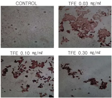
3. 천련자가 MCF-7 cell의 DNA 분절에 미치는 영향
TFE가 MCF-7 cell을 0.03-0.30 ㎎/㎖
의 농도에서 유도한 세포자멸사를 세포
의 형태학적 특징인 cendensed nuclei를
관찰하기 위하여 TUNEL assay를 실시
하였다.
대조군에서는 TUNEL stain에 양성인 MCF-7세포가 많이 나타나지 않았으나, TFE를 처치한 경우에는 TUNEL stain 에 양성인 세포가 농도에 따라 증가하였
다(Fig. 3). DNA laddering 실험에서도 TFE의 농도 의존적으로 DNA의 fragmentation이 증가하는 것으로 나타 났다(Fig. 4).
CONTROL
TFE 0.03 ㎎/㎖
TFE 0.10 ㎎/㎖
TFE 0.30 ㎎/㎖
Fig. 3. TUNEL assay of TFE-treated MCF-7 cells
MCF-7 cells treated with 0.03-0.30 ㎎/㎖ of TFE for 24 h. The TUNEL assay confirms that the TFE treatment induces apoptotic cell death. The dark violet spots are condensed nuclei.
(TFE; Toosendan Fructus Extract)
Fig. 4. DNA Laddering of MCF-7 cells exposed to TFE
TFE induces internucleousmal DNA fragmentaion.
MCF-7 cells treated with TFE for 24 h. M is the maker of size. CON is untreated control.
(TFE; Toosendan Fructus Extract)
4. 천련자가 MCF-7 cell의 PARP 분할
및 Caspase에 미치는 영향.
Flow cytometric analysis와 DNA laddering으로 확인된 TFE의 세포사멸 유도 결과와 관련하여, 세포내 apoptosis 관련 단백질중의 하나인 핵내의 PARP 의 변화를 핵분획에서 관찰하였다.
본 실험에서는 농도가 증가할수록
cleaved PARP가 유의하게 증가함을 보
여 주었고 (Fig. 5A), procaspase가 감소
하는 것을 보여 주었다(Fig. 5B). Cleaved
caspase-3 kit를 사용하여 caspase-3의
활성형을 측정한 결과에서도 TFE 농도
의존적으로 활성이 증가되었다(Fig. 5C).
C)
Fig. 5. The effects of TFE on the levels of proteins associated with apoptosis.
MCF-7 cells were exposed to TFE (0.03-0.30 ㎎/㎖), for 24 h. PARP was immunoblotted using the PARP antibodies in the nuclear fractions and procaspase-3 and -9 were immunoblotted in total cell lysate. Actin was used as a loading control (A). The density of proteins was calculated using densitometer (B). Cleaved caspase-3 was detected by sandwich ELISA kit of cleaved caspase-3 (C). The 1 is control, 2 is 0.03 ㎎/㎖ of TFE, 3 is 0.10 ㎎/㎖ of TFE, and 4 is 0.30 ㎎/㎖ of TFE. (TFE; Toosendan Fructus Extract)
5. 천련자가 MCF-7 cell의 pro/anti -apoptotic protein에 미치는 영향.
TFE가 유도하는 세포자멸사에 관여 하는 protein을 알아보기 위하여, pro- apoptotic protein인 Bad, Bax, anti- apoptotic protein인 Bcl-2, Bcl- XL 30,31) 의 발현을total lysate로 분석하였다.
실험결과 TFE는 pro-apoptotic protein
인 Bad와 Bax의 발현에는 영향을 미치
지 않았으며, anti-apoptotic protein인
Bcl-2의 발현을 농도 의존적으로 감소하
는 경향을 보였다. Bcl- XL 의 발현은 대조
군에 비해 감소했으나 유의한 수준의 결
과는 나타나지 않았다(Fig. 6).
Bad Bax Bcl-2 Bcl- XL
1 2 3 4 A)
Fig. 6. The effects of TFE on the levels of pro/anti-apoptotic proteins.
MCF-7 cells were exposed to TFE (0.03-0.30 ㎎/㎖), for 24 h. Bcl-2, Bcl-
XL
, Bax and Bad proteins were immunoblotted using the respective antibodies in the total lysate. Bad is a loading control (a). The density of proteins was calculated using densitometer (b). The 1 is control, 2 is 0.03 ㎎/㎖ of TFE, 3 is 0.10 ㎎/㎖ of TFE, and 4 is 0.30 ㎎/㎖ of TFE. (TFE;Toosendan Fructus Extract)
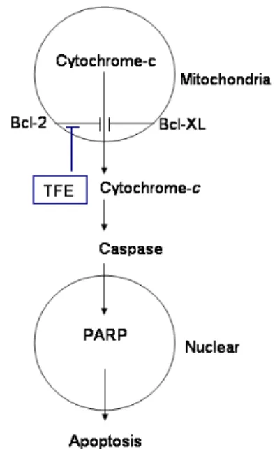
6. 천련자가 MCF-7 cell의 cytochrome-c 에 미치는 영향.
TFE가 Bcl-2의 발현을 농도 의존적으로 감소시킨 것과 관련하여 미토콘드리아를 제거한 cytosolic fraction에서 cytochrome-c 의 발현을 분석하였다.
대조군에서는 세포질내에 cytochrome-c 의 양이 적었으나, TFE를 0.30㎎/㎖을 처치한 경우는 세포질내의 cytochrome-c 가 2배 이상 증가하였다(Fig. 7).
Cytochrome-c
CON 0.03 0.10 0.30
TFE (㎎/㎖)
Fig. 7. The effects of TFE on the levels of cytochrome-c protein associated with
apoptosis.
MCF-7 cells were exposed to TFE (0.03-0.30
㎎/㎖) for 24 h. Cytochrome-c was immunoblotted using the cytochrome-c antibody in the cytosolic fraction. The density of cytochrome-c was calculated using densitometer. (TFE;