Received: November 1, 2019 Revised: January 7, 2020 Accepted: January 22, 2020 Trauma and InJury
Correspondence to Tan Jih Huei, M.R.C.S.
Department of General Surgery, Sultanah Aminah Hospital, Jalan Persiaran Abu Bakar Sultan, 80100 Johor Bahru, Johor, Malaysia
Tel: +60-17-611-3305 Fax: +60-7-353-5957 E-mail: [email protected]
The management of Pancreatic fistu- la Complicated by Gastric fistulation following Emergency Splenectomy
Tan Jih Huei, M.R.C.S, Henry Tan Chor Lip, M.D., Chow Sing Thou, Yuzaidi Mohamad, M.Med. (Surg), Rizal Imran Alwi, M.Med. (Surg) Department of General Surgery, Sultanah Aminah Hospital, Johor Bahru, Malaysia
Pancreatic and gastric fistulas are rare complications of emergency splenectomy, and it is extremely rare for a pancreatic fistula to be further complicated by a fistulation into the stomach. Here, we present a case of pancreatogastric fistula in a 60-year-old man who experienced polytrauma due to a blunt mechanism. He underwent emergency splenectomy for splenic injury and developed a pancreatic fistula as a complication. A percutaneous endoscopic procedure was performed to drain the fistula, after which he developed a pancreatogastric fistula as a further complication. A double-pigtail stent was inserted via gastroscopy into the fistula tract to allow internal drainage of the pan- creatic collection into the stomach cavity. When a pancreatic fistula is complicated by gastric fistulation, endoscopic stenting of the pancreatogastric fistula tract for internal drainage is an effective treatment option.
Keywords: Pancreas; Splenectomy; Gastric; Fistula
INTRODUCTION
Pancreatic fistula formation is a rare complication following splenectomy in patients with traumatic injuries [1]. Management and treatment approaches range from sup- portive care, endoscopic therapy, and percutaneous drainage to surgical therapy [1,2].
Gastric fistula following splenectomy is uncommon, but can be caused by gastric wall
necrosis due to suture ligation of the short gastric artery to the gastric tissue [3]. It is
extremely rare for these two complications to co-occur as a pancreatogastric fistula. To
our knowledge, no such cases have been reported in patients after emergency splenec-
tomy, although this phenomenon has been described in patients with chronic pancre-
atitis and intraductal papillary mucinous neoplasm [4]. Herein, we describe the case
of a pancreatogastric fistula in a 60-year-old man after emergency splenectomy for a splenic injury due to blunt trauma. He developed a pancreatic fistula as a complica- tion, and later, the endoscopic drainage procedure for the pancreatic fistula resulted in a pancreatogastric fistula.
We discuss the available evidence on the management of post-splenectomy pancreatic fistula and the treatment of pancreatogastric fistula, which is an unusual complica- tion.
CASE REPORT
A 60-year-old Chinese man involved in a motor vehicle accident presented with reduced consciousness, shortness of breath, and abdominal pain. His blood pressure on arrival was 166/87, his heart rate was 101 bpm, and his oxygen saturation was 94% under high-flow oxygen. His Glasgow Coma Scale score was 10/15, with unequal reac- tive pupils. Upon palpation, the abdomen was soft, but mildly distended. He was intubated due to reduced con- sciousness and poor oxygenation. Arterial blood gas anal- ysis revealed a PaO
2of 282 mmHg, a pH of 7.366, a pCO
2of 36 mmHg, a bicarbonate level of 21.1 mmol/L, and a base deficit of -4.3 mmol/L. The patient’s haemoglobin level was 14.3 g/dL and platelet count was 147×10
9/L.
Chest radiography revealed fractures of multiple left
ribs and the clavicle. An abdominal Focused Assessment with Sonography for Trauma scan showed free fluid over the perihepatic and perisplenic spaces. Computed tomography (CT) of the thorax, abdomen, and pelvis revealed grade 2 splenic injury and fracture of the left third through 10th ribs with lung contusions. Brain CT revealed right temporal acute subdural haemorrhage of 0.5 cm thickness with adjacent contusions, the largest of which was approximately 2×1 cm. There was associated right frontotemporoparietal subarachnoid haemorrhage and left parietal subgaleal haematoma. The patient re- ceived intensive care with cerebral protection.
Despite optimal resuscitation with blood transfusions, there was a gradual decrease in the patient’s haemoglobin level to 8.8 g/dL. In light of the dropping haemoglobin level and the lack of an interventional radiology service for angioembolization and severe traumatic brain injury, an emergency exploratory laparotomy was performed.
Intraoperatively, the spleen was identified to have a grade 3 splenic injury with multiple splenic lacerations measur- ing 1-3 cm at its anterior and superior borders. The spleen was mobilized from its lateral ligamentous attachment and brought medially to the wound. The hilar vessels were clamped and ligated with absorbable 2-0 sutures and stitch-tied with Prolene 2-0. A Surgicell (ETHICON, Neuchâtel, Switzerland) haemostatic device was placed around the hilar ligature. A soft drain was placed over the
Fig. 1. Computed tomography performed 9 days post-splenectomy showing a collection (arrows) at the splenic bed/pancreatic tail with a close
relationship to the greater curvature of the stomach.
splenic bed and drained 100 to 300 mL per day. The amy- lase level in the drainage fluid amylase level was 41,180 U/L.
The drain was removed on day 7 following splenectomy when it stopped flowing and the amylase level dropped to 17,506 U/L. The patient developed fever following drain removal. Repeat ultrasonography and abdominal CT revealed a left subphrenic collection (Fig. 1). The white cell count had increased to 29.2×10
9/L, with a C-reactive protein level of 242 U/L. Ultrasound-guided percutane- ous drainage of the collection was performed with a 12-Fr pigtail catheter. Tracheostomy was performed to facilitate early weaning from the ventilator. Tazobactam and pip- eracillin were prescribed empirically, as the culture yield- ed mixed patterns of microorganisms.
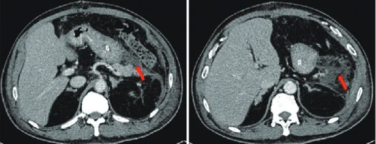
The percutaneous abdominal drain persistently drained 300 to 800 mL of yellowish fluid per day due to a pan- creatic fistula. An endoscopic retrograde cholangiopan- creatography with pancreatography was performed, and revealed contrast leakage at the pancreatic tail. A pancreatic stent was inserted during the procedure. The percutaneous pig-tail drain into the pancreatic fistula collection was upsized to 36-Fr due to poor drainage flow. Repeated abdominal CT after upsizing of the drain revealed residual collection with a close relationship of the rubber drain to the gastric fundus (Fig. 2). Two months post-splenectomy, endoscopic washout was performed with a therapeutic gastroscope via the drain track. A 36-Fr Portex (silicone-drain; Primed, Halberstadt, Germany)
tube was inserted following the procedure. Three days af- ter insertion of the Portex (Primed) tube, undigested food was present within the drainage tube.
Gastroduodenoscopy was performed and revealed that the drainage tube penetrated into the gastric body (Fig. 3).
The pancreatic stent was removed during the endosco- py, and a 10-Fr double-pigtail stent was placed at the pancreatogastric fistula opening. The rubber tube drain within the fistula was removed and the cutaneous drain opening was contained by a stoma device. Following this, the patient remained afebrile and with no peritonism for
Fig. 2. The rubber drain (blue arrows) was abutting onto the gastric fundus (red arrows). The image on the left is cephalad to the right.
Fig. 3. Gastroscopy view showing the Portex (silicone-drain; Primed,