355
http://dx.doi.org/10.4046/trd.2011.71.5.355 ISSN: 1738-3536(Print)/2005-6184(Online) Tuberc Respir Dis 2011;71:355-358
CopyrightⒸ2011. The Korean Academy of Tuberculosis and Respiratory Diseases. All rights reserved.
Histopathologic Diagnosis of Pleural Metastasis of Renal Cell Carcinoma Using Endobronchial Ultrasound-Guided Transbronchial Needle Aspiration
Yeh Rim Kang, M.D.1, Byung Woo Jhun, M.D.1, Kyeongman Jeon, M.D., Ph.D.1, Won-Jung Koh, M.D., Ph.D.1, Gee Young Suh, M.D., Ph.D.1, Man Pyo Chung, M.D., Ph.D.1, Hojoong Kim, M.D., Ph.D.1, O Jung Kwon, M.D., Ph.D.1, Joungho Han, M.D., Ph.D.2, Sang-Won Um, M.D., Ph.D.1
1Division of Pulmonary and Critical Care Medicine, Department of Medicine, 2Department of Pathology, Samsung Medical Center, Sungkyunkwan University School of Medicine, Seoul, Korea
Endobronchial ultrasound-guided transbronchial needle aspiration (EBUS-TBNA) is a useful, safe diagnostic modality for evaluating mediastinal and hilar lymphadenopathy. We report a 51-year-old male who presented with a left renal mass and multiple pleural masses without lung parenchymal lesions. The pleural masses were thought to be metastatic tumors or malignant mesothelioma. The patient underwent two percutaneous needle biopsies of the pleural mass, but the specimens were insufficient for a histopathological diagnosis. Because one pleural mass was adjacent to the right main bronchus, we decided to perform EBUS-TBNA for the pleural mass. As a result, sufficient core tissue was obtained with no complications, and the histopathological findings were consistent with metastatic papillary renal cell carcinoma. To our knowledge, this is the first case of using EBUS-TBNA for a pleural mass.
Key Words: Biopsy, Fine-Needle; Ultrasonography; Pleura; Neoplasms; Carcinoma, Renal Cell
Address for correspondence: Sang-Won Um, M.D., Ph.D.
Division of Pulmonary and Critical Care Medicine, Depart- ment of Medicine, Samsung Medical Center, Sungkyunkwan University School of Medicine, 50, Irwon-dong, Kangnam- gu, Seoul 135-710, Korea
Phone: 82-2-3410-3429, Fax: 82-2-3410-3849 E-mail: [email protected]
Received: Feb. 20, 2011 Accepted: Mar. 24, 2011
Introduction
The lung is the most frequent site of breast, renal, colorectal, or melanoma metastasis. However, isolated pleural metastasis without lung parenchymal metastasis also can occur in some cases. In such cases, specimens are usually obtained from pleural fluid aspiration cytol- ogy, percutaneous needle biopsy, or surgical resection of the pleura1,2. This case study describes metastatic re- nal cell carcinoma of the pleura that was diagnosed by endobronchial ultrasound-guided transbronchial needle aspiration (EBUS-TBNA). To our knowledge, this is the first case of EBUS-TBNA for a pleural mass.
Case Report
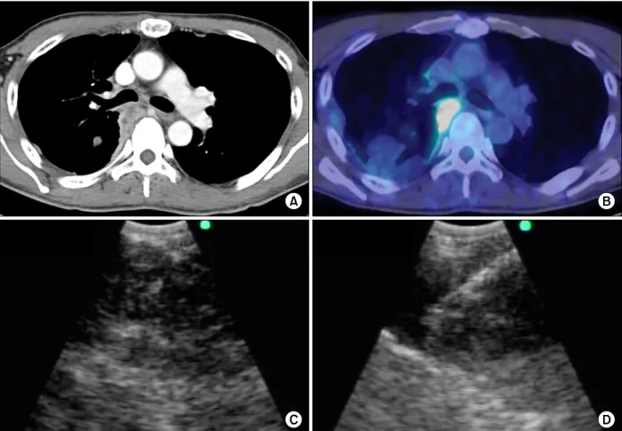
A 51-year-old non-smoking male with no chronic dis- ease presented with a cough and right-side chest pain for 1 month. He visited another institution, where a chest x-ray showed a widened mediastinum and pleural effusion in the right hemithorax. Contrast-enhanced computed tomography (CT) of the chest revealed a small pleural effusion and thickened pleura forming multiple masses (Figure 1A). There was no parenchymal lesion in either lung. On positron emission tomography (PET)/CT, 18F-fluorodeoxyglucose (FDG) uptake was noted in the pleural masses, which were suspected to be malignant mesothelioma or metastatic tumor (Figure 1B). A left renal mass with FDG uptake was suspicious of renal cell carcinoma (RCC). The patient underwent a percutaneous needle biopsy of the pleura. A few atyp- ical mesothelial cells were seen in the tissue specimen, which was insufficient for immunohistostaining. The percutaneous pleural biopsy was repeated, but again,
Case Report
YR Kang et al: Histopathologic diagnosis of pleural metastasis using EBUS-TBNA
356
Figure 1. (A) Contrast-enhanced CT of the chest showing the pleural mass adjacent to the right main bronchus. (B) PET/CT shows hypermetabolic pleural masses. (C) EBUS reveals a heterogeneous low-echogenic pleural mass. (D) The transbronchial aspiration needle was inserted into the pleural mass under EBUS guidance. CT: computed tomography;
PET: positron emission tomography; EBUS: endobronchial ultrasound.
the specimen was insufficient for a diagnosis. Therefore, the patient was referred to Samsung Medical Center (Seoul, South Korea) for a histopathological diagnosis.
Because one pleural mass was adjacent to the right main bronchus, we decided to perform EBUS-TBNA of the pleural mass using a flexible convex probe ultra- sonic puncture bronchoscope with a 7.5-MHz-frequency linear scanning transducer (CP-EBUS, BF-UC206F-OL8;
Olympus, Tokyo, Japan). A dedicated ultrasound scan- ner (EU-C2000; Olympus Tokyo, Japan) was used as the image processor. On flexible bronchoscopic examina- tion, the posterior wall of the right main bronchus was narrowed by extrinsic compression. On EBUS, a huge, heterogeneous, low-echogenic pleural mass was seen at the right main bronchus level (Figure 1C). We per-
formed EBUS-TBNA of the pleural mass and obtained a large tissues core (Figure 1D). The patient did not suf- fer any complication from the procedure such as pneu- mothorax, pulmonary infection, or hemoptysis.
The specimens from the pleural mass were sufficient for histopathological examination and showed papillae formation of cells with atypical nuclei (Figure 2A).
Immunohistochemical staining (IHC) was negative for calretinin, Wilm's tumor-1 (WT-1), and Hector Battifora mesothelial cell-1 (HBME-1), which are markers for ma- lignant mesothelioma. It was also negative for IHC staining for transcription termination factor-1 (TTF-1), indicating a low probability of peripheral adenocar- cinoma of the lung. The specimen was positive for CD10 (Figure 2B). In this case, the patient was sus-
Tuberculosis and Respiratory Diseases Vol. 71. No. 5, Nov. 2011
357 Figure 2. (A) Histopathological examination of the core tissue shows papillae formation by cells with atypical nuclei (pleura,
×200). (B) The cells were immunoreactive for CD10, consistent with papillary renal carcinoma (CD10 stain, ×200).
pected of having RCC on PET/CT, and the positive re- action for CD10 suggested metastatic malignancy from RCC3,4. Four days after the EBUS-TBNA, a renal biopsy was performed, and the pathology indicated papil- lary-type RCC. Therefore, the patient was diagnosed with papillary RCC with metastasis to the right pleura.
Discussion
Pleural metastasis without lung parenchymal spread from extrathoracic malignancy is rare and difficult to diagnose. As Mitsuhiro et al. reported, it can occur sev- eral years after curative resection, especially in patients with RCC. It is often confused with malignant meso- thelioma because the CT images are similar5,6. To dis- tinguish metastatic malignancy from malignant meso- thelioma, it is important for clinicians to obtain a suffi- cient specimen for histopathological examination.
Endobronchial ultrasound-guided transbronchial nee- dle aspiration is a very useful procedure for obtaining specimens from mediastinal and hilar lymph nodes or lung masses accessible from airways in both primary lung cancer and metastatic malignancy7. It has high di- agnostic accuracy and excellent safety profiles, with minimal invasiveness under real-time visualization8. EBUS-TBNA can also be performed in other specific
cases. Nakajima et al.9 reported a case of central airway stenosis caused by a mediastinal cyst, which improved after cyst aspiration using EBUS-TBNA. Takahiro et al.
also reported a diagnosis of spinal chondrosarcoma based on EBUS-TBNA10; as the protruding posterior me- diastinal mass was adjacent to the carina, a histopatho- logical diagnosis could be made using EBUS-TBNA.
These cases show the usefulness of EBUS-TBNA for the diagnosis of mediastinal masses and the therapeutic util- ity of EBUS-TBNA for the aspiration of mediastinal cysts. Nevertheless, there has been no report of EBUS- TBNA for the diagnosis of pleural disease.
This case study reports a renal cell carcinoma with pleural metastasis. What is more unusual about our case is that a specimen was obtained from the pleural mass using EBUS-TBNA. To our knowledge, this is the first case of EBUS-TBNA for a pleural mass. Although the patient underwent percutaneous needle biopsy twice, the specimens were insufficient for IHC staining, which is necessary for a histopathological diagnosis. We de- cided to perform another diagnostic modality rather than a third percutaneous needle biopsy. EBUS-TBNA, instead of a surgical biopsy such as video-assisted thor- acoscopic surgery, ensured that the patient was free from pain and did not require general anesthesia, and the procedure was less invasive. Furthermore, sufficient
YR Kang et al: Histopathologic diagnosis of pleural metastasis using EBUS-TBNA
358
tissues from the EBUS-TBNA enabled the IHC examina- tion of multiple markers, which helped make a definite histopathological diagnosis in this patient.
Posterior pleural masses or lesions which are located in the proximity to both main bronchi or right bronchus intermedius can be accessible by EBUS-TBNA. Respira- tory physicians should aware that it is possible to obtain an adequate specimen from a pleural mass safely using EBUS-TBNA when the mass is adjacent to an airway.
References
1. Fischer MD, Goodman PC. Pleural effusion and renal cell carcinoma: an angiographic-pathologic correlation.
Chest 1979;75:647-8.
2. Ohnishi H, Abe M, Hamada H, Yokoyama A, Hirayama T, Ito R, et al. Metastatic renal cell carcinoma present- ing as multiple pleural tumours. Respirology 2005;10:
128-31.
3. Oates J, Edwards C. HBME-1, MOC-31, WT1 and calre- tinin: an assessment of recently described markers for mesothelioma and adenocarcinoma. Histopathology 2000;36:341-7.
4. Ordóñez NG. The diagnostic utility of immunohisto- chemistry in distinguishing between mesothelioma and
renal cell carcinoma: a comparative study. Hum Pathol 2004;35:697-710.
5. Azuma T, Nishimatsu H, Nakagawa T, Tomita K, Takeuchi T, Homma Y, et al. Metastatic renal cell carci- noma mimicking pleural mesothelioma. Scand J Urol Nephrol 1999;33:140-1.
6. Kamiyoshihara M, Ibe T, Takise A, Itou H, Takeyoshi I. Pleural metastases from renal cell carcinoma 16 years after resection. J Clin Oncol 2007;25:4009-11.
7. Tournoy KG, Govaerts E, Malfait T, Dooms C. Endo- bronchial ultrasound-guided transbronchial needle bi- opsy for M1 staging of extrathoracic malignancies. Ann Oncol 2011;22:127-31.
8. Varela-Lema L, Fernández-Villar A, Ruano-Ravina A.
Effectiveness and safety of endobronchial ultrasound- transbronchial needle aspiration: a systematic review.
Eur Respir J 2009;33:1156-64.
9. Nakajima T, Yasufuku K, Shibuya K, Fujisawa T.
Endobronchial ultrasound-guided transbronchial nee- dle aspiration for the treatment of central airway steno- sis caused by a mediastinal cyst. Eur J Cardiothorac Surg 2007;32:538-40.
10. Nakajima T, Yasufuku K, Suzuki M, Sekine Y, Shibuya K, Hiroshima K, et al. Histological diagnosis of spinal chondrosarcoma by endobronchial ultrasound-guided transbronchial needle aspiration. Respirology 2007;12:
308-10.