419
The Natural Course of Endobronchial Inflammatory Polyps as a Complication after Endobronchial Ultrasound-Guided Transbronchial Needle Aspiration
Kyu Min Lee, M.D.
1,*, Sun Mi Jang, M.D.
1,*, Seo Young Oh, M.D.
1, Do Young Kim, M.D.
1, Geewon Lee, M.D.
2, Ahrong Kim, M.D.
3, Min Ji Kim, M.D.
1, Tae Hwa Kim, M.D.
1, Joon Woo Park, M.D.
1, Kwangha Lee, M.D.
1, Ki Uk Kim, M.D.
1, Min Ki Lee, M.D.
1and Jung Seop Eom, M.D.
1Departments of
1Internal Medicine,
2Radiology, and
3Pathology, Pusan National University School of Medicine, Busan, Korea
We presented a case of unusual endobronchial inflammatory polyps as a complication following endobronchial ultrasound–guided transbronchial needle aspiration (EBUS-TBNA) in a patient with tuberculous lymphadenitis. EBUS- TBNA of the right hilar lymph node was performed in a 29-year-old, previously healthy man. The patient was confirmed with tuberculous lymphadenitis and received antituberculosis medication over the course of 6 months. Chest computed tomography, after 6 months of antituberculosis therapy following the EBUS-TBNA showed nodular bronchial wall thickening of the right main bronchus. Histological and microbiological examinations revealed inflammatory polyps.
After 7 months, the inflammatory polyps regressed almost completely without need for removal.
Keywords: Needles; Complications; Lymphadenitis; Polyps; Tuberculosis
plications are occasionally reported with the recent wide- spread use of EBUS-TBNA, including hemorrhage, infection, and inflammatory polyps
4-6.
Reports have described the successful removal of inflam- matory polyps, which had developed after EBUS-TBNA, using biopsy forceps
6. However, the natural clinical course of inflam- matory polyps after EBUS-TBNA is still unclear. We report a patient with tuberculous lymphadenitis who developed inflammatory polyps 6 months after EBUS-TBNA, but did not undergo polyp removal, and discuss the natural progression of the polyps over the course of 7 months.
Case Report
A 29-year-old man visited our clinic with a 1-month history of a dry cough and night sweats. He had been healthy and had no specific family medical history related to these symptoms.
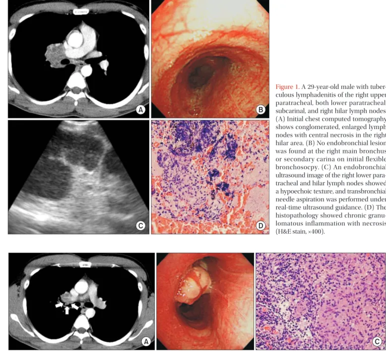
Chest computed tomography (CT) showed conglomerated, enlarged lymph nodes with central necrosis in the right upper paratracheal, both lower paratracheal, subcarinal, and right Copyright © 2015
The Korean Academy of Tuberculosis and Respiratory Diseases.
All rights reserved.
Introduction
Endobronchial ultrasound–guided transbronchial needle aspiration (EBUS-TBNA) is a useful technique for investigat- ing patients with various mediastinal diseases
1-3. Although EBUS-TBNA is generally considered to be safe, various com-
CASE REPORT
http://dx.doi.org/10.4046/trd.2015.78.4.419ISSN: 1738-3536(Print)/2005-6184(Online) • Tuberc Respir Dis 2015;78:419-422
Address for correspondence: Jung Seop Eom, M.D.
Department of Internal Medicine, Pusan National University School of Medicine, 179 Gudeok-ro, Seo-gu, Busan 49241, Korea
Phone: 82-51-240-7889, Fax: 82-51-254-3127 E-mail: [email protected]
*Kyu Min Lee and Sun Mi Jang contributed equally to this work.
Received: May 10, 2015 Revised: Jun. 11, 2015 Accepted: Jun. 29, 2015
cc