Tuberc Respir Dis 2012;72:182-186
CopyrightⒸ2012. The Korean Academy of Tuberculosis and Respiratory Diseases. All rights reserved.
뇌낭미충증과 감별이 어려웠던 폐암의 속립성 뇌전이 1예
1
전남대학교 의과대학 내과학교실,
2서남대학교 의과대학 내과학교실
이호준1, 오인재1, 박상우1, 반희정1, 김영철1, 김수옥2A Case of Miliary Brain Metastasis of Lung Cancer Mimicking Neurocysticercosis
Ho-Jun Lee, M.D.
1, In-Jae Oh, M.D.
1, Sang-Woo Park, M.D.
1, Hee-Jung Ban, M.D.
1, Young-Chul Kim, M.D.
1, Soo-Ok Kim, M.D.
2Department of Internal Medicine,
1Chonnam National University Medical School, Hwasun,
2Seonam University College of Medicine, Gwangju, Korea
Miliary brain metastasis from the lung is uncommon and has a poor therapeutic response. We report a case of pulmonary adenocarcinoma combined with multiple brain cystic lesions that were initially misdiagnosed as neurocysticercosis. A 53-year-old male who never smoked was admitted to our hospital with complaints of agitation and cognitive impairment. Brain magnetic resonance imaging showed innumerable, small nodular lesions with a central, low signal intensity in whole brain parenchyma. His symptoms were not improved by the empirical praziquantel medication for disseminated neurocysticercosis. After a transbronchial biopsy from the right middle lobe, we could diagnose the primary lung adenocarcinoma with a single nucleotide polymorphism in the epidermal growth factor receptor exon 20 at codon 787 (Q787Q). His neurologic symptoms and imaging findings have been gradually improving with a first-line Gefitinib treatment for five months. We recommend a more active diagnostic approach including biopsy in case of atypical imaging findings.
Key Words: Neurocysticercosis; Neoplasm Metastasis; Brain; Gefitinib
Address for correspondence: In-Jae Oh, M.D.
Lung and Esophageal Cancer Clinic, Chonnam National University Hwasun Hospital, 322, Seoyang-ro, Hwasun 519-763, Korea
Phone: 82-61-379-7617, Fax: 82-61-379-7619 E-mail: [email protected]
Received: Oct. 1, 2011 Revised: Oct. 27, 2011 Accepted: Nov. 27, 2011
서 론
폐암은 뇌전이를 일으키는 가장 흔한 고형암 중 하나이 나, 초기 뇌전이는 비특이적인 다양한 증상을 동반할 수 있어 의심하지 않으면 진단하기 쉽지 않은 경우가 많다.
다발성 뇌전이 특히 속립성 결절의 경우는, 뇌낭미충증을 비롯한 기생충감염, 혈관 및 염증성 질환, 기회감염 등의 신경계 감염성 질환과의 감별이 필요하다1. 폐암의 속립 성 뇌전이의 예후는 매우 좋지 않으며, 기존의 항암화학요
법 및 방사선치료에 반응이 좋지 않은 것으로 알려져 있고2, 표피세포 성장인자수용체 티로신인산화억제제(epidermal growth factor receptor tyrosine kinase inhibitor, EGFR- TKI)의 치료효과도 아직까지는 정립되어 있지 않다3-6. 저 자들은 혼돈과 초조를 주소로 내원한 환자에서 뇌낭미충 증과 유사한 영상 소견으로 인해 뇌전이를 진단하기가 쉽 지 않았으나, 1차치료제로 gefitinib을 사용하여 임상양상 의 호전을 보였던 폐선암 1예를 경험하여 문헌고찰과 함 께 보고하고자 한다.
증 례
환 자: 53세, 남자
주 소: 간헐적인 혼돈과 초조(agitation)
현병력: 3개월 전 혼돈된 의식상태를 보여 외부 병원에 서 시행한 뇌 자기공명영상에서 다발성 결절양 병소가 관
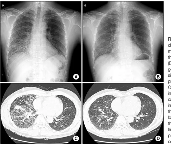
Figure 1. (A) The initial chest X-ray shows multiple miliary nodular lesions in the lower right lung fields.
(B) After 1 month of the gefitinib treatment, the le- sions are remarkably im- proved on X-ray. (C) Chest CT shows irregular nodular interstitial thickenings and consolidations combined with innumerable tiny nodules in the right middle and lower lobes. (D) After 1 month of the gefitinib treatment, the lesions are remarkably im- proved on CT scan. CT:
computed tomography.
찰되어 3차병원으로 전원을 권유 받았으나 개인적인 사정 으로 진료를 미루다가 내원 하루 전부터 의식상태 악화를 보여 응급센터로 내원하였다.
과거력: 특이 소견 없음 사회력: 비흡연자, 비음주자 가족력: 특이 소견 없음
진찰 소견: 내원 당시 측정한 혈압은 110/70 mm Hg, 맥박 84회/분, 호흡수 22회/분, 체온 36.0oC였으며, 급성 병색을 보였고 혼돈상태를 보였다. 피부에 특별한 병변은 없었고, 경부 임파절은 만져지지 않았으며 결막은 창백하 지 않았다. 흉부진찰에서 심음은 정상이었으나 우측 하폐 야에서 수포음이 청진되었다. 복부에서 간이나 비장은 만 져지지 않았고 압통과 반사압통도 없었으며 장음은 정상 이었다. 양측에 늑골척추각압통은 없었고 하지의 함요부 종도 없었다. 신경학적 검사상 Glasgow coma scale 13점 으로 경도의 뇌손상 소견을 보였으며, 전반적으로 불안감, 무기력, 인지기능 저하 등이 관찰되었다.
검사실 소견: 말초혈액 검사상 백혈구 13,500/mm3, 혈 색소 14.6 g/dL, 혈소판 315,000/mm3이었다. C-반응성 단 백은 0.5 mg/dL 미만이었다. 전해질 검사는 나트륨 138
mEq/L, 칼륨 4.0 mEq/L, 염소 108 mEq/L이었고, 생화학 검사에서 혈액요소질소 14.9 mg/dL, 크레아티닌 0.9 mg/
dL, 총단백질 7.0 g/dL, 알부민 4.2 g/dL, AST 59 IU/L, ALT 66 IU/L, LDH 431 IU/L, ALP 66 IU/L이었다. 간염표 지자 검사는 HBsAg 음성, anti-HBs 음성, anti-HCV 음성 이었고, 소변 검사는 비중 1.025, pH 5.0 WBC, RBC 음성 이었으며 당은 검출되지 않았고, 단백질이 2+ 양성을 보 였다. 객담 그람염색 및 항산균 도말 검사, 결핵 배양 검사 는 음성이었다.
방사선 소견: 흉부 방사선 검사에서 양측 폐야의 음영 증가 소견이 관찰되었으며(Figure 1A), 심전도상에서 분 당 80회의 정상 동성 리듬을 보였다. 뇌 자기공명영상에 서 다발성 석회화 음영과 함께 테두리에 상대적으로 고음 영을 보이는 원형의 다발성 병변이 관찰되어(Figure 2A, B), 뇌낭미충증과 같은 뇌신경계 감염증을 의심할 수 있 었으나, 속립성 결핵, 전이성 암 등에 대한 감별 노력이 필요할 것으로 판단되었다. 분변 및 혈액 기생충 효소면 역측정법(enzyme-linked immunosorbent assay, ELISA) 은 음성 소견을 보였다.
뇌척수액 검사 소견: 내원 당시 시행했던 뇌척수액 검
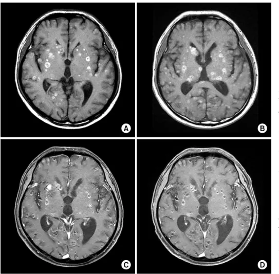
Figure 2. (A) The initial brain MRI of an outside hospital shows multiple cy- stic lesions with tiny scat- tered calcified nodules. (B) After 4 months, a follow-up MRI shows innumerable, small nodular lesions (up to 1.6 cm) with a central, low signal intensity in the whole brain parenchyma, sugge- stive of disseminated neu- rocysticercosis at the ve- sicular stage. (C) The cyst- ic and nodular lesions are improved by a gefitinib treatment for 1 month. (D) The cystic and nodular le- sions are improved by a gefitinib treatment for 5 months. MRI: magnetic re- sonance imaging.
Figure 3. Direct sequencing showing the mutation found in the kinase domain of EGFR-2361G>A transition in exon 20, but this did not alter the final amino acids of glutamine at codon 787 (Q787Q). EGFR: epidermal growth factor receptor.
사상 뇌척수 개방압력(opening pressure) 18 cm H2O, 총 핵세포(total nucleated cell) 음성, 포도당 66 mg/dL, 단백 질 58 mg/dL, adenosine deaminase 1.6 IU/L로 정상 소 견을 보여 중추신경계 감염증을 배제할 수 있었고, 뇌척수 액 세포 검사에서 비정상세포도 관찰되지 않았다.
임상경과: 환자는 혼돈과 초조로 내원하여, 방사선학적 검사상 뇌낭미충증이 강력히 의심되어 신경외과에서 경 험적 항기생충요법(praziquantel 3,600 mg/일) 2주 시행 후 추적하였으나 증상의 호전을 보이지 않았으며, 흉부 X-ray상 양측 폐야의 음영 또한 악화되어 시행한 흉부 전 산화 단층촬영 결과 불규칙적인 결절성 간질 비후 및 다수 의 작은 속립성 결절들이 주로 우측 폐에서 관찰되었다 (Figure 1C). 기관지내시경에서는 저명한 기관지 내 폐쇄 병소는 보이지 않았으나, 경기관지 조직 생검상 폐선암으 로 진단되었고, EGFR 유전자 엑손 20번의 단일유전자변 이(2361G>A 이행, Figure 3) 소견을 보여 gefitinib 250 mg을 경구 복용하였고, 1개월째 추적한 흉부 방사선 검사
(Figure 1B) 및 흉부 전산화 단층촬영(Figure 1D)에서 속 립성 결절의 호전을 보였다. 치료 1개월째 뇌 자기공명검 사상 병변의 수와 크기가 현저히 감소하였으며(Figure 2C), 의식상태도 호전을 보였다. 현재 환자는 5개월째 ge- fitinib치료를 하고 있고 뇌병변도 호전이 지속되고 있다
(Figure 2D).
고 찰
폐암의 뇌전이 소견은 흔하고 치명적인 예후를 보이는 질환이지만, 속립성 뇌전이를 보이는 경우 신경계 감염증 특히 뇌낭미충증과 같은 기생충 질환과 감별이 어려워 진 단이 어려울 수 있다7-9. 본 증례 또한 뇌 자기공명영상에 서 뇌낭미충증을 시사하는 소견 때문에 진단이 지연되었 으나, EGFR-TKI를 1차치료제로 사용하여 신경학적 증상 및 폐병변의 호전을 보였다.
뇌낭미충증의 영상 소견은 기생충의 생존단계에 따라 각각 다르게 보일 수 있다. 뇌낭미충증 초기(소포단계)에 는 컴퓨터 단층촬영상 뇌실질과 잘 경계되는 저음영의 둥 근 작은 병변으로 보이고, 뇌 자기공명영상에서 뇌척수액 과 비슷한 신호강도의 낭포가 형성되어 보인다. 낭포 내 에는 살아있는 촌중의 두절(scolex)이 고음영의 결절로 보 이기도 하며, 기생충 낭포의 퇴화가 연속적으로 진행하면 서 조영제 주입 후 반지 모양으로 소포 테두리에 조영 증 강 되어 보이는 교질소포 단계에 이르게 된다. 성충과 심 한 면역반응이 진행하면 T1, T2 강조 영상에서 저신호강 도를 보이면서, 주변에 부종이 동반되거나, 교세포 증다증 (gliosis)으로 인한 저신호강도 병변 테두리에 고신호강도 로 보이는 결절-과립 단계로 보이기도 하며, 최종적으로는 다발성 석회 병변이 보일 수 있다7. 전이성 뇌암의 경우 뇌낭미충증처럼 다양한 형태의 영상 소견을 보일 수 있고 다발성 전이의 형태가 매우 흔하지만, 5개 병소 이상의 다발성 병변은 5% 이하에서 관찰되며, 특히 속립성 전이 는 매우 드문 것으로 보고되고 있다8. 본 증례의 환자는 신경학적 증상을 주소로 내원한 환자였으며, 뇌 영상검사 상 뇌낭미충증을 의심할 수 있는 병적 단계에 따른 영상 소견(소포 단계, 교질소포 단계, 결절-과립 단계, 석회 단 계)이 저명하였고, 뇌실질 내에 수많은 결절이 동반된 속 립성 형태를 보이고 있어 초기에 전이성 뇌암 보다는 뇌낭 미충증으로 오인하였고, 폐병변에 대해서도 정밀검사를 하지 않아 진단이 지연되었다.
뇌전이는 폐암을 포함한 성인 고형암환자의 10∼30%
에서 나타난다10. 노인인구의 증가, 영상기법의 향상에 따 른 전 임상단계에서의 진단 및 전신질환치료의 발달로 암 환자의 생존기간이 길어지게 되고 그에 따라 뇌전이 소견 의 발견 또한 증가추세에 있다1. 널리 알려진 뇌전이를 잘 하는 일차성 암이 있는 환자에게 뇌의 다발성 병변이 발견
된다면 일차암의 뇌전이 소견을 확인하기 위한 뇌병변 조 직 검사를 추천하지 않으나, 일차암이 확인되지 않은 경우 나 일차암의 전신치료가 효과가 있어 장기생존이 기대되 는 경우, 뇌농양이 의심되는 경우, 뇌 자기공명검사상 비 특이적인 병변이 의심되는 경우에는 뇌 조직 검사가 필요 하다11. 본 증례에서도 뇌병변에 대한 조직 검사가 필요할 것으로 생각되었으나 경기관지 폐생검으로 원발성 폐암 이 진단 되었고, gefitinib 전신치료에 반응을 보여 전이 병소로 강력히 의심되었으며, 치료시작 후 시행된 추적 뇌 척수액 세포 검사에서 전이성 암세포를 확인할 수 있었다.
뇌전이를 동반한 폐암환자에서는 낮은 기대여명, 치료 에 따른 독성, 부작용, 혈액-뇌관 장벽 통과 저하 등의 이 유로 전신항암요법은 큰 효과가 없는 것으로 알려져 있으 며, 전뇌 방사선치료나 정위고정 방사선수술(stereotactic radiosurgery) 등이 표준치료로 추천되고 있지만 치료성 적은 좋지 않은 것으로 보고 되고 있다9,12. 본 증례에서는 EGFR 돌연변이가 있는 선암의 일차치료로 제안13되고 있 는 gefitinib을 경구 복용하였고, 1개월 후 추적검사상 흉 부의 다수 속립성 결절들의 크기와 수가 현저히 감소되었 을 뿐 아니라, 뇌병변의 수와 크기가 다소 감소하였으며, 의식상태도 호전을 보였다. 본 환자의 선암조직은 기관지 내시경에서 얻어진 것으로 검체의 양은 많지 않았으나 직 접 염기서열분석(direct sequencing)법으로 EGFR 엑손20 번의 2361G>A 단일유전자변이(또는 Q787Q)를 발견하 였는데, 이는 EGFR-TKI에 민감한 전형적인 변이가 아닌 silent mutation으로 알려져 있다14. 이 단일유전자변이만 있는 환자에서 EGFR-TKI 치료반응이 있는 보고는 거의 없고, 엑손19번 결손이나 L858R처럼 민감한 돌연변이가 동반된 경우만 치료반응이 있는 것으로 보고되어 있으나14, EGFR 단일유전자변이의 임상적 의의는 아직 불명확하다.
일반적으로 민감한 EGFR 돌연변이가 없는데도 EGFR-TKI 에 반응이 있는 경우 직접 염기서열분석법의 낮은 민감도 때문에 변이를 찾지 못하는 것이 원인이다. 본 증례는 PNA-mediated real-time PCR clamping법으로 재검한 결 과에서도 추가적인 EGFR 변이를 발견하지 못하였지만 환 자는 5개월 이상 gefitinib치료에 반응을 보이고 있다. 현 재 gefitinib 표준용량으로도 뇌와 폐병소가 조절되고 있 으나, 최근 보고에서는 표준용량으로 조절이 힘든 뇌전이 병소에 대해 고용량 gefitinib나 최대 내성용량이 더 높은 것으로 알려진 erlotinib을 시도하기도 하였다4-6. 결론적으로, 뇌낭미충증과 유사한 속립성 뇌전이와 같 이 영상 검사에서 비특이적 소견이 있으면 뇌 또는 가능한
원발병소에서의 조직검사 등 정확한 진단적 접근을 위한 노력이 필요할 것으로 생각한다.
참 고 문 헌
1. Klos KJ, O'Neill BP. Brain metastases. Neurologist 2004;10:31-46.
2. Ruppert AM, Stankoff B, Lavolé A, Gounant V, Milleron B, Seilhean D. Miliary brain metastases in lung cancer.
J Clin Oncol 2010;28:e714-6.
3. Porta R, Sánchez-Torres JM, Paz-Ares L, Massutí B, Reguart N, Mayo C, et al. Brain metastases from lung cancer responding to erlotinib: the importance of EGFR mutation. Eur Respir J 2011;37:624-31.
4. Katayama T, Shimizu J, Suda K, Onozato R, Fukui T, Ito S, et al. Efficacy of erlotinib for brain and lep- tomeningeal metastases in patients with lung ad- enocarcinoma who showed initial good response to gefitinib. J Thorac Oncol 2009;4:1415-9.
5. Kim JE, Lee DH, Choi Y, Yoon DH, Kim SW, Suh C, et al. Epidermal growth factor receptor tyrosine kinase inhibitors as a first-line therapy for never-smokers with adenocarcinoma of the lung having asymptomatic syn- chronous brain metastasis. Lung Cancer 2009;65:351-4.
6. Jackman DM, Holmes AJ, Lindeman N, Wen PY, Kesari S, Borras AM, et al. Response and resistance in a non-small-cell lung cancer patient with an epidermal growth factor receptor mutation and leptomeningeal metastases treated with high-dose gefitinib. J Clin Oncol 2006;24:4517-20.
7. García HH, Del Brutto OH. Imaging findings in neurocysticercosis. Acta Trop 2003;87:71-8.
8. Iguchi Y, Mano K, Goto Y, Nakano T, Nomura F, Shimo- kata T, et al. Miliary brain metastases from adenocar- cinoma of the lung: MR imaging findings with clinical and post-mortem histopathologic correlation. Neurora- diology 2007;49:35-9.
9. Ruppert AM, Stankoff B, Lavolé A, Gounant V, Milleron B, Seilhean D. Miliary brain metastases in lung cancer.
J Clin Oncol 2010;28:e714-6.
10. Barnholtz-Sloan JS, Sloan AE, Davis FG, Vigneau FD, Lai P, Sawaya RE. Incidence proportions of brain meta- stases in patients diagnosed (1973 to 2001) in the Metropolitan Detroit Cancer Surveillance System. J Clin Oncol 2004;22:2865-72.
11. Soffietti R, Cornu P, Delattre JY, Grant R, Graus F, Grisold W, et al. EFNS Guidelines on diagnosis and treatment of brain metastases: report of an EFNS Task Force. Eur J Neurol 2006;13:674-81.
12. Kvale PA, Simoff M, Prakash UB; American College of Chest Physicians. Lung cancer. Palliative care. Chest 2003;123(1 Suppl):284S-311S.
13. Cataldo VD, Gibbons DL, Pérez-Soler R, Quintás- Cardama A. Treatment of non-small-cell lung cancer with erlotinib or gefitinib. N Engl J Med 2011;364:947- 55.
14. Mu XL, Li LY, Zhang XT, Wang MZ, Feng RE, Cui QC, et al. Gefitinib-sensitive mutations of the epidermal growth factor receptor tyrosine kinase domain in chi- nese patients with non-small cell lung cancer. Clin Cancer Res 2005;11:4289-94.