학 술 논 문
107
경피적 전기 신경 자극의 적용 부위에 따른 자율신경계 활성도 변화 비교 및 평가
박승원
1ǂ· 최준원
2ǂ· 정면규
3· 이강인
3· 김한성
2*
1연세대학교 의공학과, 2연세대학교 의공학부, 3현대자동차
Comparison and Evaluation of Autonomic Nervous System (ANS) Activity According to the Stimulus Position of Transcutaneous
Electrical Nerve Stimulation (TENS)
Seung Won Park
1ǂ, Jun Won Choi
2ǂ, Myeon Gyu Jeong
3, Kang In Lee
3and Han Sung Kim
2*
1
Department of Biomedical Engineering, Yonsei University, Korea
2
Department of Biomedical Engineering, Yonsei University College of Health Sciences, Yonsei University, Korea
3
Hyundai Motor Company
(Manuscript received 6 May 2021 ; revised 22 June 2021 ; accepted 25 June 2021)
Abstract: Purpose: The aim of this study is to find the location of acupoints that are effective for increasing para- sympathetic nervous system(PNS) activity among acupoints used to relieve symptoms such as dizziness and nausea.
Materials and Methods: Twenty healthy adult men participated in this experiment. Transcutaneous electrical nerve stimulation(TENS) was applied to P6(Neiguan), ST36(Zusanli) and BL20(Pishu) for 30 minutes. For 5 minutes before and after stimulation, Electrocardiogram(ECG), Electrodermal activity(EDA), Respiration, and Skin tempera- ture(SKT) were measured using biosignalsplux Professional(biosignalsplux; Portugal). Paired t-test was performed for the results before and after stimulation, and a one-way ANOVA test between stimulations was performed for the rate of change before and after stimulation. Results: When each acupoint was stimulated with TENS, it was found that the PNS was generally activated. In addition, when considering the change in biosignals after stimulation, the participants stimulated with P6 showed the most consistent results. Conclusion: As a result of stimulating each acu- point, it was found that stimulating P6 can effectively increase PNS activity. These results indicate that methods of stimulating P6 may be most effective in alleviating symptoms of motion sickness.
Key words: Transcutaneous electrical nerve stimulation (TENS), Autonomic nervous system (ANS), Biosignal, Acupoint
I. 서 론
침술은 재활 및 물리치료 영역, 다양한 급만성 질병에 대 한 치료 효과가 밝혀졌으며, 이에 대한 기전 연구가 활발하
게 진행되어 왔다[1-6]. 침술은 수천 년 전부터 중국을 포함한 다양한 동양 국가에서 사용되어왔으며, 특정 신체 부위를 선 택하는 것을 침술 치료의 첫번째 단계로 받아들여지고 있다[7-9].
중국 전통 의학에서는 이러한 신체 부위를 경혈(Acupoint) 이라 부른다[10]. 경혈에 대한 분명한 정의는 아직까지 존 재하지 않지만, 감각 신경이 있는 신체 부위에 존재하는 신 경원성 점(Neurogenic Point)으로 받아들여지고 있다[11].
경혈로 알려진 지점과 그 주위의 지점 간에는 해부학적으로 구조적인 차이가 없는 것으로 나타났으며, 이것은 경혈이 신 체에서 구조적 영역이 아닌 기능적 영역이라는 것을 시사한
*Corresponding Author : Han Sung Kim
1, Yeonsedae-gil, Heungeop-myeon, Wonju-si, Gangwon-do, Republic of Korea
Tel: +82-33-760-2913
E-mail: [email protected]
ǂContributed equally to this work
본 연구는 ㈜현대자동차의 지원을 받아 수행하였음.
108
다[12-14].
경혈을 자극하기 위해 물리적인 힘을 가하는 지압과 기계 적인 자극을 이용하는 전통적인 방식의 침술에 더해, 바늘에 전 류를 인가하여 자극의 효과를 높이는 전기 침술(Electro- acupuncture) 등이 주로 사용되어 왔다[15-18]. 그 외에도, 경 피적 전기 신경 자극(Transcutaneous Electrical Nerve Stimulation; TENS) 과 같이 침술의 개념을 이용한 방법이 시도되었다[19-21].
경피적 전기 신경 자극은 주로 감각 신경을 자극하여 통 증을 완화시키는 것을 목표로 사용되는 전기 자극 방법이며, 비침습적인 방법으로 기흉이나 감염과 같은 합병증없이 안 전하게 자극을 가할 수 있다는 장점이 있다[22,23]. 이전 연 구들에서 경혈에 대한 TENS는 우울증이나 불안 장애와 같은 심리적 질환을 완화하고, 운동 기능, 시공간 능력, 자세 조절 및 인지 기능을 향상시키는 효과가 보고되었다[24-26]. 그 밖 에도, 자율신경계(Autonomic Nervous System)의 활성도를 변화시켜 기능 장애를 포함한 다양한 질병을 완화시킬 수 있는 것으로 알려졌다[27,28].
이에 더해, 경혈에 대한 경피적 전기 신경 자극은 멀미 (Motion Sickness)를 완화하기 위해서 사용된다[27]. 멀미가 발 생하면, 교감 신경계(Sympathetic Nervous System)의 활 성도가 증가하고, 부교감 신경계(Parasympathetic Nervous System)의 활성도가 감소하는 자율신경계 변화로 인한 위 장계 증상이 나타난다[29-32]. 이러한 신체 반응 메커니즘을 TENS 자극을 통해 억제하고 멀미 증상을 완화할 수 있는 것으로 나타났다[27].
멀미 증상을 완화하기 위해 다양한 경혈에 대해 자극을 적용하는 연구가 진행되어왔다. Stern, Robert M et al.(2001) 의 경우, 경혈 중 P6(Neiguan)에 대한 지압 자극을 통해 회전으로 인해 유발된 멀미를 완화시켰다[33]. 그 외에, ST36(Zusanli), BL20(Pishu) 에 대한 자극을 통해 멀미 관 련 증상을 완화시키려는 시도가 있었다[34,35]. 그러나, 각 경혈 부위를 자극하는 방법이 상이하며, 그 효과를 정량적 으로 비교한 연구가 부족한 실정이다.
본 연구에서는 멀미 증상 완화에 사용되는 대표적인 경혈 부위인 P6, ST36, BL20 부위에 경피적 전기 신경 자극을 적용하고, 생체 신호의 변화를 평가하여 부교감신경계 활성 도 변화에 효과적인 경혈 부위를 평가하고자 하였다.
II. 연구 방법
1. 실험 참가자
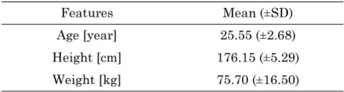
본 연구에는 경피적 전기 신경 자극으로 인한 부작용 발생 가능성이 낮은 건강한 성인 남성 20 명이 참여하였다(표 1).
본 연구는 국제 통증 연구협회(International Association
on Study of Pain) 내 윤리 문제위원회의 통증 연구를 위한 윤리 지침을 준수하는 범위에서 진행되었다[36]. 또한, 연세 대학교 미래캠퍼스 생명윤리심의위원회에서 본 실험 과정에 대해 승인받았다(1041849-202012-BM-176-02). 모든 실 험 참가자들은 실험 과정에 대한 내용을 안내받았다. 더불 어, 경피적 전기 신경 자극으로 인해 근수축이 유발될 수 있고, 경우에 따라 고통이나 불쾌감이 느껴질 수 있음을 안내하였 으며, 이후 자발적으로 실험 참여에 동의하였다.
2. 실험 과정
본 연구에서 자극 효과를 평가하고자하는 경혈들은 사전 연구들을 토대로 P6(Neiguan), ST36(Zusanli), BL20(Pishu) 와 같은 경혈을 선정하였다(그림 1). 각 실험 참가자는 1 일 1 회 실험에 참여하였으며, 각 자극 부위의 종류에 따라 총 3 회의 실험을 진행하였다.
각 실험에서 실험 참가자는 경피적 전기 신경 자극 전(t0)에 생체신호를 5분 동안 측정하고, 경피적 전기 신경 자극 후 (t1) 에 생체신호를 5분 동안 측정했다(그림 2). 이전 연구에 따라 총 30분 동안의 경피적 전기 신경 자극을 적용하였으 며, 모든 실험 과정에서 실험 참가자는 의자에 착석한 상태를 유지했다[37]. 실험에서 위약 효과를 제외하기 위해서, 실험 참가자들에게 실험 목적을 경피적 전기 신경 자극을 적용한
그림 1. 경혈 위치(P6, ST36, BL20)
Fig. 1. The position of acupoints(P6, ST36, BL20) 표 1. 실험 참가자 정보
Table 1. The information of participants
Features Mean (±SD)
Age [year] 25.55 (±2.68)
Height [cm] 176.15 (±5.29)
Weight [kg] 75.70 (±16.50)
그림 2. 실험 과정
Fig. 2. The design of experiment
109 부위별 안락감 평가로 안내하였으며, 안락감에 대한 가짜 설
문을 진행하였다.
3. 경피적 전기 신경 자극
각 경혈에 경피적 전기 신경 자극을 적용하기 위해, 30 mm의 일회용 Ag/AgCl 전극(Medi-Trace 100, Kendall Healthcare, Canada)을 부착하였다(그림 3). 경혈을 찾는 방법으로는 가 장 정확하게 받아들여지는 세계보건기구(WHO)에서 발표한 해부학적 방법을 사용하였으며, 이를 통해 전극을 부착하였 다[38].
각 경혈 지점에 Stimio TENS(TC32, Stimio, France)를 통해 10 Hz의 경피적 전기 신경 자극을 적용하였다. 자극 에 대한 적응 가능성을 고려하여, 자극 부위에 따른 실험 순 서를 실험 참가자마다 임의로 정하여 진행하였다. 자극 강 도는 실험 참가자가 충분히 느낄 수 있지만, 고통스럽거나 불쾌하지 않은 범위에서 가장 강한 강도로 자극을 적용하였 다. 실험 참가자가 고통스럽거나, 심리적 불안감 및 불쾌감을 호소할 경우, 실험을 즉각 중단하고 휴식을 취했다.
4. 생체신호
본 연구에서는 실험 참가자의 자율신경계 활성도 변화를 관찰하기 위해, 다중 생체 신호 측정 장비(biosignalsplux Professional, biosignalsplux; Portugal) 를 사용하여 심전 도(Electrocardiogram; ECG), 피부전도도(Electrodermal Activity; EDA), 호흡(Respiration), 피부 온도(Skin Temperature;
SKT) 와 같은 생체 신호를 측정하였으며, 1000 Hz의 샘플링 레 이트(Sampling Rate)로 데이터를 수집하였다(그림 4). 생체 신 호를 측정하기 위해 지름 30 mm의 일회용 Ag/AgCl 전극 (Medi-Trace 100, Kendall Healthcare, Canada) 을 사용 하였으며, 각각의 전극은 biosignalsplux에서 제공한 사용자 매 뉴얼에 명시된 위치에 부착하였으며, ECG를 측정하기 위한 전극 위치는 Einthoven leads 방법이 적용되었다[39,40](그림 5).
심전도 신호에 대한 시간 영역에서의 분석을 위해 3-45 Hz 의 4 차 대역 통과 필터(Band Pass Filter)와 80 ms의 이동 윈도우 적분(Moving Window Integration)을 적용하
여 전처리하였다(그림 6). 전처리된 심전도 신호를 통해, 심 박변이도(Heart Rate Variability; HRV) 분석을 수행하였 다[41]. 시간 영역에서의 분석에 사용되는 R-peak 검출을 위해, 판-톰킨스 알고리즘(Pan & Tomkins Algorithm)을 적용하였으며, 이를 통해 심박수(Heart Rate; HR)를 분석 하였다[42]. 주파수 영역에서의 전력 스펙트럼(Power Spectrum) 분석을 위해, Welch’s 방법을 활용하였다. 0.04-0.15 Hz 영 역의 저주파 전력 성분(Low Frequency Power; LF)과 0.15-0.40 Hz 영역의 고주파 전력 성분(High Frequency Power; HF) 을 검출하였으며, 식 (1)과 (2)를 통해 저주파 비율(Normalized Low Frequency; normLF)과 고주파 비 율(Normalized High Frequency; normHF)를 분석하였 다[41].
그림 3. 경혈 자극을 위한 전극 부착 위치
Fig. 3. Position of electrode attachment for acupuncture stimulation
그림 4. 다중 생체 신호 측정 장비
Fig. 4. Multi-biosignals collection equipment
그림 5. 생체신호 측정을 위한 전극 부착 위치
Fig. 5. Electrode attachment positions for measuring biosignals
그림 6. 생체신호 전처리 과정
Fig. 6. Biosignals preprocessing process
110
Normalized low frequency (normLF) = (1)
Normalized high frequency (normHF) = (2)
피부전도도는 5 Hz의 4 차 저역 통과 필터(Low Pass Fiter) 로 필터링하여 분석하였다[43](그림 6). 피부전도도를 통 해 교감신경계 활성도의 변화를 평가하기 위한 생리학적 지 표인 피부 전기 전도도 수준(Skin Conductance Level;
SCL) 을 관찰하였다[44-46].
호흡은 스트레인 게이지(Strain Gauge)가 내장된 호흡 센서를 사용하여 측정하였으며, 0.1-0.35 Hz의 1 차 대역 통과 필터(Band Pass Filter)를 통해 전처리하였다(그림 6). 제로-크로 싱(Zero-Crossing) 방법을 통해 호기와 흡기를 구분하여, 1 분 동안의 호흡수(Respiratory Rate; RR)을 분석하였다[43].
실험 참가자의 손목에서 피부 온도를 측정하였다[47]. 측
정된 신호는 0.25 Hz의 4 차 저역 통과 필터(Low Pass Fiter) 를 통해 필터링하였다[43](그림 6).
5. 통계 분석
실험 결과에 대해 SPSS statistics 25(SPSS, IBM, USA)를 사용하여 분석을 진행하였다. 자극군 내에서 자극의 효과를 평가하고자, 유의수준 0.05에서 자극 전/후에 대해 대응표 본 t-검정(Paired t-test)을 진행하였다. 자극 부위별 효과를 비 교하고자, 자극 전/후의 변화율을 분석하여, 유의수준 0.05 에서 자극군 간 일원배치 분산분석(One-way ANOVA test)을 수행하였다.
III. 연구 결과
1. 심박 변이도(HRV) 평가
시간 영역에서의 심박변이도 분석을 통해 심박수를 분석 LF
LF HF + ---
LF HF HF + ---
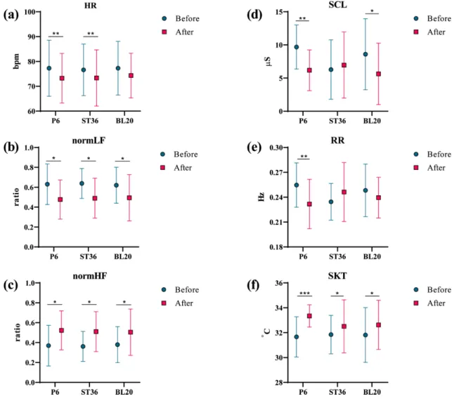
그림 7. 경피적 전기 신경 자극 전/후의 생체신호 분석 결과(*p<.05, **p<.01, ***p<.001)
Fig. 7. Biosignal analysis results before and after applying transcutaneous electrical nerve stimulation(*p<.05, **p<.01,
***p<.001)
111
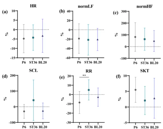
하였다. 모든 경혈 지점에서 자극 이후 실험 참가자의 심박 수가 감소하는 경향을 보였다(그림 7a). 자극 전후에 따른 대응표본 t-검정 결과에서 BL20를 제외한 나머지 자극 부 위에서 통계적으로 유의한 결과가 나타났다(표 2). 자극 부 위별 자극에 따른 심박수 변화율을 비교하였으며, P6(-4.81%), ST26(-4.26%), BL20(-3.29%) 순으로 심박수가 감소하였다 ( 그림 8a). 일원배치 분산분석 결과, 통계적으로 유의한 결 과가 나타나지 않았다.
주파수 영역에서의 심박변이도 분석을 통해 normLF와 normHF 를 분석하였다. P6, ST36, BL20은 normLF가 감 소하고 nomHF가 증가하는 추세를 보였다(그림 7b, c). 자 극 전후에 따른 대응표본 t-검정 결과에서 모든 자극 부위 에서 통계적으로 유의한 결과가 나타났다(표 2). 자극 부위 별 자극에 따른 normLF, normHF 변화율을 평가하였으며, ST36(normLF: -24.57%, normHF: 62.94%), BL20(normLF:
-20.83%, normHF: 47.76%), P6(normLF: -18.84%, normHF:
81.75%) 에서 normLF가 감소하고 normHF가 증가하는 경 향을 보였다(그림 8b, c). 일원배치 분산분석 결과, 통계적 으로 유의한 결과가 나타나지 않았다(표 3).
2. 피부 전기 전도도 수준(SCL) 평가
자율신경계 활성도 변화를 평가하기 위해 자극 전후에 대한 피부 전기 전도도 수준을 분석하였다(그림 7d). ST36을 제 외한 P6, BL20에서 피부 전기 전도도 수준이 감소하는 경 향을 보였으며, 자극 전후에 따른 대응표본 t-검정 결과에서 P6 와 BL20에서 통계적으로 유의한 차이를 보였다(표 2).
자극 부위별 자극에 따른 피부 전기 전도도 수준 변화율을 비교하였으며, ST36(41.47%)은 증가하였고, P6(-29.68%), BL20(-26.31%) 순으로 감소하였다(그림 8d). 일원배치 분 산분석 결과, 통계적으로 유의한 결과가 나타나지 않았다(표 3).
3. 호흡수(RR) 평가
분당 호흡수를 관찰하기 위해 시간 영역에서 호흡수 분석 하였다(그림 7e). 대응표본 t-검정 결과, P6에서 통계적으로 유의한 결과가 나타났다(표 2). 자극 부위별 자극에 따른 호 흡수 변화율을 비교하였을 때, ST36(4.86%)을 제외하고 P6(-8.55%), BL20(-2.79%), 순으로 감소하는 추세가 나타 났다(그림 8e). 일원배치 분산분석 결과, P6와 ST36 간에 통계적으로 유의한 차이가 나타났다(표 3).
표 2. 경피적 전기 신경 자극 전/후 생체신호에 대한 대응표본 t검정 결과(*p<.05, **p<.01, ***p<.001)
Table 2. Paired t-test results of biosignals before and after applying transcutaneous electrical nerve stimulation(*p<.05,
**p<.01, ***p<.001)
Features P6 ST36 BL20
Before After Before After Before After
HR [bpm]
Mean (SD)
77.28 (11.30)
73.26 (9.96)
76.61 (10.36)
73.34 (11.30)
77.28 (10.80)
74.30 (9.01) t, df
p-value
3.051, 19
**.007
2.925, 18
**.009
2.028, 19 .057
normLF [ratio]
Mean (SD)
0.63 (0.20)
0.48 (0.20)
0.64 (0.15)
0.49 (0.20)
0.62 (0.18)
0.49 (0.23) t, df
p-value
2.641, 19
*.020
2.464, 18
*.034
2.925, 19
*.011
normHF [ratio]
Mean (SD)
0.37 (0.20)
0.52 (0.20)
0.36 (0.15)
0.51 (0.20)
0.38 (0.18)
0.51 (0.23) t, df
p-value
2.641, 19
*.020
2.464, 18
*.034
2.925, 19
*.011
SCL [µS]
Mean (SD)
9.69 (0.85)
6.18 (3.07)
6.28 (4.47)
6.96 (4.99)
8.59 (5.35)
5.63 (4.62) t, df
p-value
3.476, 19
**.004
0.7147, 18 .485
2.584, 19
*.020
RR [Hz]
Mean (SD)
0.25 (0.03)
0.23 (0.03)
0.23 (0.02)
0.25 (0.04)
0.25 (0.03)
0.24 (0.02) t, df
p-value
3.203, 19
**.006
1.904, 18 .076
1.362, 19 .193
SKT [°C]
Mean (SD)
31.66 (1.62)
33.35 (0.90)
31.84 (1.55)
32.51 (2.13)
31.81 (2.20)
32.62 (1.97) t, df
p-value
6.6064, 19
***<.001
2.169, 18
*.043
2.170, 19
*.043
112 4. 피부 온도(SKT) 평가
모든 자극 부위에서 자극 이후 피부 온도가 증가하는 추 세를 보였다(그림 7f). 대응표본 t-검정 결과, 자극 부위에서 통계적으로 유의한 결과가 나타났다(표 2). 자극 부위별 자
극에 따른 피부 온도 변화율을 비교하였을 때, P6(5.49%), BL20(2.79%), ST36(2.08%) 순으로 피부 온도가 증가한 것 으로 나타났다(그림 8f). 일원배치 분산분석 결과, 통계적으로 유의한 결과가 나타나지 않았다(표 3).
그림 8. 자극 부위간 경피적 전기 신경 자극 전/후의 생체신호 변화율 분석 결과(*p<.05, **p<.01, ***p<.001)
Fig. 8. Analysis of the rate of change of biosignals before and after applying transcutaneous electrical nerve stimulation between stimulation positions(*p<.05, **p<.01, ***p<.001)
표 3. 경피적 전기 신경 자극 전/후 생체신호에 대한 일원배치 분산분석 결과(*p<.05, **p<.01, ***p<.001)
Table 3. One-way ANOVA results of the rate of change of biosignals before and after applying transcutaneous electrical nerve stimulation between stimulation positions(*p<.05, **p<.01, ***p<.001)
Features SS(Sum of Square) df(degrees of freedom) MS(Mean Square) F p-value
HR
Between Within Total
23.49 3460 3483
2 57 59
11.74
60.70 0.194 .825
normLF
Between Within Total
51.96 32096 32148
2 57 59
25.98
867.4 0.030 .971
normHF
Between Within Total
8379 395362 403741
2 57 59
4190
10685 0.392 .678
SCL
Between Within Total
55656 353042 408698
2 57 59
27828
7512 3.705 *.032
RR
Between Within Total
1510 5548 7057
2 57 59
754.9
120.6 6.259 **.004
SKT
Between Within Total
120.9 1438 1559
2 57 59
60.46
26.15 2.312 0.109
113
IV. 고찰 및 결론
본 연구의 목적은 멀미 증상 완화를 위해 주로 사용되는 경혈 부위들에 경피적 전기 신경 자극을 적용하고, 자율신 경계 활성도 변화를 생체 신호를 통해 비교 및 평가하는 것에 있다.
시간 영역에 대한 심박변이도 분석은 자율신경계 활성도를 평가하기 위한 방법으로 널리 사용되는 방법 중 하나이다.
심박수의 변화는 자율신경계에 의해 조절되는 굴심방 결절 (Sinus node) 에 의해 조절되는데, 심박수의 감소는 일반적 으로 부교감신경계 활성도가 증가하는 현상과 관련이 깊은 것으로 알려져 있다[48]. 본 연구 결과에서는 모든 경혈 부 위에 대한 자극 이후, 심박수가 감소하여 부교감신경계 활 성도가 증가한 결과를 보였다. 주파수 영역에서의 심박변이도 분석에서는 normLF과 normHF을 분석하였다. 이전 연구에 따르면, normLF는 교감신경계 활성도를 반영하고, normHF 은 교감신경계와 부교감신경계의 활성도 모두를 반영한다[49].
본 연구에서 P6, ST36, BL20를 자극하였을 때에는 normLF이 감소하고 normHF이 증가하였다. 이러한 결과를 고려해보면, 경혈 자극 이후 교감신경계보다 부교감신경계가 큰 폭으로 변화하는 것을 알 수 있으며, P6, ST36, BL20에 대한 자 극은 부교감신경계 활성도를 증가시키는 것으로 보인다.
시간 영역에서의 심박변이도 분석 결과와 주파수 영역에 서의 심박변이도 분석 결과를 고려해보면, P6, ST36에 대 한 자극 이후 부교감신경계 활성도가 증가하는 일관된 양상을 보였다.
일반적으로 교감신경계와 부교감신경계의 활성도는 상보 적인 관계를 갖는 것으로 알려져있다[50]. 이를 바탕으로 교 감신경계에 의해 조절되는 땀샘 운동성(Sudomotor)의 활 성을 정량적으로 평가하기 위해서 피부 전기 전도도 수준을 평가하였다[51-55]. 이전 연구에 따르면, 피부 전기 전도도 수준은 교감신경계 활성도와 관련이 있는 것으로 나타났다 [45]. 본 실험에서는 P6와 BL20에 대한 자극 이후, 교감신 경계 활성도가 감소한 것을 고려해보면, 해당 경혈 자극으로 인해 실험 참가자의 부교감신경계 활성도가 증가된 것으로 사료된다.
이전 연구에 따르면, 부교감신경계 활성도가 증가하면 호 흡수가 감소하는 것으로 나타났다[56]. 본 실험에서는 P6에 경피적 전기 신경 자극을 적용하였을 때, ST36을 제외한 나 머지 실험군에서 호흡수가 감소하는 것으로 이전 연구 결과 와 동일한 양상을 보였다. 그 중, P6에 대한 자극이 부교감 신경계 활성도 증가에 효과적인 것으로 나타났다.
멀미가 유발되면 부교감신경계가 활성화되는 현상과 더불 어, 혈관이 확장되고 혈류가 증가하여 신체의 열 손실이 감 소하는 것으로 알려져 있다[57]. 본 실험에서는 모든 경혈
부위에서 피부 온도가 증가하며, P6에서 가장 두드러진 변 화를 보였다
이전 연구에서는 P6와 ST36을 동시에 자극하는 것이 P6 를 단독으로 자극하는 것보다 더욱 효과적인 것으로 밝혀졌 기 때문에, 여러 자극 위치에 대한 복합 자극을 고려한 추 가 연구가 필요할 것으로 보인다[58].
P6, ST36, BL20 에 대한 경피적 전기 신경 자극은 교감 신경계 활성도를 감소시키고, 부교감신경계 활성도를 증가 시키는 것을 확인하였으며, 그 중에서 P6를 자극하였을 때 가장 일관적인 변화가 나타났다. 이러한 결과는 멀미 증상 완화를 위해서 P6를 자극하는 방법이 부교감신경계를 활성 화시키는 데에 가장 효과적일 수 있는 가능성을 보였다.
References
[1] Nager AL, et al. Effects of acupuncture on pain and inflam- mation in pediatric emergency department patients with acute appendicitis: a pilot study. The Journal of Alternative and Complementary Medicine. 2015;21(5):269-272.
[2] Sok SR, Erlen JA, Kim KB. Effects of acupuncture therapy on insomnia. Journal of Advanced Nursing. 2003;44(4): 375- 384.
[3] Zhang ZJ, et al. The effectiveness and safety of acupuncture therapy in depressive disorders: systematic review and meta- analysis. Journal of affective disorders. 2010;124(1-2):9-21.
[4] Galantino ML, et al. Acupuncture as an adjuvant modality with physical therapy for patients with knee osteoarthritis.
Medical Acupuncture. 2009;21(3):157-166.
[5] Ma T, et al. A study on the clinical effects of physical therapy and acupuncture to treat spontaneous frozen shoulder. The American journal of Chinese medicine. 2006;34(05):759- 775.
[6] Wu P, et al. Acupuncture in poststroke rehabilitation: a sys- tematic review and meta-analysis of randomized trials.
Stroke. 2010;41(4):e171-e179.
[7] Xinnong C. Acupuncture analgesia. Chinese acupuncture and moxibustion. Beijing: Foreign Languages Press. 1987:513- 23.
[8] Tan H, et al. Understanding acupoint sensitization: a narra- tive review on phenomena, potential mechanism, and clinical application. Evidence-Based Complementary and Alterna- tive Medicine. 2019.
[9] Li F, et al. What is the Acupoint? A preliminary review of Acupoints. Pain Medicine. 2015;16(10):1905-1915.
[10] Xing JJ, et al. Acupuncture point specificity. International review of neurobiology. 2013;111:49-65.
[11] Zhou W, Benharash P. Effects and mechanisms of acupunc- ture based on the principle of meridians. Journal of acupunc- ture and meridian studies. 2014;7(4):190-193.
[12] Lu Y, et al. Anatomic structure of Shaoze (SI 1), Qiangu (SI 2), Houxi (SI 3), Yanggu (SI 5) and Xiaohai (SI 8) regions of Hand-Taiyang Meridian in the rabbit's forelimb. Zhen ci yan jiu= Acupuncture research. 2013;38(6):473-477.
[13] Sun XG, et al. Macroscopical anatomic structure of acupoint Shangyang (LI 1), Erjian (LI 2), Sanjian (LI 3), Hegu (LI 4),
114
and Quchi (LI 11) regions in the forelimb in rabbits. Zhen ci yan jiu= Acupuncture research. 2012;37(1):31-37.
[14] Lou XF, Jiang SH. Anatomical characters and classification of acupoint. Zhongguo zhen jiu=Chinese acupuncture &
moxibustion. 2012;32(4):319-323.
[15] Napadow V, et al. Effects of electroacupuncture versus man- ual acupuncture on the human brain as measured by fMRI.
Human brain mapping. 2005;24(3):193-205.
[16] Fydanaki O, et al. Electroacupuncture could reduce motion sickness susceptibility in healthy male adults: a double-blinded study. Medical acupuncture. 2017;29(6):377-382.
[17] Shiao SYPK, Dune LS. Metaanalyses of acustimulations:
effects on nausea and vomiting in postoperative adult patients.
Explore. 2006;2(3):202-215.
[18] Tsui P, Leung MC. Comparison of the effectiveness between manual acupuncture and electro-acupuncture on patients with tennis elbow. Acupuncture & electro-therapeutics research.
2002;27(2):107-117.
[19] Lv JQ, Feng RZ, Li N. P6 acupoint stimulation for preven- tion of postoperative nausea and vomiting in patients under- going craniotomy: study protocol for a randomized controlled trial. Trials. 2013;14(1):1-7.
[20] Cheung, MC, Chan AS, Yip J. Microcurrent stimulation at shenmen acupoint facilitates EEG associated with sleepiness and positive mood: a randomized controlled electrophysio- logical study. Evidence-Based Complementary and Alterna- tive Medicine. 2015;2015.
[21] Kim SB, et al. Comparison of 2 methods of non-invasive treatment between transcutaneous electrical stimulation and pulsed electromagnetic field stimulation as replacement of invasive manual acupuncture. Acupuncture & electro-thera- peutics research. 2013;37(4):247-261.
[22] Wang B, et al. Effect of the intensity of transcutaneous acu- point electrical stimulation on the postoperative analgesic requirement. Anesthesia & Analgesia. 1997;85(2):406-413.
[23] White A, et al. Survey of adverse events following acupunc- ture (SAFA): a prospective study of 32,000 consultations.
Acupuncture in Medicine. 2001;19(2):84-92.
[24] Sanderson JE, et al. The effect of transcutaneous electrical nerve stimulation (TENS) on autonomic cardiovascular reflexes.
Clinical Autonomic Research. 1995;5(2):81-84.
[25] Van Dijk, KR, et al. Effects of transcutaneous electrical nerve stimulation (TENS) on non-pain related cognitive and behavioural functioning. Reviews in the Neurosciences. 2002;13(3):257- 270.
[26] Agelink M, et al. Does acupuncture influence the cardiac autonomic nervous system in patients with minor depression or anxiety disorders? Fortschritte der Neurologie-psychiatrie.
2003;71(3):141-149.
[27] Chu H, et al. Effects of transcutaneous electrical nerve stim- ulation on motion sickness induced by rotary chair: a cross- over study. The Journal of Alternative and Complementary Medicine. 2012;18(5):494-500.
[28] Sakatani K, et al., Effects of acupuncture on autonomic ner- vous function and prefrontal cortex activity, in Oxygen Transport to Tissue XXXI. Springer. 2010. pp. 455-460.
[29] Wibirama S, Hamamoto K. Investigation of visually induced motion sickness in dynamic 3D contents based on subjective judgment, heart rate variability, and depth gaze behavior. 2014 36th annual international conference of the IEEE engineering
in medicine and biology society. 2014;4803-4806.
[30] Yokota Y, et al. Motion sickness susceptibility associated with visually induced postural instability and cardiac auto- nomic responses in healthy subjects. Acta oto-laryngologica.
2005;125(3):280-285.
[31] Cowings PS, Naifeh KH, Toscano WB. The stability of indi- vidual patterns of autonomic responses to motion sickness stimulation. Aviation, space, and environmental medicine.
1990;61(5):399-405.
[32] Cowings PS, et al. General autonomic components of motion sickness. Psychophysiology. 1986;23(5):542-551.
[33] Stern RM, et al. Acupressure Relieves the Symptons of Motion Sickness and Reduces Abnormal Gatric Activity. Alternative therapies in health and medicine. 2001;7(4):91.
[34] Takahashi T. Acupuncture for functional gastrointestinal dis- orders. Journal of gastroenterology. 2006;41(5):408-417.
[35] Kotani N, et al. Preoperative intradermal acupuncture reduces postoperative pain, nausea and vomiting, analgesic require- ment, and sympathoadrenal responses. The Journal of the Amer- ican Society of Anesthesiologists. 2001;95(2):349-356.
[36] Charlton E. Ethical guidelines for pain research in humans.
Committee on Ethical Issues of the International Association for the Study of Pain. Pain. 1995;63(3):277-278.
[37] Stein C, et al. Transcutaneous electrical nerve stimulation at different frequencies on heart rate variability in healthy sub- jects. Autonomic Neuroscience. 2011;165(2):205-208.
[38] Lim S. WHO standard acupuncture point locations. Evidence- Based Complementary and Alternative Medicine. 2010;7(2):167- 168.
[39] PLUX Wireless Biosignals S.A.: ‘Biosignalsplux user manual’.
Available from: https://biosignalsplux.com/downloads/docs/
manuals/biosignalsplux_User_Manual.pdf.
[40] Electrocardiography (ECG) Sensor User Manual. Available from:
https://biosignalsplux.com/downloads/docs/manuals/Electrocar- diography_(ECG)_User_Manual.pdf.
[41] Camm AJ, et al. Heart rate variability. Standards of measure- ment, physiological interpretation, and clinical use. Circula- tion. 1996;93(5):1043-1065.
[42] Pan J, Tompkins WJ. A real-time QRS detection algorithm.
IEEE transactions on biomedical engineering. 1985;(3):230- 236.
[43] Heinisch JS, Hübener I, David K. The Impact of Physical Activities on the Physiological Response to Emotions. 2018 IEEE International Conference on Pervasive Computing and Communications Workshops (PerCom Workshops). 2018;
824-829.
[44] Posada-Quintero HF, et al. Power spectral density analysis of electrodermal activity for sympathetic function assessment.
Annals of biomedical engineering. 2016;44(10):3124-3135.
[45] Critchley HD. Electrodermal responses: what happens in the brain. The Neuroscientist. 2002;8(2):132-142.
[46] Freeman R, Chapleau MW. Testing the autonomic nervous system. Handbook of clinical neurology. 2013;115:115-136.
[47] Blazquez A, et al. Wrist skin temperature, motor activity, and body position as determinants of the circadian pattern of blood pressure. Chronobiology international. 2012;29(6):747- 756.
[48] Kitney R, Rompelman O, The study of heart-rate variability.
1980: Oxford University Press, USA.
[49] Mackersie CL, Calderon-Moultrie N. Autonomic nervous sys-
115
tem reactivity during speech repetition tasks: Heart rate variabilityand skin conductance. Ear and Hearing. 2016;37:118S-125S.
[50] Muth ER. Motion and space sickness: intestinal and autonomic correlates. Autonomic Neuroscience. 2006;129(1-2):58-66.
[51] Benedek M, Kaernbach C. A continuous measure of phasic electrodermal activity. Journal of neuroscience methods.
2010;190(1):80-91.
[52] Ellaway P, et al. Sweat production and the sympathetic skin response: Improving the clinical assessment of autonomic function. Autonomic Neuroscience. 2010;155(1-2):109-114.
[53] Healey JA, Picard RW. Detecting stress during real-world driving tasks using physiological sensors. IEEE Transactions on intelligent transportation systems. 2005;6(2):156-166.
[54] Illigens BM, Gibbons CH. Sweat testing to evaluate auto- nomic function. Clinical Autonomic Research. 2009;19(2):79.
[55] Setz C, et al. Discriminating stress from cognitive load using a wearable EDA device. IEEE Transactions on information technology in biomedicine. 2009;14(2):410-417.
[56] Lin HP, et al. Effects of stress, depression, and their interac- tion on heart rate, skin conductance, finger temperature, and respiratory rate: sympathetic-parasympathetic hypothesis of stress and depression. Journal of clinical psychology. 2011;67(10):1080- 1091.
[57] Nobel G, et al. Motion sickness increases the risk of acciden- tal hypothermia. European journal of applied physiology.
2006;98(1):48-55.
[58] Sallam HS, Song J, Chen JZ. Electroacupuncture via chron- ically implanted electrodes: potential treatment for chemo- therapy-induced delayed emesis. Neuromodulation: Technology at the Neural Interface. 2015;18(6):494-498.