pISSN: 0378-6471 eISSN: 2092-9374
DOI : 10.3341/jkos.2010.51.2.180
= 증례보고 =
굴절교정술 후 백내장 수술 환자에서 인공수정체 도수계산법 비교
이명옥⋅정태영⋅정의상⋅기창원 성균관대학교 의과대학 삼성서울병원 안과학교실
목적: 굴절교정술을 받았던 환자의 백내장 수술 시 인공수정체도수계산법들의 예측도를 후향적으로 비교하고 가장 적합한 도수계산법 을 알아보고 지침을 제시하고자 한다.
대상과 방법: 16명 18안을 대상으로 1) 각막곡률산출법, 변형 Maloney법, 술전굴절력보정법 2) 단일 K값과 이중 K값 공식 3) 인공수정 체도수계산공식(SRK/T, Holladay 1, Hoffer Q)에 따른 예측오차를 비교하였다.
결과: 각막곡률산출법, 단일 K값의 SRK/T와 Holladay 1 공식의 절대예측오차가 0.60±0.63D, 0.74±0.60D로 가장 작았다. 술전굴절 력보정법은 예측오차가 근시경향을 보였고, 변형 Maloney법은 예측오차가 원시경향을 보였으며, 이는 굴절교정술 전 근시가 7D 이상 인 경우 더욱 유의하였다. 이중 K값 공식은 단일 K값 공식보다 각막곡률산출법과 변형 Maloney법에서 0.5D 이상의 원시 발생 안의 수를 감소시켰다.
결론: 각막곡률산출법과 SRK/T 혹은 Holladay 1 방법을 이용한 경우 예측오차가 가장 작았다. 이전의 근시가 7D 이상인 경우, 변형 Malone법은 술 후 원시 발생 가능성이 있으며 이중 K값 공식을 통한 보정이 필요하다.
<대한안과학회지 2010;51(2):180-187>
■ 접 수 일: 2009년 10월 21일 ■ 심사통과일: 2009년 12월 15일
■ 책 임 저 자: 기 창 원
서울특별시 강남구 일원동 50 성균관의과대학 삼성서울병원 안과 Tel: 02-3410-3564, Fax: 02-3410-0074 E-mail: ckee@skku.edu
굴절교정수술은 Fyodorov and Durnev1에 의해 전부 방 사상각막절개술(RK)이 시술 된 이래로 굴절교정레이저각 막절제술(photorefractive keratectomy, PRK), 레이저각 막절삭성형술(laser in situ keratomileusis, LASIK), 레이 저각막상피절삭성형술(laser-assisted subepithelial ker- atectomy, LASEK)이 시행되고 있으며 현재 국내에서도 굴절교정수술이 보편화 되면서 수술을 받은 사람의 수가 급증하고 있다. 그리고 굴절교정수술을 받았던 환자들의 연 령이 증가함에 따라 백내장 수술을 받은 사람의 수가 증가 하고 있다. 앞으로 그 수는 급속히 늘어날 것으로 예상되므 로 굴절교정수술을 받은 환자의 백내장 수술을 위한 정확 한 인공수정체도수계산법이 필요하다.
Koch2는 1989년 방사상각막절개술 후 백내장 수술을 받 은 환자에서 원시가 발생하였음을 처음으로 보고하였다. 이 후 이를 보정하기 위해 각막곡률산출법(clinical history method),3 경성콘택트렌즈법(contact lens method),4,5 술 전굴절력보정법(Feiz-Mannis method),6,7 변형 Maloney 방법(modified Maloney method)8-11 등 다양한 방법들이
보고되었다. 각막곡률산출법은 Holladay3에 의해 처음으로 보고 된 이후로 가장 정확한 방법 중 하나로 보고되고 있지 만 수술 전의 각막곡률값과 굴절력을 알 수 없는 경우는 사 용이 불가능하므로 이를 대신하기 위한 다양한 방법들이 소개 되었다. 그러나 아직도 백내장 수술 후 굴절력의 예측 도가 굴절교정수술을 받지 않은 사람에 비해 정확하지 않 고 술 후 예측도에 대한 평가가 아직 충분히 이루어지지 못 하였으며 발표된 논문도 제한적이다. 더욱이 국내에서는 이 러한 방법에 대한 평가가 전무하다. 이에 본 연구에서는 굴 절수술 후 백내장이 발생한 환자에게서 백내장 수술을 시행 한 후 다양한 도수 계산법에 따른 굴절력과 실제 굴절력의 차이를 역으로 비교함으로써 가장 적합한 도수 계산법을 알 아보고 인공수정체도수 선택의 지침을 제시하고자 하였다.
대상과 방법
굴절교정수술 후 2005년도 1월에서 2009년도 5월 사이 에 본원에서 백내장 수술을 받은 환자 중 굴절 수술 전의 각막곡률값과 굴절력을 알고 있는 16명 18안을 대상으로 후향적으로 분석하였다. 안질환이나 안내 수술의 병력이 있 는 경우는 대상에서 제외하였으며 18안 중 4안에서는 PRK 를 받았고, 13은 LASIK, 1안은 LASEK 수술을 받은 환자였 다. 백내장 수술 전에 조절마비굴절검사, 현성굴절검사, 초
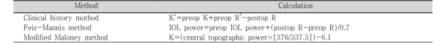
Table 1. Methods of intraocular lens power calculation after refractive surgery
Method Calculation
Clinical history mothod K*=preop K+preop R†-postop R
Feiz-Mannis method IOL power=preop IOL power+(postop R-preop R)/0.7 Modified Maloney method K=(central topographic power×[376/337.5])-6.1
*K=keratometry; †R=refraction
Table 2. Demographic data for eyes with cataract surgery after refractive surgery
Eyes
Age (years) 52.56±11.38
Axial length (mm) 27.82±1.84
Implanted Intraocular lens power (D*) 18.41±2.33
K-value before refractive surgery by manual keratometer (D) 43.28±1.47 K-value after refractive surgery by manual keratometer (D) 38.49±1.36 3-mm zone K-value after refractive surgery by Orbscan (D) 38.65±1.23
Spherical equivalent before refractive surgery (D) -7.83±3.76
Spherical equivalent after refractive surgery (D) -2.51±2.34
Spherical equivalent after cataract surgery (D) -0.89±1.39
*D=diopter
음파를 이용한 안축장 길이(Ultrasonic Pachometer, Hu- mphrey Instruments, Inc. San Leandro, California, U.S.A.), 각막곡률검사(KR 7100P, Topcon Corp, Japan)와 각막지 형도검사(Orbscan II, Bausch & Lomb, Rochester, New York, USA)를 시행하였다. 백내장 수술은 3 mm 크기의 투 명각막절개 후 초음파수정체유화술을 통해 수정체를 제거 하고 인공수정체를 삽입하였다.
굴절력의 예측도를 알아보기 위해 백내장 수술 후 굴절 력이 안정되는 1달~3달 사이의 현성굴절검사값과 환자에 게 삽입한 인공수정체 도수의 예측값의 차이인 예측오차 (prediction error)를 비교하였다. 예측오차는 음의 값과 양 의 값이 서로 상쇄되기 때문에 정확한 비교를 위하여 절대 예측오차(absolute prediction error)를 함께 비교하였다.
각막 절삭 후 발생하는 각막곡률값의 오차를 보정하는 방 법 중 널리 사용되고 있는 각막곡률산출법, 변형 Maloney 방법, 술전굴절력보정법을 비교하였다(Table 1). 효과적인 렌즈 위치(effective lens position, ELP)를 계산하기 위해 굴절교정수술 전의 K값과 술 후의 K값을 이용하는 이중 K값 공식과 단일 K값 공식의 차이를 비교하였다. 이중 K 값 계산 법은 기존의 논문에서 소개된 방법을 사용하였다(Appendix 1,2,3).12-15 아울러 인공수정체도수계산공식(SRK/T, Holladay 1, Hoffer Q)에 따른 예측도의 차이도 비교하였다. 변형 Maloney 방법은 굴절교정수술 전의 각막곡률값과 굴절력 을 알 수 없는 경우 많이 사용되는 것을 감안하여, 이중 K 값 공식을 이용하여 계산시 굴절교정수술 전의 각막곡률값 (pre refractive surgery K-value; Kpre)을 43.5로 대신하 여 계산하였다.
통계적 분석은 통계처리 프로그램 SPSS 12.0을 이용하 였다. 각막곡률산출법, 변형 Maloney 방법, 술전굴절력보 정법 세 그룹간의 예측도와 인공수정체도수계산공식(SRK/T, Holladay 1, Hoffer Q)간의 예측도를 비교하기 위해 이원분산 분석(two-way ANOVA)을 사용하였고, 단일 K값 공식과 이중 K값 공식의 예측도를 비교하기 위해 대응표본 t-test (paired-sample t-test)를 사용하였다. 굴절교정수술 전 의 근시값을 두 그룹으로 나눌 때 경계값(cut-off value) 산출은 최소유의확률법(minimum P-value approach)을 사용하여 구하였는데, 경계값의 범위는 5디옵터(D)에서 9D 범위에서 검토하였다. P-value의 유의수준은 0.05 미 만으로 하였다.
결 과
대상 환자 16명 18안의 평균 연령은 52.56±11.38세 였 고, 남자는 7명 8안, 여자는 9명 10안이었다. 백내장 수술 후 1.65±0.63개월째의 평균 구면렌즈 대응치는 -0.89±1.39 디옵터(D)였다(Table 2). 나안시력은 평균 LogMAR 0.26
±0.44, 교정시력은 평균 LogMAR 0.06±0.14이었다.
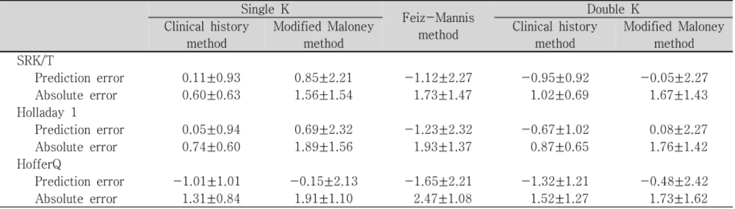
각막곡률산출법과 변형 Maloney 방법, 술전교정력보정 법을 비교하였을 때 각막곡률산출법의 단일 K값의 SRK/T 와 Holladay 1 공식의 절대예측오차가 0.60±0.63D, 0.74±
0.60D로 가장 좋았다(Table 3). 단일 K값 공식 변형 Maloney 방법은 다른 방법들에 비해 가장 큰 원시 예측오차를 보였 고, 술전굴절력보정법은 다른 방법들에 비해 가장 큰 근시 예측오차를 보였다. 단일 K값 공식 각막곡률산출법은 단일
Table 3. Mean prediction error and absolute prediction error (in diopters) according to different methods for cataract surgery after refractive surgery
Single K
Feiz-Mannis method
Double K
Clinical history
method
Modified Maloney method
Clinical history method
Modified Maloney method
SRK/T
Prediction error 0.11±0.93 0.85±2.21 -1.12±2.27 -0.95±0.92 -0.05±2.27
Absolute error 0.60±0.63 1.56±1.54 1.73±1.47 1.02±0.69 1.67±1.43
Holladay 1
Prediction error 0.05±0.94 0.69±2.32 -1.23±2.32 -0.67±1.02 0.08±2.27
Absolute error 0.74±0.60 1.89±1.56 1.93±1.37 0.87±0.65 1.76±1.42
HofferQ
Prediction error -1.01±1.01 -0.15±2.13 -1.65±2.21 -1.32±1.21 -0.48±2.42
Absolute error 1.31±0.84 1.91±1.10 2.47±1.08 1.52±1.27 1.73±1.62
Table 4. P-value of prediction error and absolute prediction error (in diopters) according to different methods for cataract surgery after refractive surgery
CHM*vs.
MMM† (with SK)
CHM* (with SK)
vs. FMM‡
MMM* (with SK)
vs. FMM‡
CHM* vs.
MMM† (with DK)
CHM* (with DK)
vs. FMM‡
MMM* (with DK)
vs. FMM‡
SK vs. DK (with CHM*)
SK vs. DK (with MMM†) SRK/T
P§ error 0.03 0.01 0.01 0.05 0.67 0.03 0.03 0.03
A∏error 0.02 0.03 0.63 0.13 0.17 0.85 0.31 0.81
Holladay 1
P§ error 0.03 0.01 0.01 0.07 0.31 0.02 0.04 0.04
A∏error 0.01 0.02 0.92 0.05 0.08 0.87 0.74 0.73
HofferQ
P§ error 0.02 0.03 0.01 0.03 0.56 0.02 0.43 0.13
A∏error 0.23 0.02 0.59 0.63 0.05 0.72 0.58 0.76
*CHM=clinical history method; †MMM=modified Maloney method; ‡FMM=Feiz-Mannis method; §P=prediction error; ∏A=
absolute prediction error.
The mean prediction errors and absolute prediction errors produced by different methods were compared using the paired t test and two-way ANOVA.
K값 공식 변형 Maloney 방법과 술전굴절력보정법에 비해 SRK/T, Holladay 1공식에서 예측오차 및 절대예측오차가 통계학적으로 유의하게 작은 값을 보였다(Table 3, 4).
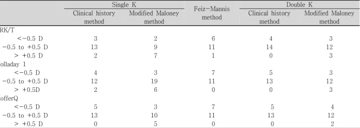
단일 K값 공식과 이중 K값 공식을 비교 했을 때 각막곡 률산출법과 변형 Maloney 방법 모두 이중 K값 공식에서 통계학적으로 유의한 근시 예측오차를 보였다. 각막곡률산 출법은 단일 K값 SRK/T와 Holladay 1 공식에서 0.5D 이상 의 원시가 각각 2안에서 관찰되었으나, 이중 K값 공식에서 는 0.5D 이상의 원시를 보인 안은 없었다. 변형 Maloney 방법의 단일 K값 SRK/T 공식은 7안에서 0.5D 이상의 원시 가 관찰되었으나 이중 K값 공식에서는 3안에서만 관찰되 어 원시 경향이 감소 함을 알 수 있었다(Table 6).
굴절교정수술 전의 근시를 7D 이상인 그룹과 이하인 그 룹으로 나누었을 때 술 전 근시가 7D 이상인 안은 8안, 7D 미만인 안은 10안이었다. 두 그룹 사이에서 변형 Maloney 방법은 7D 이상인 그룹이 7D 미만인 그룹보다 통계학적으 로 유의한 원시 예측오차를 보였다(Table 7).
Hoffer Q공식은 SRK/T와 Holladay 1공식에 비해 큰 근 시 예측오차를 보였다. 단일 K값 공식의 각막곡률산출법과 변형 Maloney 방법에서 Hoffer Q와 SRK/T의 차이는 통계 학적으로 유의하였으나(p=0.04, 0.04), 이를 제외한 인공 수정체도수계산공식에 의한 차이는 유의하지 않았다 (Table 3, 5).
고 찰
굴절교정수술 후의 인공수정체의 도수 산출시의 오차는 기구의 측정 오차(instrument error), 굴절지수에 의한 오 차(index of refraction), 그리고 공식에 의한 오차(formula error) 3가지로 크게 나눌 수 있다. 기구측정의 오차는 굴 절교정수술을 받은 후 백내장 발생시 인공수정체의 도수를 계산하기 위해 수동 또는 자동각막굴절력계의 측정치를 이 용 할 때 각막주변부를 측정하게 되는데 이 영역은 각막의 굴절교정수술 후 편평해진 중심부보다 가파르기 때문에 실
Table 5. P-value of prediction error and absolute prediction error (in diopters) according to intraocular lens (IOL) formulas for cataract surgery after refractive surgery
Single K
Feiz-Mannis method
Double K
Clinical history
method
Modified Maloney method
Clinical history method
Modified Maloney method
SRK/T vs. Holladay 1
Prediction error 0.77 0.68 0.79 0.53 0.86
Absolute error 0.76 0.63 0.72 0.71 0.85
Holladay 1 vs. Hoffer Q
Prediction error 0.08 0.06 0.59 0.26 0.39
Absolute error 0.41 0.87 0.53 0.29 0.95
HofferQ vs. SRK/T
Prediction error 0.04 0.04 0.36 0.38 0.31
Absolute error 0.32 0.61 0.41 0.35 0.81
The mean prediction errors and absolute prediction errors produced by IOL formulas were compared using the two-way ANOVA.
Table 6.Range of prediction error (in diopters) according to different methods for cataract surgery after refractive surgery
Single K
Feiz-Mannis method
Double K
Clinical history
method
Modified Maloney method
Clinical history method
Modified Maloney method
SRK/T
<-0.5 D 3 2 6 4 3
-0.5 to +0.5 D 13 9 11 14 12
> +0.5 D 2 7 1 0 3
Holladay 1
<-0.5 D 4 3 7 5 3
-0.5 to +0.5 D 12 19 11 13 12
> +0.5D 2 6 0 0 3
HofferQ
<-0.5 D 5 3 7 5 4
-0.5 to +0.5 D 13 10 11 13 12
> +0.5 D 0 5 0 0 2
Table 7. Comparison of mean prediction error and absolute prediction error (in diopters) according to degree of myopia before refractive surgery
SRK/T
Single K
p-value Feiz-Mannis method
Double K
p-value Clinical
history method
Modified Maloney methods
Clinical history method
Modified Maloney methods Prediction error
Myopia <-7 D (n=10) 0.15±0.91 -0.28±0.58 0.68 -0.73±1.37 -0.85±1.15 -1.05±1.19 0.69 Myopia ≥-7 D (n=8) -0.21±1.71 2.45±1.93 0.01 -0.07±3.52 -0.97±0.67 2.06±2.83 0.01
p-value 0.54 0.01 0.44 0.76 0.01
Absolute error
Myopia <-7 D (n=10) 0.44±0.63 0.58±0.47 0.78 1.28±1.04 0.99±0.83 1.13±0.61 0.71 Myopia ≥-7 D (n=8) 0.75±1.35 2.61±1.76 0.01 2.32±1.76 1.07±0.57 2.31±2.31 0.02
p-value 0.46 0.01 0.12 0.83 0.18
The cut-off value of myopia was based on a minimum P-value approach.
The prediction errors and absolute prediction errors were compared using the paired t test and two-way ANOVA.
제 중심부 보다 높은 굴절력을 가진 것처럼 측정이 되어 백 내장 수술 후 원시가 되는 것이다.2두 번째는 현재 사용되 고 있는 각막곡률측정계는 Gullstrand의 모형안을 기초로 하여 각막의 전면과 후면을 하나의 유효굴절지수(effective index of refraction)를 가정하고 각막전면의 곡률반경을
측정하여 각막의 굴절력을 구하는 것인데, 굴절교정술 후에 는 각막전면의 곡률반경이 증가하며 각막 후면과 전면 사 이의 거리도 줄어든다. 또 각막 후면의 변화가 고려되지 않 기 때문에 굴절교정수술 후의 환자의 각막굴절력의 산출은 고전적인 방법을 따르면 부정확 할 수 있다는 것이다. 약
7D의 근시를 굴절교정수술을 이용하여 교정하였을 때 각막 power는 약 1 D 정도 과측정 하게 된다.16-18세 번째는 인 공수정체도수계산공식에서(SRK/T, Holladay 1, Hoffer Q) 굴절교정수술 후 전방의 깊이는 변하지 않았음에도 불구하 고 작아진 K값에 따른 잘못된 인공수정체의 위치 예측 때 문에 발생한다. 이러한 문제점을 해결하기 위해 굴절교정수 술 후 백내장이 발생한 환자의 인공수정체도수계산을 위한 많은 방법이 보고되었다.6,7,10-12,14,15,19
본 연구에서 인공수정체도수계산법 간의 예측도를 비교 하였을 때 각막곡률산출법은 변형 Maloney 방법과 술전굴 절력보정법 보다 정확도를 의미하는 절대예측오차가 가장 작은 값을 보여 예측도가 가장 좋음을 알 수 있었으며 기존 의 연구와 일치된 결과를 보였다.10,20그러나 이 방법은 백 내장이 많이 진행된 경우 백내장으로 인한 수정체의 굴절 력 변화로 현성굴절검사 결과가 부정확하게 될 수 있다는 단점이 있으므로 굴절교정술 직후의 굴절력을 알아야 한다.
굴절교정술 전의 각막곡률값, 굴절력 그리고 굴절교정술 직 후의 굴절력을 알고 있는 경우 첫 번째로 선택할 수 있는 계산법으로 생각된다.
술전굴절력보정법은 각막곡률산출법과 변형 Maloney 방 법에 비해 가장 큰 근시값을 보였고 이는 이전의 보고와 비 슷하였다.6,10이 방법은 수술 후 원시 발생을 최소화 할 수 있으므로 삽입할 인공수정체 도수의 상한값(upper limit)으 로 이용될 수 있을 것이다.
변형 Maloney 방법은 Maloney8에 의해 수술 전 기록을 가지고 있지 않은 경우의 굴절교정수술 후의 각막굴절력을 계산하기 위해 처음 고안된 방법으로 굴절교정수술 전의 기록이 없는 경우에도 이용할 수 있다는 장점을 가지나 원 시 경향을 보이는 것으로 알려져있다.9,10본 연구에서도 알 려진 바와 같이 단일 K값 변형 Maloney 방법은 큰 원시 오 차를 보였다. 변형 Manloney 방법의 단일 K값 SRK/T공식 에서 7안에서 0.5D 이상의 원시를 보였는데 7안 모두 굴절 교정수술 전에 7D 이상의 근시를 가지고 있었다. 따라서 변 형 Maloney 방법은 환자가 굴절교정술 전 7D 이상의 근시 를 가지고 있었던 것으로 기억한다면 더욱 술 후 원시가 남 을 수 있음을 고려하여야 할 것이다.
이중 K값 공식은 효과적인 렌즈 위치(effective lens position, ELP)를 위한 수술 전의 K값과 굴절교정술 후의 K값 두 가지 모두를 사용하는 방법으로 수술 후의 원시를 교정 할 수 있다고 알려져있다.12,21,22본 연구에서도 기존의 보고와 같이 이중 K값 공식은 단일 K값 공식과 비교하였을 때 통계학적으로 유의한 근시 경향을 보였다. 따라서 7D 이 상의 근시를 가진 환자에서 변형 Manloney 방법을 이용하 는 경우 이중 K값 공식을 이용하여 원시의 발생을 줄일 수
있을 것이다.
Hoffer는 IOL 공식에서 안축장의 길이에 따라 안축장이 24.5에서 26.0 mm인 경우에는 Holladay 1 공식이 가장 정 확하고, 26 mm 이상인 경우에는 SRK/T 공식이 더 정확하 며 Hoffer Q 공식은 안축장이 22 mm 이하인 경우에 가장 정확하다고 보고 하였다.23,24본 연구의 환자들은 안축장의 길이가 평균 27.82±1.84(범위 24.86~31.45)이었다. 안축 장이 긴 근시를 가졌던 환자들을 대상으로 하는 본 연구에 서도 기존의 보고와 같이 Hoffer Q 공식이 SRK/T 와 Holladay 1 공식에 비해 큰 근시 오차를 보였다. 따라서 굴 절교정수술을 받은 환자에서 백내장 수술을 하는 경우 Hoffer Q 공식은 적합하지 않을 것으로 생각된다.
요약하면 굴절교정술 후 백내장이 발생한 환자에 있어서 인공수정체도수 산출 시 굴절교정술 전의 정보를 가지고 있는 경우 각막굴절력산출법과 함께 SRK/T 혹은 Holladay 1을 사용하는 것이 좋을 것으로 생각된다. 단일 K값 공식은 예측도가 더 좋고, 이중 K값 공식은 예측도는 감소하나 원 시 경향을 줄일 수 있으므로 단일 K값 공식을 사용하되 이 중 K값 공식을 참고 하는 것이 좋을 것이다. 이때 술전굴절 력보정법은 근시 예측을 하므로 렌즈 선정의 상한치(upper limit)로 참고할 수 있을 것이다. 하지만 굴절교정술 전 자 료가 없어 변형 Maloney 방법을 이용할 때에는 원시의 경 향을 줄이기 위해 이중 K값 공식으로 원시의 보정이 필요 하고, 특히 근시가 7D 이상인 경우는 더욱 그러할 것이다.
참고문헌
1) Fyodorov SN, Durnev VV. Operation of dosaged dissection of corneal circular ligament in cases of myopia of mild degree. Ann Ophthalmol 1979;11:1885-90.
2) Koch DD, Liu JF, Hyde LL, et al. Refractive complications of cat- aract surgery after radial keratotomy. Am J Ophthalmol 1989;
108:676-82.
3) Holladay JT. Consultations in refractive surgery. J Refract Corneal Surg 1989;5:202-203.
4) Ridley F. Development in contact lens theory-moulding, compu- tation, and veiling. Trans Ophthalmol Soc U K 1948;68:385-401.
5) Han JW, Kim JH, Joo CK. Comparison for the measuring meth- ods of intraocular lens power to calculate for eyes after LASIK. J Korean Ophthalmol Soc 2000;41:2191-7.
6) Feiz V, Mannis MJ, Garcia-Ferrer F, et al. Intraocular lens power calculation after laser in situ keratomileusis for myopia and hyper- opia: a standardized approach. Cornea 2001;20:792-7.
7) Moon SH, Kwon KL, Kee CW. Intraocular lens power calcu- lation in cataract surgery after excimer laser photorefractive keratectomy. J Korean Ophthalmol Soc 2000;41:60-6.
8) Seitz B, Langenbucher A. Intraocular lens power calculation in eyes after corneal refractive surgery. J Refract Surg 2000;16:349-61.
9) Smith RJ, Chan WK, Maloney RK. The prediction of surgically
Appendix 1. Aramberri double-K SRK formula
Double-K SRK/T Formula Equation 1: Preoperative corneal radius of curvature:
rpre=337.5/Kpre
Equation 2: Corrected axial length (LCOR):
If L≤24.2, LCOR=L
If L>24.2, LCOR=-3.446+1.716×L-0.0237×L2 Equation 3: Computed corneal width (Cw):
Cw=-5.41+0.58412×LCOR+0.098×Kpre Equation 4: Corneal height (H):
H=rpre_Sqrt [rpre2_(Cw2/4)]
Equation 5: Offset value:
Offset=ACDconst-3.336
Equation 6: Estimated postoperative ELP (ACD):
ACDest=H+Offset Equation 7: Constants:
V=12; na=1.336; nc=1.333; ncm1=0.333
Equation 8: Retinal thickness (RETHICK) and optical axial length (LOPT):
RETHICK=0.65696-0.02029×L LOPT=L+RETHICK
Equation 9: Postoperative corneal radius of curvature:
rpost=337.5/Kpost
Equation 10: Emmetropia IOL power (IOLemme):
IOLemme=[1000×na×(na×rpost-ncm1×LOPT)]/[(LOPT_ACDest)×(na×rpost_ncm1×ACDest)]
Equation 11: Conversion from IOL A-constant to IOL ACD constant:
ACDconst=0.62467×A-constant-68.747 Variables
L=axial length; Kpre=pre refractive surgery K-value; Kpost=post refractive surgery K-value; ACDconst=IOL constant (can be computed from A-constant).
induced refractive change from corneal topography. Am J Ophth- almol 1998;125:44-53.
10) Wang L, Booth MA, Koch DD. Comparison of intraocular lens power calculation methods in eyes that have undergone LASIK.
Ophthalmology 2004;111:1825-31.
11) Kim JH, Lee DH, Joo CK. Measuring corneal power for intra- ocular lens power calculation after refractive surgery. Compari- son of methods. J Cataract Refract Surg 2002;28:1932-8.
12) Aramberri J. Intraocular lens power calculation after corneal re- fractive surgery: double-K method. J Cataract Refract Surg 2003;
29:2063-8.
13) Retzlaff JA, Sanders DR, Kraff MC. Development of the SRK/T intraocular lens implant power calculation formula. J Cataract Refract Surg 1990;16:333-40.
14) Awwad ST, Dwarakanathan S, Bowman RW, et al. Intraocular lens power calculation after radial keratotomy: estimating the re- fractive corneal power. J Cataract Refract Surg 2007;33:1045-50.
15) Awwad ST, Kelley PS, Bowman RW, at al. Corneal refractive power estimation and intraocular lens calculation after hyperopic LASIK. Ophthalmology 2009;116:393-400.
16) Mandell RB. Corneal power correction factor for photorefractive keratectomy. J Refract Corneal Surg 1994;10:125-8.
17) Lyle WA, Jin GJ. Intraocular lens power prediction in patients who undergo cataract surgery following previous radial kerat- otomy. Arch Ophthalmol 1997;115:457-61.
18) Celikkol L, Pavlopoulos G, Weinstein B, et al. Calculation of in- traocular lens power after radial keratotomy with computerized videokeratography. Am J Ophthalmol 1995;120:739-50.
19) Kang JH, Park JI, Lee KH. A New Method for Measuring Cor- neal Refractive Power after Refractive Surgery. J Korean Ophth- almol Soc 2005;46:859-64.
20) Lee D, Hong S, Kim JS. Intraocular lens power calculation for cat- aract surgery in patients who had previous refractive surgery (RK 1 case, PRK 3 case, LASIK 1 case). J Korean Ophthalmol Soc 2000;41:2268-75.
21) Koch DD, Wang L. Calculating IOL power in eyes that have had refractive surgery. J Cataract Refract Surg 2003;29:2039-42.
22) Savini G, Barboni P, Zanini M. Intraocular lens power calculation after myopic refractive surgery: theoretical comparison of differ- ent methods, Ophthalmology 2006;113:1271-82.
23) Hoffer KJ. The Hoffer Q formula: a comparison of theoretic and regression formulas. J Catarct Refract Surg 1993;19:700-12.
24) Hoffer KJ. Clinical results using the Holladay 2 intraocular lens power formula. J Cataract Refract Surg 2000;26:1233-7.
Appendix 2. Double-K Holladay 1 formula
Double-K Holladay 1 Formula Equation 1:
Rag=Rpre; if Rpre<7.0 mm, Rag=7.0 mm:
Equation 2: Anterior chamber diameter, angle to angle
AG=1.25×(AL/23.45); If AG>13.5 mm, AG=13.5 mm Equation 3: Anatomic anterior chamber depth
ACD=Z 0.56+Rag-(SQRT{ABS[RAG2-(AG2/4)]}) Equation 4: Optical anterior chamber depth
OACD=ACD+SF
Equation 5: Intraocular lens power from desired postoperative refraction
IOL=1000×na×{na×Rpost-(nc-1)×Alm-0.001×Ref×[V×(na×Rpost-(nc-1)×Alm)+(Alm×Rpost)]}/
(Alm-OACD)×{(na×Rpost)-(nc-1)×OACD-0.001×Ref×[V×(na×Rpost-(nc-1)×OACD)+OACD×Rpost]}
Equation 6: Resultant refraction from IOL power
Ref=[1000×na×[na×Rpost-(nc-1)×Alm]-IOL×(Alm-OACD)×{(na×Rpost)-[(nc-1)×OACD]} /
na×{V×[na×Rpost-(nc-1)×Alm]+(Alm×Rpost)}-0.001×IOL×(Alm-OACD)×{[V×(na×Rpost-(nc-1)×
OACD)]+(OACD×Rpost)}
Measured Values
Kpre=average K-reading before radial keratotomy (RK) (diopters[D]) Kpost=average K-reading after RK and before cataract surgery (diopters) Rpre=average corneal radius (mm)=337.5/Kpre
Rpost=average corneal radius (mm)=337.5/Kpost AL=average axial length (AL) (mm)
Chosen Values
V=vertex distance of pseudophakic spectacles (mm); default=12.0 mm Ref=desired postoperative spheroequivalent refraction (D)
SF=surgeon factor (mm) SF=0.5663×A constant-65.6 Other Variables
AG=anterior chamber diameter from angle to angle (mm) ACD=anatomic anterior chamber depth (mm)
Alm=modified AL (mm)=measured AL(AL)+retinal thickness factor (RT)
Appendix 3. Double-K Hoffer Q Formula
Double-K modification of the Hoffer Q Formula Equation 1: Anterior chamber depth
ACD=pACD+0.3(AL-23.5)+tan(Kpre-LASIK)2+(0.1×M×[23.5_ AL]2_[tan{0.1(G-AL)2}])-0.99166 M: if AL≤23.00, M=1; if AL>23mm, M=-1
G: if AL≤23.00, G=28.00 mm; if AL>23 mm, G=23.5 mm If AL>31, AL=31.0; if AL<18.5, AL=18.5
Equation 2: Refractive error at corneal plane R=Rx⁄(1-0.012 Rx)
Equation 3: Intraocular lens power
P=(1336⁄[AL-ACD-0.05])-(1.336⁄[{1.336⁄(Kpost-LASIK +R)}-{(ACD+0.05)⁄1000}]) Equation 4: Refractive error
R=(1.336⁄[1.336⁄{1336 ⁄(AL-ACD-0.05)-P}+{ACD+0.05}⁄1000])-Kpost-LASIK
Rx=R⁄(1+0.012R) Recommended constants:
Refractive index of cornea=1.336 Retinal thickness factor=0 Measured and extrapolated values:
Kpre-LASIK=average K-reading before LASIK (D)
Kpost-LASIK=estimated refractive corneal power after LASIK (D) AL=measured axial length (mm)
Chosen values
V=vertex distance of pseudophakic spectacles (mm), default=12 mm pACD=personalized anterior chamber depth constant
pACD=0.58357×A constant-63.896 Calculated variables
P=power of the IOL (D)
R=refractive error at corneal plane (D)
Rx=target refractive error at spectacle plane (D)
=ABSTRACT=
Comparison of Intraocular Lens Power Calculation Methods for Cataract Surgery after Refractive Surgery: A Retrospective Surgery
Myoung-Ok Lee, MD, Tae-Young Chung, MD, PhD, Eui-Sang Chung, MD, PhD, Chang Won Kee, MD, PhD
Department of Ophthalmology, Samsung Medical Center, Sungkyunkwan University School of Medicine, Seoul, Korea
Purpose: To investigate the predictability of and propose guidelines for intraocular lens (IOL) power calculation in post-cataract surgery patients with prior corneal refractive surgery and suggest the guideline.
Methods: Medical records of 18 eyes of 16 patients were retrospectively evaluated for IOL power calculation predictability using three combinations of methods: 1) clinical history method, modified Maloney method, and the Feiz-Mannis method; 2) single-K formula versus double-K formula; and 3) Three IOL formulas (SRK/T, Holladay 1, and Hoffer Q).
Results: The clinical history method using the single-K formula with the SRK/T and Holliday 1 formula showed the best predictability, with an absolute error of 0.60±0.63 D and 0.74±0.60 D, respectively. The Feiz-Mannis method showed a tendency of myopic prediction, whereas the modified Maloney method showed a tendency of hyperopic prediction, especially in the patients with myopia more than 7 D prior to the refractive surgery. The double-K formula, when compared to the single-K formula, prevented hyperopic prediction when used with the clinical history method or modified Maloney method.
Conclusions: IOL power calculation using the clinical history method with SRK/T or Holliday 1 formula showed the best predictability in patients after corneal refractive surgery. IOL power calculation using the modified Maloney method, however, because of the hyperopic prediction tendency, should be used cautiously, especially for patients with myopia of 7 D or more prior to the refractive surgery.
J Korean Ophthalmol Soc 2010;51(2):180-187
Key Words: Cataract, Corneal refractive surgery, IOL power calculation
Address reprint requests to Chang Won Kee, MD, PhD
Department of Ophthalmology, Samsung Medical Center, Sungkyunkwan University School of Medicine
#50 Ilwon-dong, Gangnam-gu, Seoul 135-710, Korea
Tel: 82-2-3410-3564, Fax: 82-2-3410-0074, E-mail: ckee@skku.edu