doaneurysm in a patient with normal palpable pulses, but with swollen knee and ankle with a functional disability, intractable pain in the popliteal fossa, and weakness of ankle dorsiflexors.
Case Report
A 61-year-old woman who underwent bilateral TKA at a dif- ferent institution, presented to our emergency orthopedic de- partment on the third postoperative day with right knee and calf swelling, intolerable pain in the right lower limb, and weakness of right ankle dorsiflexors. Based on her surgery records, it was determined that the procedure involved an anterior midline skin incision followed by medial subperiosteal release along the proximal tibia via a medial parapatellar approach and implanta- tion of a cemented knee prosthesis without tourniquet control (Fig. 1). There were no records available on the thromboembolic prophylaxis employed during the surgery, and the procedure was uneventful. She had no previous history of arterial disease or cardiovascular risk factors and trauma. However, the patient ex- perienced weakness of right ankle dorsiflexors in the immediate postoperative period, which was diagnosed as common peroneal nerve palsy. The overall condition of the patient and the status of the right lower limb were good. Moreover, postoperative limb pulsation was present. The patient was immediately transferred to our emergency department and opioid analgesics were adminis- tered intravenously for pain relief. A thorough examination of the patient revealed a stable medical status and maintenance of vital
Popliteal Artery Pseudoaneurysm Following Primary Total Knee Arthroplasty
Young-Soo Shin, MD, Yeok-Gu Hwang, MD, Abhijit Prakash Savale, MD, and Seung-Beom Han, MD
Department of Orthopedic Surgery, Korea University Anam Hospital, Korea University College of Medicine, Seoul, Korea
An early diagnosis of popliteal artery pseudoaneurysm-a sequela of popliteal artery trauma-is difficult owing to its late presentation following total knee arthroplasty. The incidence of a popliteal artery pseudoaneurysm with a hematoma presenting only a peripheral nerve injury after total knee arthroplasty is also uncommon in the absence of common diagnostic features such as a pulsatile swelling with an audible bruit on auscultation. In the present report, we describe popliteal artery pseudoaneurysm following total knee arthroplasty.
Keywords: Popliteal artery, Pseudoaneurysm, Arthroplasty
Case Report
Knee Surg Relat Res 2014;26(2):117-120 http://dx.doi.org/10.5792/ksrr.2014.26.2.117 pISSN 2234-0726 · eISSN 2234-2451
Knee Surgery & Related Research
Received July 19, 2013; Revised (1st) September 16, 2013;
(2nd) October 22, 2013; Accepted October 30, 2013 Correspondence to: Seung-Beom Han, MD
Department of Orthopedic Surgery, Korea University Anam Hopital, Korea University College of Medicine, 73 Inchon-ro, Seongbuk-gu, Seoul 136-705, Korea
Tel: +82-2-920-5692, Fax: +82-2-924-2471 E-mail: [email protected]
Complications involving injury to neurological and vascular structures are infrequently reported after total knee arthroplasty (TKA). Popliteal artery pseudoaneurysm has been reported in various case studies with an incidence ranging from 0.03% to 0.17%1). In addition, certain studies have indicated that the indi- vidual incidence of peroneal nerve palsy after TKA varies from 0%
to 9.5%2). Conditions associated with peroneal nerve injury after TKA include flexion contracture, valgus deformity, postoperative epidural analgesia, external leg compression, increased tourni- quet duration, postoperative hematoma, and history of nerve root compression2). The current clinical diagnostic criteria for popliteal artery pseudoaneurysm are the presence of a pulsatile hematoma with an audible bruit and swelling over the popliteal region and the entire lower leg3).
Thus, in the present report, we describe popliteal artery pseu-
117
This is an Open Access article distributed under the terms of the Creative Commons Attribution Non-Commercial License (http://creativecommons.org/licenses/by-nc/3.0/) which permits unrestricted non-commercial use, distribution, and reproduction in any medium, provided the original work is properly cited.
Copyright © 2014 KOREAN KNEE SOCIETY www.jksrr.org
118
Shin et al. Popliteal Artery Pseudoaneurysm after TKAparameters. On auscultation, there were no signs of adventitious sounds such as crepitus or rhonchi. Local examination revealed swelling over the entire right lower limb with painful restriction of motion of the right knee. In addition, we also noted swelling over the popliteal region, which was non pulsatile and without any signs of auscultation bruit. The postoperative status of the wound was good with no fresh discharge. On distal neurovas- cular examination, the right lower limb was found to be warm, and the pulsation of both the dorsalis pedis artery and posterior tibial artery was found to be normal. Neurologically, the ankle dorsiflexors were found to have motor grade 1, and there was a sensory deficit noted in the common peroneal nerve distribu- tion. Based on these findings, a provisional diagnosis of common peroneal nerve palsy was made and a foot drop splint was applied
immediately. The patient was then admitted to a ward for further observation. During the course of treatment, the patient reported decreased pain in the right lower limb with the use of analgesics, but the swelling of the entire right lower limb remained. A duplex examination of the right lower limb was performed as deep vein thrombosis was suspected; however, no evidence of deep vein thrombosis was noted. A computed tomographic (CT) angiog- raphy revealed a large popliteal artery pseudoaneurysm arising from the popliteal artery just above the knee with a large defect in the arterial wall, explaining the neurological symptoms (Fig. 2).
The patient was immediately referred to a vascular surgeon for intervention. The lesion was treated with placement of a covered endovascular stent graft. After obtaining ante grade percutane- ous access across the lesion, a covered 8×30 mm wall graft was placed. This resulted in a satisfactory sealing of the pseudoaneu- rysm (Fig. 3).
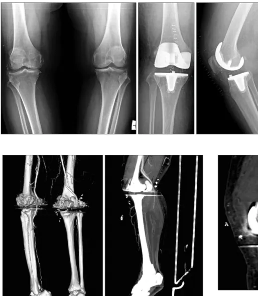
Fig. 1. Preoperative anteroposterior ra- diographs of both knees in a 61-year-old woman showing severe tricompartmental osteoarthritis of both knees (right>left). Im- mediate postoperative anteroposterior ra- diograph following total knee replacement of the right knee and lateral radiograph showing a well-aligned right knee with ad- equate cementing of components without any residual deformity.
Fig. 2. Computed tomographic angiography of the right lower limb. A large pseudoaneurysm of the right popliteal artery. The popliteal artery was occluded distal to the pseudoaneurysm and was reconstituted at the tibioperoneal trunk. No significant stenosis was noted in both crural ar- teries with good distal runoff.
Fig. 3. A covered endovascular stent graft was placed in the right pop- liteal artery, and in-stent restenosis was not observed in the artery. No significant stenosis was noted in both crural arteries with good distal runoff.
Knee Surg Relat Res, Vol. 26, No. 2, Jun. 2014
119
However, even 2 weeks after the onset, the peripheral neu- ropathies were not completely resolved and were assessed with electromyography and somatosensory evoked potential studies to document the extent of the lesion. This examination indicated evidence of denervation, thus confirming the presence of pero- neal and posterior tibial nerve dysfunction; the findings were used as a baseline for subsequent studies to track recovery.
At a 6-month follow-up examination, the patient remained asymptomatic without pain or swelling; however, the patient con- tinued to exhibit a right foot drop with normal distal pulsation.
Discussion
Popliteal artery injury most commonly occurs during the removal of osteophytes from the proximal aspect of posterior femoral condyles, during release of the posterior capsule, and during resection of the proximal tibia with an oscillating saw. The knee should be flexed during these maneuvers, thus allowing the popliteal neurovascular structures to fall posteriorly, away from the posterior capsule. The posterior midline structures should be protected, with appropriately placed retractors, when performing these maneuvers. A lever-type retractor should be placed at a suf- ficient distance, distally on the tibia, to facilitate protection of the neurovascular structures during translation of the proximal tibia.
During resection of the posterior femoral condyles, an increased margin of safety can be gained by using a narrower saw blade and placing the retractors between the femoral condyles and collat- eral ligaments.
If a tourniquet is used, it should be deflated before closure. If brisk arterial bleeding is noted or if signs of distal ischemia are present, immediate consultation with a vascular surgeon is war- ranted. Intraoperative angiography and direct arterial repair may be necessary in such cases4). In general, any pseudoaneu- rysm necessarily implies a trauma to the artery (from retractors, oscillating saw, drill, rongeur, heat from bone cement, or from repeated local trauma)5,6). In our case, the cause of the pseudoan- euryms also may be intraoperative trauma to the popliteal artery.
However, we could not exactly reveal the apparent reasons based on her surgery records documented in another institution.
An early diagnosis of the aneurysm is very essential as it could prevent development of complications that may have serious consequences for the limb. Duplex ultrasonography is a cheap and safe diagnostic tool and is recommended, but CT angiogra- phy remains the definitive investigation to confirm the diagno- sis7). Therefore, we performed only CT angiography on the third postoperative day. However, many of these injuries are initially
subclinical and remain undetected or may be misdiagnosed as postoperative hematoma or deep vein thrombosis, which are commonly occurring complications following total knee replace- ment8).
Geertsema et al.5), in their study of a 67-year-old woman under- going TKAs, found that two days postoperatively, she complained of pain at the medial part of the left ankle and plantar sensory loss of the foot explaining partial neuropathy of the peroneal and posterior tibial nerves, possibly caused by hematoma or trac- tion. Therefore, early diagnosis is essential to avoid fatal ischemic changes of the lower extremities due to a false aneurysm and to avoid the development of persistent, irreversible, and neurologi- cal damage.
In conclusion, our study emphasized that the different presen- tations of popliteal artery pseudoaneurysm should be carefully considered in the absence of common diagnostic features, such as a pulsatile swelling with an audible bruit on auscultation.
Conflict of Interest
No potential conflict of interest relevant to this article was re- ported.
References
1. Rand JA. Vascular complications of total knee arthroplasty:
report of three cases. J Arthroplasty. 1987;2:89-93.
2. Schinsky MF, Macaulay W, Parks ML, Kiernan H, Nerces- sian OA. Nerve injury after primary total knee arthroplasty.
J Arthroplasty. 2001;16:1048-54.
3. Sloan K, Mofidi R, Nagy J, Flett MM, Chakraverty S. Endo- vascular treatment for traumatic popliteal artery pseudoa- neurysms after knee arthroplasty. Vasc Endovascular Surg 2009;43:286-90.
4. Rama KR, Apsingi S, Poovali S, Jetti A. Timing of tourniquet release in knee arthroplasty. Meta-analysis of randomized, controlled trials. J Bone Joint Surg Am. 2007;89:699-705.
5. Geertsema D, Defoort KC, van Hellemondt GG. Popliteal pseudoaneurysm after total knee arthroplasty: a report of 3 cases. J Arthroplasty. 2012;27:1581.
6. Sandoval E, Ortega FJ, Garcia-Rayo MR, Resines C. Popliteal pseudoaneurysm after total knee arthroplasty secondary to intraoperative arterial injury with a surgical pin: review of the literature. J Arthroplasty. 2008;23:1239.
7. Boutchichi A, Ciornohac J, Daubresse F. Pseudoaneurysm after total knee arthroplasty: a rare complication with dif-
120
Shin et al. Popliteal Artery Pseudoaneurysm after TKAferent possible clinical presentations. Acta Orthop Belg.
2013;79:16-9.
8. Kobayashi S, Isobe K, Koike T, Saitoh S, Takaoka K. Acute
arterial occlusion associated with total knee arthroplasty.
Arch Orthop Trauma Surg. 1999;119:223-4.