Introduction
The popularity of cone-beam computed tomography (CBCT) has increased in recent years, with an increasing use for diagnosis and treatment planning in different fields of dentistry.1The incorporation of cone-beam technology into clinical practice took place thanks to two main facts, namely the conversion to digital imaging and the progress in image acquisition, allowing detailed three-dimensional (3D) images. Images are now held on an axial matrix and can be displayed in axial, sagittal, or coronal views. They
can also be seen in the traditional panoramic view or as 3D reconstructions.2
CBCT has different applications in dentistry and oral and maxillofacial surgery with some advantages: it allows the modification of the visual field, provides a high resolution thanks to a voxel size range of 0.076-0.4 mm, reduces arti- facts caused by metals in conventional computed tomogra- phy, and generates a very low amount of radiation (partic- ularly when compared with conventional computed tomog- raphy for medical use). The equipment design is becoming more ergonomic and easier to use, image distortion is min- imal, and the images are compatible with other planning and simulation software due to their DICOM format.3,4
CBCT has recently begun to be used in dental root canal morphology analysis.5Some applications in this field are as follows: anatomical variability of roots and root canals,
Received October 7, 2013; Revised April 2, 2014; Accepted May 26, 2014
*Correspondence to: Dr. Carmen Llena
Department of Stomatology, Universitat de València, Gascó Oliag 1, 46010 Valencia, Spain
Tel) 34-963-86-41-75, Fax) 34-963-86-41-42, E-mail) [email protected]
Copyright ⓒ 2014 by Korean Academy of Oral and Maxillofacial Radiology
This is an Open Access article distributed under the terms of the Creative Commons Attribution Non-Commercial License (http://creativecommons.org/licenses/by-nc/3.0) which permits unrestricted non-commercial use, distribution, and reproduction in any medium, provided the original work is properly cited.
Imaging Science in Dentistry∙pISSN 2233-7822 eISSN 2233-7830
Cone-beam computed tomography analysis of root and canal morphology of mandibular premolars in a Spanish population
Carmen Llena1,*, Jaime Fernandez2, Pablo Sebastián Ortolani1, Leopoldo Forner1
1Department of Stomatology, Universitat de Valencia, Valencia, Spain
2Fernández Ugedo y Chaves Clinic, Alicante, Spain
ABSTRACT
Purpose: This study aimed to investigate the clinical anatomy of lower premolar roots in a Spanish population by using cone-beam computed tomography (CBCT), correlating findings with patient gender and tooth type.
Materials and Methods: Using 70 CBCT images, we evaluated 126 healthy, untreated, well-developed lower premolars. The number and morphology of roots and root canals, and the foramina number were assessed. Results for gender and tooth type were compared using the chi-squared and ANOVA tests.
Results: The average length of teeth and roots was significantly higher in men (p==0.00). All 126 premolars had a single root. One canal was found in 83.3% of the premolars, with no gender or tooth type differences; Vertucci configuration types I and V were the most prevalent. The first premolars showed significantly greater variability than the second premolars (p==0.03). A single apical foramen was found in 89.7% of the premolars, with no differences by tooth type. Women had a significantly higher prevalence of two apical foramina than men (p==0.04).
Some degree of curvature was observed in 65% of the premolars, with no differences by gender or tooth type. A root angle of more than 20�was found in 12.98% of the premolars, without any differences by gender or tooth.
Conclusion: All premolars were single-rooted. One canal had the most prevalent morphology. More variability in canal anatomy was found in the first premolars. Curvatures greater than 20�were found at less than 5 mm from the apex. (Imaging Sci Dent 2014; 44: 221-7)
KEY WORDS: Bicuspid; Mandible; Anatomy, Regional; Cone-Beam Computed Tomography
diagnosis of dental traumatic injuries, periapical pathology, differential diagnosis with other pathologies, nonsurgical treatment of root canals, and presurgical treatment plan- ning.6,7
Root canal anatomy of mandibular premolars and mor- phological differences between the first and the second premolars have been evaluated by using different proce- dures.8-14 Several studies have reported a large variation in the number of roots, root canal type, and apical foram- ina in mandibular premolars.8,11-19Only a few studies, lim- ited to the western Chinese and Korean populations, have evaluated the root anatomy of mandibular premolars using CBCT.14,20 According to Micheti et al.21 CBCT is a very interesting, reliable, noninvasive measuring technique that can be used in all spatial planes to study dental anatomy.
This study aimed to investigate the external and internal morphology of human mandibular premolar roots in a Spanish population by using CBCT and to correlate the findings with the patient’s gender and the tooth type.
Materials and Methods
Sample selection
This study was approved by the Ethics Committee of the Faculty of Medicine and Dentistry of the University of Valencia (Valencia, Spain), and informed consent was obtained from patients whose scans were used.
This study consisted of 200 CBCTs taken from October 2011 to February 2012 for different diagnostic reasons, such as bone absence for implant placement, assessment of tooth root relationships with clinically relevant anatom- ical structures, dental surgery, and diagnosis of mandibu-
lar radiolucent lesions. The inclusion criteria were as fol- lows: full view of one or more mandibular premolars, com- plete root formation, no resorption signs, no endodontic treatment performed, and no posts or coronal restorations.
Finally, 70 CBCTs with 126 premolars were included in the study.
Image acquisition and assessment
For image acquisition, a Kodak 9000 3D unit (Carestream Dental, Atlanta, GA, USA) was used. Radiographic images were obtained by a radiodiagnostic technician according to the manufacturer’s specifications (70 kV, 10 mA, and 10.8 s), which guaranteed the quality of the images with the lowest irradiation for patients.
Measurements were performed with CS 3D Imaging Software 3.1. (Carestream Dental, Atlanta, GA, USA), in the oblique cut, as it is the most appropriate for canal visu- alization, thereby obtaining a clear view of the three planes.
Sections were performed at 76μm (voxel size: 76 μm×76 μm×76 μm), with a screen resolution of 1366×768 pixels.
One dentist and one radiologist evaluated the samples sep- arately, and in cases where differences were found after discussion, another dentist was asked to perform a third evaluation and further discussion was conducted to reach a final consensus.
In order to select the images using which the measure- ments would be made, two reference planes, namely mesi- odistal and buccolingual, were chosen. Therefore, mesio- distal root curvatures were assessed in the first evaluation and buccolingual curvatures were evaluated in the second evaluation. The axial plane was sliding from the chamber to the apical foramen, facilitating the observation of the
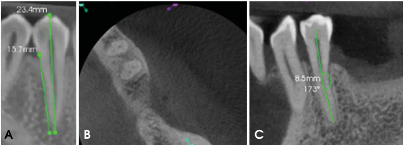
Fig. 1.A. Total tooth length from the cuspid to the apex and root length from the cemento-enamel junction to the apex are measured using a mesiodistal cone-beam computed tomography (CBCT) image. B. Axial CBCT image shows two premolars with two canals. C. Root curvature and distance from the angle vertex to the apex are measured using a mesiodistal CBCT image.
A B C
possible canal divisions and forms. The contrast was adjust- ed when needed, and a zoom of between 0.4 and 1.1 was used.
Measurements
The evaluated variables were as follows: patient’s age and gender, tooth position in the arch, number of roots, length of the tooth and root, number of canals, canal sys- tem configuration according to the Vertucci criteria22dis- tance from the cemento-enamel junction to the canal bifur- cation and canal reunification (when it was the case), num- ber of foramina, root curvature, angle location, and dis- tance of the angle vertex to the apex.
The tooth length in the mesiodistal plane (maximum crown-apex distance) as well as the root length (from the
mesial cemento-enamel junction to the apex) was measur- ed with the linear measurement tool (Fig. 1A). A number of canals were observed by moving the coronal plane, checking them in the axial plane (Fig. 1B), and covering the tooth from the pulp chamber to the apex in order to confirm the observed configuration. With respect to the angles, each tooth was observed in the mesiodistal and buccolingual planes to check them and their orientation.
Root curvature was measured in degrees, and the comple- mentary angle was calculated to be 180�; the distance from the angle vertex to the apex in millimeters was also mea- sured (Fig. 1C).
Statistical analysis
Data were tabulated and analyzed using IBM SPSS sta-
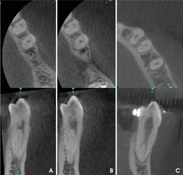
Fig. 2.A. Axial (up) and buccolingual (down) CBCT images show the Vertucci type II configuration. B. Shapes not included in Vertucci’s classification. One canal in the coronal third of the root, three canals in the middle third, and one canal in the apical third. C. Axial (up) and buccolingual (down) CBCT images show the Vertucci type V configuration.
A B C
tistical package 19.0 (IBM Co., Chicago, IL, USA). A de- scriptive analysis of the data was performed. Qualitative variables were compared using a chi-squared test, and an analysis of variance (ANOVA) test was used for compar- ing the qualitative and the quantitative variables. The sig- nificance level was p⁄0.05 in all the cases.
Results
The final sample size was 70 patients who underwent CBCT - 31 (43.7%) women and 39 (56.3%) men - with an average age of 45.26 years (confidence interval (CI): 32.12- 58.71). The number of mandibular premolars was 126. The number of root canals is given in Table 1. All the elements in the sample were single-rooted teeth. The mean length of the teeth was 22.18 mm ranging from 21.87 to 22.49 mm (men: 22.79 mm, CI: 22.39-23.18; women: 21.39 mm, CI:
20.98-21.89). The teeth of the men were significantly long- er than those of the women (p==0.00). The mean length of the first premolars was 22.3 mm (CI: 21.83-22.97) and that of the second premolars was 21.8 mm (CI: 21.23-22.51).
The mean length of the roots was 15.56 mm (CI: 15.25- 15.86) (men: 16.05 mm, CI: 15.64-16.46; women: 14.91 mm, CI: 14.50-15.32). The roots of the males were signi- ficantly longer than those of the women (p==0.00). The mean length of the roots was 15.60 mm (CI: 15.18-16.14) for the first premolars and 15 mm (CI: 14.62-16.11) for the second premolars, without any statistically significant differences between them (p¤0.05).
There were 105 teeth (83.3%) with one canal, 20 (15.9%) with two canals (Fig. 2A), and just 1 (0.8%) with three canals (Fig. 2B). In this case, the differences between men and women were not significant (p==0.34). There were no statistically significant differences between the first and the second premolars (p==0.21), but the first premolars showed more than one canal in more cases than the second premolars (Table 1).
Canal configurations according to the Vertucci criteria are listed in Table 2. Most of the teeth were type I, and the following most prevalent situation was type V distribution (Fig. 2C). One tooth showed a root canal configuration, not included in Vertucci’s classification (one canal in the coronal third of the root, three canals in the middle third, and one canal in the apical third). Variability was signifi- cantly higher in the first premolars than in the second pre- molars (p==0.03). No difference by gender was detected (p
==0.29). In cases where bifurcation was found, it was locat- ed at 6.87 mm (CI: 3.77-10.01) from the cemento-enamel junction. Canal confluence, when present, was found at 10.07 mm (CI: 9.37-12.77) of the cemento-enamel junction.
89.7% of the teeth showed a single apical foramen (113 teeth), and the remaining 10.3%, two apical foramina (13 teeth, 9 of them in women, 69.2%). A significant associa- tion was found between the number of foramina and the female gender (p==0.04). 12.3% of the first premolars and 7.5% of the second premolars showed two foramina (p== 0.55).
Data for root curvature are presented in Table 3. They
Table 1.Number of canals according to gender and tooth
Tooth 1 canal 2 canals 3 canals Total
Left first premolar 28 (82.4%) 6 (17.6%) 0 (0.0%) 34 (46.6%)
Right first premolar 29 (74.4%) 9 (23.1%) 1 (2.6%) 39 (53.4%)
Total 57 (78.0%) 15 (20.5%) 1 (1.5%) 73 (100.0%)
Left second premolar 14 (82.4%) 3 (17.8%) 0 (0.0%) 17 (32.1%)
Right second premolar 34 (94.4%) 2 (5.6%) 0 (0.0%) 36 (67.9%)
Total 48 (90.5%) 5 (4.5%) 0 (0.0%) 53 (100.0%)
Gender
Male 62 (87.3%) 8 (11.3%) 1 (1.4%) 71 (56.3%)
Female 43 (78.2%) 12 (60%) 0 (0.0%) 56 (43.7%)
Total 105 (83.3%) 20 (15.9%) 1 (0.8%) 126 (100.0%)
Table 2.Number and distribution of root canals according to the Vertucci classification22
Type I Type II Type V Type VI Unclassified Total
First premolar 57 (78.1%) 6 (8.2%) 8 (12.3%) 1 (1.3%) 1 (1.3%) 73 (57.9%)
Second premolar 48 (90.6%) 1 (1.8%) 4 (7.5%) 0 (0.0%) 0 (0.0) 53 (42.1%)
Total 105 (83.3%) 7 (5.5%) 12 (9.6%) 1 (0.8%) 1 (0.8) 126 (100%)
were detected in 65% of the sample with no significant gen- der association (p==0.06), although 62.3% of the angulated teeth belonged to men. There were no significant differ- ences between the first and the second angulated premolar percentages (p==0.32). Distal curvature was the most pre- valent for the first premolars (30.1%), whereas buccal cur- vature was the most prevalent for the second premolars (22.6%), without any significant differences (p==0.67).
With respect to the curvature position, there was no signi- ficant difference between men and women (p==0.21).
In the angulated teeth, an angle lower than 20�was found in 67 teeth (87.0%) and an angle greater than 20�in 10 cases (13.0%), without any significant differences for gen- der (p==0.23) or tooth. When the curvature was greater than 20�, the angle vertex was found at less than 5 mm from the apex (Table 4).
Discussion
The complex anatomy of the canal system in the mandi- bular premolars makes it difficult to assess their clinical morphology with conventional radiography.23In most clin- ical situations, when using conventional radiological pro- cedures such as periapical radiographs, the presence of morphological variations in the anatomy of the root canals can only be supposed when a change in shape or direction is detected, or when the root canal is not completely seen.24 With the use of the CBCT equipment, it is possible to obtain images with sufficient resolution to allow the study of the canal system anatomy, as performed in the present work. Although no specific tools for a root canal system study have been developed to date, the ones available for measurements in other dental applications have turned out to be suitable for this study. Micheti et al21found an excel- lent correlation between CBCT reconstructions obtained with Kodak 9000 3D and the corresponding histological
sections, claiming that CBCT is a reliable technique for the analysis of the root canal system anatomy.
Although there are few studies in this regard, some au- thors have used CBCT to study variations in dental anato- my in different series of teeth.25-31In particular, the anatomy of the mandibular premolars has been studied using CBCT in the Chinese and Korean populations.14,20
The mean length of premolars as found in this study was 22.18 mm. The premolars of males were significantly long- er than those of women, and the same trend was observed for the root length; this gender difference has not been an- alyzed in other studies reported thus far. The length of the second premolars was somewhat lower than that of the first premolars, but without any significant differences, these data are consistent with the findings of a study performed on the extracted teeth of a Jordanian population by Awaw- deh et al.25However, other studies32,33found almost iden- tical lengths for both teeth, possibly because these studies used radiographs and the present study used CBCT.
The most frequent morphology found in our study was one root and one canal, in accordance with the findings of other researchers.11,12 The results of studies in different ethnic populations with different procedures also agreed with these results; nevertheless, cases of other situations including C-shaped canals, more than one root, or more than two canals have described very rarely.12,14,34,35Althou- gh no significant difference was found between the first and the second premolars with respect to the number of canals, the prevalence of two canals was higher in the first premolars than in the second premolars, 20.5% versus 4.5
%, as described by Yu et al.20
The most prevalent root canal configuration observed in our study was Vertucci type I, followed by type V, with a higher morphological variability in the first premolars.
21% of the canals showed some kind of bifurcation, locat- ed at 3.77-10.01 mm from the cemento-enamel junction
Table 3.Angulation presence and its orientation
No angulation Buccal Lingual Mesial Distal Total
First premolar 25 (34.2%) 7 (9.6%) 9 (12.3%) 10 (13.7) 22 (30.1%) 73 (57.9%)
Second premolar 24 (45.3%) 12 (22.6%) 4 (7.5%) 4 (7.5%) 9 (17.0%) 53 (42.1%)
Total 49 (38.9%) 19 (15.1%) 13 (10.3%) 14 (11.1%) 31 (24.6%) 126 (100%)
Table 4.Number and distribution of teeth according to root angulation degrees and distance from the angle vertex to the apex
⁄20�and ‹5 mm ⁄20�and ¤5 mm ›20�and ‹5 mm Total
First premolar 29 (60.4%) 15 (31.3%) 4 (8.3%) 48 (62.3%)
Second premolar 16 (55.2%) 7 (24.1%) 6 (20.7%) 29 (37.7%)
Total 45 (58.4%) 22 (28.6%) 10 (13.0%) 77 (100%)
(middle/apical root thirds). These results are consistent with those found in the literature by other researchers.20,23,35
Root curvature was more prevalent in the first premolars than in the second premolars, with distal curvature being the most observed in the first premolars, a situation simi- lar to that described by Khedmat et al in an Iranian pop- ulation23although the prevalence of deviation found in our population was higher, 30.1% versus 17.5%. Further, in our study, the curvature angle was lower than 20�in the first and the second premolars in 91.7% and 79.3% of the cases, respectively. In the few cases where the angle was larger than 20�, it was always found at less than 5 mm from the apex (Table 4). These results are in accordance with those obtained by Willershausen et al in 200936 wherein the angle ranged between 13�and 19�and the last curva- ture was done at less than 5 mm from the apex. This con- figuration is important as it is a factor increasing the diffi- culty of the root canal treatment in the teeth with the high- est curvatures close to the tooth apex.37
In the present study, we found a higher prevalence of two foramina in women than in men, something not detect- ed in the literature review. The higher prevalence of more than one foramen in the first premolars (12.3%) than in the second premolars (7.6%) found in our work, is in agree- ment with the results presented in an extensive series pub- lished in two literature reviews, but there, the percentages were higher for both tooth types.11,12
It can be concluded that CBCT was useful for evaluating the anatomy of mandibular premolars. Premolar lengths for men were greater than those for women. The first pre- molars showed a higher anatomical variability than the second premolars. The most prevalent Vertucci configura- tion was type I, followed by type V, with a higher variabi- lity in the first premolars than in the second premolars.
Most teeth had a single foramen, and women had a higher prevalence of two foramina than men.
References
1. Miracle AC, Mukherji SK. Conebeam CT of the head and neck, part 2: clinical applications. AJNR Am J Neuroradiol 2009; 30:
1285-92.
2. Yajima A, Otonari-Yamamoto M, Sano T, Hayakawa Y, Oto- nari T, Tanabe K, et al. Cone-beam CT (CB Throne) applied to dentomaxillofacial region. Bull Tokyo Dent Coll 2006; 47:
133-41.
3. Scarfe WC, Farman AG, Sukovic P. Clinical applications of cone-beam computed tomography in dental practice. J Can Dent Assoc 2006; 72: 75-80.
4. Madrigal C, Ortega R, Meniz C, López-Quiles J. Study of avai- lable bone for interforaminal implant treatment using cone-
beam computed tomography. Med Oral Patol Oral Cir Bucal 2008; 13: E307-12.
5. Scarfe WC, Levin MD, Gane D, Farman AG. Use of cone beam computed tomography in endodontics. Int J Dent 2009; 2009:
634567.
6. Patel S. New dimensions in endodontic imaging: part 2. Cone beam computed tomography. Int Endod J 2009; 42: 463-75.
7. Patel S, Horner K. The use of cone beam computed tomogra- phy in endodontics. Int Endod J 2009; 42: 755-6.
8. Zillich R, Dowson J. Root canal morphology of mandibular first and second premolars. Oral Surg Oral Med Oral Pathol 1973; 36: 738-44.
9. Slowey RR. Root canal anatomy. Road map to successful en- dodontics. Dent Clin North Am 1979; 23: 555-73.
10. Glassman GD. Flare-up with associated paresthesia of a man- dibular second premolar with three root canals. Oral Surg Oral Med Oral Pathol 1987; 64: 110-3.
11. Cleghorn BM, Christie WH, Dong CC. The root and root canal morphology of the human mandibular first premolar: a litera- ture review. J Endod 2007; 33: 509-16.
12. Cleghorn BM, Christie WH, Dong CC. The root and root canal morphology of the human mandibular second premolar: a lite- rature review. J Endod 2007; 33: 1031-7.
13. Rahimi S1, Shahi S, Yavari HR, Manafi H, Eskandarzadeh N.
Root canal configuration of mandibular first and second pre- molars in an Iranian population. J Dent Res Dent Clin Dent Prospects 2007; 1: 59-64.
14. Park JB, Kim N, Park S, Kim Y, Ko Y. Evaluation of root an- atomy of permanent mandibular premolars and molars in a Korean population with cone-beam computed tomography. Eur J Dent 2013; 7: 94-101.
15. Trope M, Elfenbein L, Tronstad L. Mandibular premolars with more than one root canal in different race groups. J Endod 1986; 12: 343-5.
16. Hulsmann M. Mandibular first premolar with three root canals.
Endod Dent Traumatol 1990; 6: 189-91.
17. Yang ZP. Multiple canals in a mandibular first premolar. Case report. Aust Dent J 1994; 39: 18-9.
18. Nallapati S. Three canal mandibular first and second premo- lars: a treatment approach. J Endod 2005; 31: 474-6.
19. Kakkar P, Singh A. Mandibular first premolar with three roots:
a case report. Iran Endod J 2012; 7: 207-10.
20. Yu X, Guo B, Li KZ, Zhang R, Tian YY, Wang H, et al. Cone- beam computed tomography study of root and canal morpho- logy of mandibular premolars in a western Chinese population.
BMC Med Imaging 2012; 12: 18.
21. Michetti J, Maret D, Mallet JP, Diemer F. Validation of cone beam computed tomography as a tool to explore root canal anatomy. J Endod 2010; 36: 1187-90.
22. Vertucci FJ. Root canal anatomy of the human permanent teeth.
Oral Surg Oral Med Oral Pathol 1984; 58: 589-99.
23. Khedmat S, Assadian H, Saravani AA. Root canal morphology of the mandibular first premolars in an Iranian population using cross-sections and radiography. J Endod 2010; 36: 214-7.
24. Tzanetakis GN, Lagoudakos TA, Kontakiotis EG. Endodontic treatment of a mandibular second premolar with four canals using operating microscope. J Endod 2007; 33: 318-21.
25. Awawdeh LA, Al-Qudah AA. Root form and canal morpho-
logy of mandibular premolars in a Jordanian population. Int Endod J 2008; 41:240-8.
26. Kim Y, Lee SJ, Woo J. Morphology of maxillary first and second molars analyzed by cone-beam computed tomography in a Korean population: variations in the number of roots and canals and the incidence of fusion. J Endod 2012; 38: 1063-8.
27. Zhang R, Yang H, Yu X, Wang H, Hu T, Dummer PM. Use of CBCT to identify the morphology of maxillary permanent molar teeth in a Chinese subpopulation. Int Endod J 2011; 44:
162-9.
28. Zheng Q, Zhang L, Zhou X, Wang Q, Wang Y, Tang L, et al.
C-shaped root canal system in mandibular second molars in a Chinese population evaluated by cone-beam computed tomo- graphy. Int Endod J 2011; 44: 857-62.
29. Reis AG, Grazziotin-Soares R, Barletta FB, Fontanella VR, Mahl CR. Second canal in mesiobuccal root of maxillary molars is correlated with root third and patient age: a cone-beam com- puted tomographic study. J Endod 2013; 39: 588-92.
30. Demirbuga S, Sekerci AE, Dinçer AN, Cayabatmaz M, Zorba YO. Use of cone-beam computed tomography to evaluate root and canal morphology of mandibular first and second molars
in Turkish individuals. Med Oral Patol Oral Cir Bucal 2013;
18: e737-44.
31. Silva EJ, Nejaim Y, Silva AV, Haiter-Neto F, Cohenca N. Eva- luation of root canal configuration of mandibular molars in a Brazilian population by using cone-beam computed tomogra- phy: an in vivo study. J Endod 2013; 39: 849-52.
32. Kim E, Fallahrastegar A, Hur YY, Jung IY, Kim S, Lee SJ.
Difference in root canal length between Asians and Caucasians.
Int Endod J 2005; 38:149-51.
33. Vertucci FJ. Root canal morphology of mandibular premolars.
J Am Dent Assoc 1978; 97: 47-50.
34. Green D. Double canals in single roots. Oral Surg Oral Med Oral Pathol 1973; 35: 689-96.
35. Lu TY, Yang SF, Pai SF. Complicated root canal morphology of mandibular first premolar in a Chinese population using the cross section method. J Endod 2006; 32: 932-6.
36. Willershausen B, Kasaj A, Röhrig B, Briseno B. The determi-~ nation of the initial straight length in root canals of mandibular premolars - an in vitro study. Eur J Med Res 2009; 14: 85-9.
37. Lombart B, Michonneau JC. Premolar anatomy and endodontic treatment. Rev Belge Med Dent (1984) 2005; 60: 322-36.