REPO RT
http://dx.doi.org/10.11106/ijt.2015.8.2.216Received May 16, 2015 / Revised August 5, 2015 / Accepted September 20, 2015
Correspondence: Eun Kyung Lee, MD, Department of Internal Medicine, Center for Thyroid Cancer, National Cancer Center, 323 Ilsan-ro, Ilsandong-gu, Goyang 10408, Korea
Tel: 82-31-920-1743, Fax: 82-31-920-2798, E-mail: [email protected]
Copyright ⓒ 2015, the Korean Thyroid Association. All rights reserved.
This is an open-access article distributed under the terms of the Creative Commons Attribution Non-Commercial License (http:// creative- commons.org/licenses/by-nc/4.0/), which permits unrestricted non-commercial use, distribution, and reproduction in any medium, provided the original work is properly cited.
Rare Concurrence of Triple Primary Thyroid Cancer: A Patient of Papillary Carcinoma,
Follicular Carcinoma, and Primary Lymphoma of the Thyroid
Eun Jeong Ko
1, Eun Kyung Lee
1,2, Si Won Lee
1and Sang Il Choi
1Department of Internal Medicine1, Center for Thyroid Cancer2, National Cancer Center, Goyang, Korea
We report a rare case of co-occurrence of papillary thyroid carcinoma (PTC), follicular thyroid carcinoma (FTC) and primary thyroid lymphoma. A 55-year-old woman presented with a large mass in left lobe of thyroid, biopsy confirmed diffuse large B-cell lymphoma. After 4 cycles of rituximab, cyclophosphamide, doxorubicin hyd- rochloride, vincristine sulfate, and prednisolone chemotherapy, positron emission tomography scan revealed markedly decreased in size, but still present. Repeated ultrasonography-guided gun biopsies of 2 lesions indicated Hurthle cell neoplasm. After total thyroidectomy and bilateral central lymph node dissection, residual hypermetabolic lesion of left lobe was determined to be FTC and right lower lesion to be nodular hyperplasia.
Besides, a PTC was incidentally detected in left lobe. If there are multiple nodular lesions at diagnosis or there is insufficient response after 1st line chemotherapy for primary thyroid lymphoma, each lesion should be biopsied to confirm its pathological type.
Key Words: Thyroid lymphoma, Papillary thyroid cancer, Follicular thyroid cancer, Triple primary thyroid cancer
Introduction
Papillary thyroid carcinoma (PTC) and follicular thyroid carcinoma (FTC) account for 70-80% and 15% of all thyroid cancers, respectively.1) Primary thyroid lympho- ma is a rare cancer, accounting for an estimated 1-5%
of thyroid malignancies and 2% of all extranodal lymphomas. The simultaneous occurrence of these 3 malignancies in the same patient is extremely rare.
Case Report
A 55-year-old woman visited in our clinic because of shortness of breath and the progressive enlarge-
ment of a mass in her neck. She had felt the mass in her neck growing for the past 3 months and lost approximately 3-4 kg in weight without fever or night sweating. She denied taking any pills, had no history of thyroid disease, goiter, or radiation therapy to the head or neck, and had no family history of thyroid cancer or other malignancy. On physical examination, a firm nodule about 6 cm in size was palpable in her left thyroid gland moved with deglutition. There was no palpable cervical lymphadenopathy.
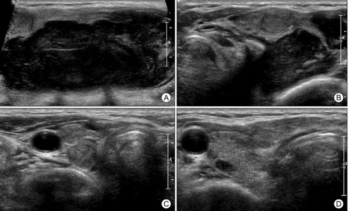
Initial thyroid ultrasonography (US) showed a huge mass that was accompanied by 3 small nodules in both lobes with underlying diffuse thyroiditis. The huge 6.4×5.4×3.0 cm lobulated contoured hypoechoic solid mass located in the left thyroid gland and an isoechoic
Fig. 1. Initial ultrasonography of thyroid. A huge lobulated contoured hypoechoic solid mass in left thyroid gland, measured 6.4×
5.4×3.0 cm (A), isoechoic solid mass with calcifications in left thyroid gland upper pole, measured 1.1×1.1×0.6 cm (B), isoechoic solid nodule in right thyroid gland lower pole, measured 0.9×0.8×0.7 cm (C), hypoechoic solid nodule in right thyroid middle lobe, measured 0.7×0.5×0.3 cm (D).
1.1×1.1×0.6 cm nodule with calcification in the left upper lobe appeared mostly suspicious. Another iso- echoic solid nodule was located in the right lower lobe and measured 0.9×0.8×0.7 cm, appeared likely to be benign. Finally, a 0.7×0.5×0.3 cm hypoechoic nodule was evident in the right thyroid middle lobe (Fig. 1). US-guided gun biopsy of the huge mass in the left lobe revealed a diffuse large B cell lymphoma (DLBL), stained positively for CD20 (+), Bcl-6 (+), and Ki-67 in 60% of cells (Fig. 2).
An initial positron emission tomography (PET) scan detected a hypermetabolic lesion (standardized uptake value [SUV] 27.5) in the left lower neck, otherwise unremarkable. Accordingly, the lesion was classified as Ann Arbor stage IE-A, with an International Pro- gnostic Index of 0. The initial lactate dehydrogenase (LDH) was 181 U/L (normal range, 101-202 U/L), the free T4 1.36 ng/dL (normal range, 0.89-1.79 ng/dL) and the thyroid stimulating hormone (TSH) level 2.79 μIU/mL (normal range, 0.17-4.05 μIU/mL) were both
within normal ranges.
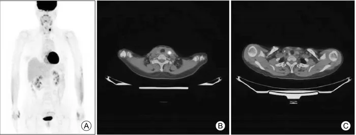
After 4 cycles of rituximab, cyclophosphamide, dox- orubicin hydrochloride, vincristine sulfate, and pre- dnisone (R-CHOP) chemotherapy, PET scanning re- vealed that the hypermetabolic nodule in the left thy- roid had decreased in size, but still present (SUV 8.4).
Scanning also showed the mild hypermetabolic lesion in the right lower lobe, which appeared to have a non-specific uptake. Otherwise, no gross abnormal uptake was evident, suggesting no additional malig- nant processes. After 8 cycles of R-CHOP chemo- therapy, PET scanning showed no major changes from the previous status (Fig. 3). The diminished, re- sidual hypermetabolic nodular lesion (SUV 6.6) in the left thyroid and the mild hypermetabolic lesion (SUV 3.9) in the right lower lobe remained evident. We re- checked thyroid function and autoantibody levels.
Thyroid function remained within the normal ranges (free T4 1.4 ng/dL, TSH 1.61 μIU/mL), but the micro- somal antibody titer was 101 U/mL (normal range,
Fig. 3. PET scan image after 8th chemotherapy of rituximab, cyclophosphamide, doxorubicin hydrochloride, vincristine sulfate, and prednisone (R-CHOP) (A), residual hypermetabolic nodular lesion (SUV 6.6) in the left thyroid (B), mild hypermetabolic lesion (SUV 3.9) in the right lower lobe (C).
Fig. 2. Pathologic examination of thyroid lymphoma obtained by core needle biopsy (Hematoxylin & Eosin stain) (A) and immunohistochemical examination of CD20 of primary thyroid lymphoma (B); (×400), follicular thyroid cancer (C) and papillary thyroid carcinoma (D) collected by surgical resection (Hematoxylin & Eosin stain; ×400).
0-100 U/mL), suggesting Hashimoto’s thyroiditis.
The repeated US result revealed the 3 small nod- ules showed no change: the 1.1×1.0×0.8 cm hy- poechoic solid nodule with microcalcification in the left upper lobe, the 0.9×0.8×0.6 cm isoechoic nodule in the right lower lobe, and the small 0.5×0.3×0.2 cm hypoechoic nodule in the left upper pole medial aspect. The 2 nodules in the left upper and right lower lobes were pathologically confirmed as Hurthle cell neoplasms using gun biopsy. A total thyroidectomy with bilateral central lymph node (LN) dissection was performed. During the operation, a moderate degree of fibrotic change was observed in the left para- tracheal area, related to Hashimoto’s thyroiditis or a chemotherapy-induced change. In addition, several regional LNs were enlarged.
Histopathological examination identified the 1.2×0.9
×0.7 cm minimally invasive follicular carcinoma in the left upper lobe, which had minimal capsular invasion (pT1bN0; delphian LN: 0/2, left level VI LN: 0/4, right level VI LN: 0/4) and did not involve hemorrhage or necrosis (Fig. 2C). The nodule in the left upper pole medial aspect was confirmed to be a 0.3×0.2×0.2 cm occult papillary microcarcinoma, without vascular or lymphatic invasion and extrathyroidal extension (Fig.
2D). Finally, the hypoechoic nodule in the right lower lobe, thought to be a Hurthle cell neoplasm based on a gun biopsy specimen, was confirmed to be nodular hyperplasia.
Immunohistochemical staining of the FTC of left lobe for galectin-3, cyclin D1, and anti-human mesothelial cell antibody were all positive. The BRAF mutation wasn’t detected in the PTC tumor on reverse tran- scription-polymerase chain reaction. The patient dis- charged with 0.15 mg/day levothyroxine, and didn’t undergo remnant iodine ablation.
Discussion
This patient was at first diagnosed with primary thy- roid lymphoma alone, but additionally diagnosed with FTC and PTC after chemotherapy. The co-occur- rence of lymphoma and other thyroid cancers is rare.
One case has been reported a 59-year-old man with
known Hashimoto’s thyroiditis, presented with a rapid enlargement of the thyroid gland over few months.2,3) In this case, pathology indicated a PTC and an extra- nodal marginal zone B-cell lymphoma of the muco- sa-associated lymphoid tissue (MALT) type. Another case involved a 61-year-old man, presented with a rapid enlargement of the thyroid gland over 3 months2,3) Pathology showed multicentric PTCs with a concomitant thyroid MALT lymphoma. In the present report, we have described the first case of a triple pri- mary thyroid cancer, of concomitant PTC, FTC, and primary thyroid lymphoma.
The treatment of choice for primary thyroid lympho- ma has evolved over the past few decades. Graff- Baker et al.4) analyzed 1408 cases of primary thyroid lymphoma, found that surgery and radiation for primary thyroid lymphoma have declined over the years, while chemotherapy has recently become more preferred.
Current treatment recommendations for primary thyroid lymphoma are histology-specific. For example, sur- gery or radiation is preferred for the initial treatment of MALT lymphomas, whereas R-CHOP chemotherapy is recommended as the first treatment for DLBL. Doria et al.5) suggested that combined chemoradiation ther- apy should be the mainstay of treatment for primary thyroid lymphoma. The choice of chemotherapy regi- men has, for primary thyroid lymphomas, been ex- trapolated from studies of extranodal lymphomas.
However, controversy remains concerning the role of thyroid surgery for early stage thyroid lymphoma especially when it occurred with thyroid carcinoma. A study of 62 patients from the Mayo Clinic concluded that thyroidectomy with adjuvant chemotherapy re- sulted in long-term cure in patients with primary thy- roid lymphoma. Meyer-Rochow et al.6) suggested that operations should only be performed to establish the diagnosis of primary thyroid lymphoma or to manage severe obstruction of the airway. Some practitioners7) have recommended that fine needle aspiration and adjuncts should be the initial tests that are used to di- agnose thyroid lymphoma, although open surgical bi- opsy may still be required in many cases. To date, however, there have been no published investigations regarding the optimal management of multiple thyroid
nodules adjacent to primary thyroid lymphoma. In the present case, we incidentally found differentiated thy- roid carcinoma during chemotherapy for DLBL, which was localized within the thyroid. Because the biopsy specimen indicated Hurthle cell neoplasm, which sug- gests neither malignancy nor benign nodule can be differentiated by biopsy, we performed surgical resec- tion. Based on our experience with this case, the pre- cise pathologic confirm of multiple nodules should be done first, and if there is any suspicious finding, sur- gery can be performed before chemotherapy. Hashi- moto’s thyroiditis increases an incidence of the pri- mary thyroid lymphoma.8) Moreover, Kim et al.9) re- ported that Hashimoto’s thyroiditis is a strong risk fac- tor for differentiated thyroid cancer (DTC), specifically with PTC. So in case of Hashimoto’s thyroiditis pa- tients, if multiple nodules are found, biopsies for each nodular lesion should be performed to confirm if there are any possibility of hidden malignancy.
The management of co-occurring cancers should be considered separately, and the patient’s prognosis is probably dominantly affected by the cancer that has the worst prognosis.3,10-14) If there is insufficient re- sponse after 1st line chemotherapy for primary thyroid lymphoma, each residual lesion should be biopsied to confirm its pathological type, since the response rate of R-CHOP chemotherapy to DLBL is excellent, over 80% in stage I. In cases that involve the simultaneous occurrence of multiple thyroid neoplasms, surgery can be considered as standard of care.3)
Conflict of Interest
Conflict of interest relevant to this article was not reported.
Acknowledgments
This study was supported by a research grant from the National Cancer Center No. 1310690-1 and 1410640-1. The funding agencies did not have any role in the design or conduct of the study; collection,
management, analysis, or interpretation of the data; or preparation, review, or approval of the manuscript. No potential conflicts of interest relevant to this article were reported.
References
1) Sipos JA, Mazzaferri EL. Thyroid cancer epidemiology and prognostic variables. Clin Oncol (R Coll Radiol) 2010;22(6):
395-404.
2) Cheng V, Brainard J, Nasr C. Co-occurrence of papillary thyroid carcinoma and primary lymphoma of the thyroid in a patient with long-standing Hashimoto's thyroiditis. Thyroid 2012;22(6):647-50.
3) Cakir M, Celik E, Tuncer FB, Tekin A. A rare coexistence of thyroid lymphoma with papillary thyroid carcinoma. Ann Afr Med 2013;12(3):188-90.
4) Graff-Baker A, Roman SA, Thomas DC, Udelsman R, Sosa JA. Prognosis of primary thyroid lymphoma: demographic, clinical, and pathologic predictors of survival in 1,408 cases.
Surgery 2009;146(6):1105-15.
5) Doria R, Jekel JF, Cooper DL. Thyroid lymphoma. The case for combined modality therapy. Cancer 1994;73(1):200-6.
6) Meyer-Rochow GY, Sywak MS, Reeve TS, Delbridge LW, Sidhu SB. Surgical trends in the management of thyroid lymphoma. Eur J Surg Oncol 2008;34(5):576-80.
7) Mack LA, Pasieka JL. An evidence-based approach to the treatment of thyroid lymphoma. World J Surg 2007;31(5):
978-86.
8) Noureldine SI, Tufano RP. Association of Hashimoto's thy- roiditis and thyroid cancer. Curr Opin Oncol 2015;27(1):21-5.
9) Kim KW, Park YJ, Kim EH, Park SY, Park do J, Ahn SH, et al. Elevated risk of papillary thyroid cancer in Korean patients with Hashimoto's thyroiditis. Head Neck 2011;33(5):
691-5.
10) Mian M, Capello D, Ventre MB, Grazio D, Svaldi M, Rossi A, et al. Early-stage diffuse large B cell lymphoma of the head and neck: clinico-biological characterization and 18 year fol- low-up of 488 patients (IELSG 23 study). Ann Hematol 2013 [Epub ahead of print].
11) Stein SA, Wartofsky L. Primary thyroid lymphoma: a clinical review. J Clin Endocrinol Metab 2013;98(8):3131-8.
12) Eccles A, Challapalli A, Khan S, Barwick T, Mangar S.
Thyroid lymphoma incidentally detected by 18F-fluorocholine (FCH) PET/CT. Clin Nucl Med 2013;38(9):755-7.
13) Szczepanek-Parulska E, Szkudlarek M, Majewski P, Brebo- rowicz J, Ruchala M. Thyroid nodule as a first manifestation of Hodgkin lymphoma-report of two cases and literature review. Diagn Pathol 2013;8:116.
14) Diaconescu MR, Costea I, Glod M, Grigorovici M, Diaco- nescu S. Unusual malignant tumors of the thyroid gland.
Chirurgia (Bucur) 2013;108(4):482-9.