129 Received: November 5, 2010, Accepted for Publication: November 26, 2010
*Corresponding author: Weon Kim, 130-701, Department of Internal Medicine, Kyung Hee University Hospital, Kyung Hee University, Phone: +82-2-958- 8176, FAX: +82-2-958-8160, E-mail: [email protected]
Chonnam Medical Journal Vol. 46, No. 3, pp. 129∼139 DOI: 10.4068/cmj.2010.46.3.129
Mechanism of Ischemia and Reperfusion Injury to the Heart:
From the Viewpoint of Nitric Oxide and Mitochondria
Sang-Jin Ha and Weon Kim*
Cardiology Division, Department of Internal Medicine, Kyung Hee University Hospital, Seoul, Korea
After an acute myocardial infarction, early and successful myocardial reperfusion is the most effective strategy for reducing the size of a myocardial infarct and improving the clinical outcome. However, the process of restoring blood flow to the ischemic myocardium can induce injury. This phenomenon,termed myocardial reperfusion injury, can paradoxically reduce the beneficial effects of myocardial reperfusion and lead to lethal damage to myocardium. During cardiac ischemia-reperfusion (IR) injury, excessive generation of reactive oxygen species (ROS), overload of intracellular Ca2+, H+ leakage at the mitochondrial level, inflammation, and metabolic modulations lead to opening of the mitochondrial permeability transition pore(PTP) on reperfusion. This can result in the depletion of ATP, irreversible oxidation of proteins, lipids, and DNA within the cardiomyocyte, and can trigger apoptosis. In contrast, mitochondria also plays an important role in the cardioprotective signaling processes of ischemic preconditioning (IPC), to prevent IR injury. Nitric oxide (NO) generated constitutively within the heart has long been known to influence myocardial function. But, Nitric oxide (NO) has emerged as a potent effector molecule for a variety of cardioprotective strategies, including IPC. Whereas NO is most noted for its activation of the "classic" soluble guanylate cyclase (sGC) signaling pathway, emerging evidence indicates that NO can directly act on mitochondria, independent of the sGC pathway, affording acute cardioprotection against IR injury. These effects of NO on mitochondria and mitochondrial role during IR injury are the focus of this review.
Key Words: Myocardial infarction; Ischemia-reperfusion; Nitric oxide; Mitochondria
Introduction
Ischemic heart disease (IHD) secondary to acute myocar- dial infarction is among the most prevalent health problems in the world and is a major cause of morbidity and mortality.
In view of this, a major research effort in cardiovascular medicine has been to develop approaches to salvage the myocardium at risk of necrosis in patients with acute my-
ocardial infarction (AMI). After an AMI, early and success- ful myocardial reperfusion with the use of pharmacological thrombolytic agents or mechanical revascularization, i.e., primary percutaneous coronary intervention (PCI), is the most effective strategy for reducing the damage to the my- ocardium and improving clinical outcome. However, reper- fusion of the ischemic myocardium can induce some injury.
This phenomenon, termed myocardial reperfusion injury, can blunt the beneficial effects of myocardial reperfusion.
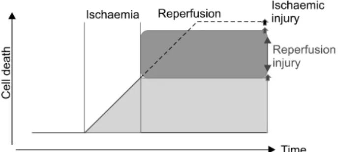
Reperfusion contributes to lethal injury following prolonged periods of ischemia (Fig. 1).
Myocardial reperfusion injury was first postulated in 1960
Fig. 1. The concept of lethal reperfusion injury. During ischemia, irreversible cell injury leading to cell death occurs within the ischemic risk zone in a time-dependent manner. In the absence of reperfusion, ischemic injury progressively kills more and more cells, eventually accounting for near total cell death (broken line). Reperfusion halts the process of ischemic cell death but in its early stages imposes injury that results in further cell death, beyond that due to the ischemic period: this is lethal reperfusion injury. The net result, however, is that the reperfused tissue sustains less cell death than would occur in ischemic tissue without reperfusion. Hence, targeting cell death due to reperfusion has the potential to maximize cell salvage. Adapted from Garcia-Dorado and Piper.1
by Jennings et al.2 as significant morphological alterations appearing after the onset of reperfusion, including cell swel- ling, contracture of myofibrils, disruption of the sarcolem- ma, and the appearance of intramitochondrial calcium phosphate particles. The very existence of lethal reperfusion injury was vigorously debated3 by those who maintained that lethal injury was expressed or hastened by reperfusion on the one hand4 and those who advocated reperfusion as a contributor to de novo injury.5
Mitochondria make up 30% of the volume of a single cardiomyocyte, underlining the importance of their tradi- tional role as the ATP producing 'powerhouse' of the cell.
However, it is clear that this complex organelle plays a variety of roles within the cardiomyocyte that extend be- yond its established function as the cellular powerhouse.
In this regard, the most significant discovery was its criti- cal role as an arbitrator of cell death6 in addition to its function in cellular survival.
Administration of nitric oxide (NO) or NO donors prior to ischemia attenuates the consequences of myocardial is- chemia-reperfusion (IR); that is, it reduces infarct size and endothelial dysfunction.7 NO synthesized by essentially all cardiac cell types is an ubiquitous cellular messenger that plays a key role in regulating cardiac function.8-12 NO is
a highly diffusible gas that spreads very rapidly from its site of synthesis and is a free radical highly reactive with other species, notably oxygen, superoxide, and iron-con- taining heme groups that act as NO scavengers. For this reason, the half-life of NO is limited to seconds and its effects are localized to where it is synthesized. NO generated within the cardiomyocytes can exert intracrine effects or modify the functional properties of adjacent cardiomyo- cytes. NO generated from noncardiomyocyte sources (coro- nary, endocardial, and endothelial cells; autonomic nerves and ganglia; and blood-formed elements) can exert direct effects on cardiomyocytes and indirect effects by modulating coronary blood flow or autonomic transmission.8-12 The heart produces NO on a beat-to-beat basis in response to changes in coronary flow and myocardial loading. In rabbit hearts, NO levels reach peak values during diastole (∼2.7 mM in the endocardium and ∼0.93 μM in the myocardium) and are lowest during systole (0.67 and 0.26 μM, respec- tively); NO concentrations are 15% lower in rat hearts.13
Molecular Events during Lethal Reperfusion Injury
1. Oxidative stress
Although low levels of oxygen radicals and oxidants are normally formed in cells and play important roles in cellular homeostasis, mitosis, differentiation, and signaling,14 follo- wing ischemia and reperfusion, radical formation is greatly increased, thus triggering cellular injury. Although mammalian cells including cardiomyocytes express endo- genous free radical scavenging enzymes,15 such as super- oxide dismutase (SOD), catalase, and glutathione peroxi- dase, these antioxidative defenses are overwhelmed after ischemia and reperfusion. The oxygen radical hypothesis is of particular importance because it potentially can explain each of the other mechanisms of reperfusion injury. It has been demonstrated that exogenous free radicals cause cellular calcium loading with inhibition of the sarcoplasmic reticulum calcium ATPase and inhibition of the sodium potassium ATPase leading to sodium-mediated calcium
gain.16 Oxygen radicals cause lipid peroxidation that can result in cell membrane breakdown causing cell swelling.
It has been suggested that oxygen radicals result in the chemotaxis of neutrophils that in turn can lead to white cell plugging of capillaries and microvascular compression.
In addition, white cells that are chemotaxed and activated are potent sources of further oxygen radical generation.17 Oxidative stress during myocardial reperfusion also reduces the bioavailability of the intracellular signaling molecule, nitric oxide, thereby removing its cardioprotective effects.
These effects include the inhibition of neutrophil accumu- lation, inactivation of superoxide radicals, and improve- ment of coronary blood flow.18
2. Calcium paradox
At the time of myocardial reperfusion, there is an abrupt increase in intracellular Ca2+ that is secondary to sarco- lemmal-membrane damage and oxidative stress-induced dysfunction of the sarcoplasmic reticulum. These two forms of injury overwhelm the normal mechanisms that regulate Ca2+ in the cardiomyocyte; this phenomenon is termed the calcium paradox.
The result is intracellular and mitochondrial Ca2+ over- load, and this excess of Ca2+ induces cardiomyocyte death by causing hypercontracture of the heart cells and mito- chondrial permeability transition pore (PTP) opening.19 3. pH paradox
The rapid restoration of physiologic pH during myo- cardial reperfusion, which follows the washout of lactic acid and the activation of the sodium-hydrogen exchanger and the sodium-bicarbonate symporter, contributes to lethal reperfusion injury. This phenomenon is termed the pH paradox.20 In neonatal rat cardiomyocytes, experimental studies have shown that reoxygenation with acidic buffer is cardioprotective;21 this effect may be mediated by the inhibition of mitochondrial PTP opening.22
4. Inflammation
After an acute myocardial infarction, the release of chemoattractants draws neutrophils into the infarct zone
during the first 6 hours of myocardial reperfusion, and during the next 24 hours they migrate into the myocardial tissue. This process is facilitated by cell-adhesion molecules.
These neutrophils cause vascular plugging and release degradative enzymes and reactive oxygen species.23 5. Metabolic modulation
Several experimental and numerous clinical studies have examined the cardioprotective potential of therapy with glucose, insulin, and potassium administered as an adjunct to myocardial reperfusion.24,25 These studies have been con- ducted on the premise that ischemic myocardium benefits more from metabolizing glucose than from fatty acids.26 A recent very large randomized controlled study from several centers reported no cardioprotective benefit from therapy with glucose, insulin, and potassium (GIK) as an adjunct to myocardial reperfusion in patients with acute myocardial infarction.27 The delay in initiating this therapy, the prolonged period of myocardial ischemia, and high and potentially damaging glucose levels have all been cited as reasons for the lack of cardioprotection. As alternatives to GIK solution, exenatide, an analog to glucagon-like peptide 1, reduces infarct size and improves cardiac function in a porcine model of ischemia and reperfusion injury.28
6. Mitochondrial K+ATP channel
One of the most important mitochondrial proteins recognized to be involved in preconditioning is the K+ATP channel. The actual mechanism by which K+ATP channel opening elicits cardioprotection is not yet clear, and debate surrounds the order of events with respect to reactive oxygen species (ROS) generation (i.e., evidence suggests both that ROS generation is an upstream trigger for K+ATP channel opening and that K+ATP channel opening is a trigger for ROS generation).29 Nevertheless, from the standpoint of NO signaling, several studies have shown that NO can directly affect the K+ATP channel via S-nitrosation.30 In addition, the mito-K+ATP channel is thought to be a downstream target for PKG in classic cGMP/NO signaling.31 However, in the latter case, the
mechanism by which PKG signaling is transmitted into the mitochondrion remains unclear.32 Furthermore, all studies on the K+ATP channel are subject to several caveats. First, much controversy surrounds the actual existence of bona fide K+ATP channel subunits (KIR, SUR) in mitochondria, with conflicting results surrounding the use of antibodies and Western blots.33,34 Second, many of the pharmacologic tools used to probe K+ATP channel function are nonspecific. Most notably, the K+ATP channel opener diazoxide is a known inhibitor of complex II,35 and a protonophoric uncoupler,36 whereas the K+ATP channel 5-HD is a β-oxidation substrate.37 The non-K+ATP channel effects of both diazoxide and 5-HD were recently reviewed extensively,37-39 with the result that a large proportion of the literature on this topic may require reassessment. The mechanism of K+ATP-mediated pro- tection is thought to include mild mitochondrial swelling, which may affect the formation of the PTP.40 Although it has been argued that the small K+ fluxes that would result from opening of K+ATP channels (and K+Ca channels) in ischemic preconditioning (IPC) would mildly uncouple mitochondria, it should be recognized that the magnitude of such K+ fluxes is not large enough to account for the magnitude of H+ leak seen in IPC.41 In a human study, sildenafil was shown to prevent endothelial dysfunction induced by ischemia and reperfusion via opening of ATP- sensitive potassium channels, and the proposed mechanism of the effect of sildenafil was direct and cGMP-mediated opening of K-ATP channels.42
7. Mitochondrial permeability transition pore
The mitochondrial PTP is a nonselective channel of the inner mitochondrial membrane. Opening the channel colla- pses the mitochondrial membrane potential and uncouples oxidative phosphorylation, resulting in ATP depletion and cell death.43 During myocardial ischemia, the mitochondrial PTP remains closed, only to open within the first few minutes after myocardial reperfusion in response to mito- chondrial Ca2+ overload, oxidative stress, restoration of a physiologic pH, and ATP depletion.22,44 The overpro- duction of ROS and mitochondrial Ca2+ overload are
known to trigger PTP formation. Downstream effects as a result of PTP opening include cytochrome c release, mito- chondrial swelling, membrane depolarization, inhibition of ATP production, caspase activation, apoptosis or necrosis or both, and additional ROS production, which initiates mitochondrial and cellular dysfunction.
At pathologically high levels, NO can induce apoptosis both via the activation of p53, and via release of cyto- chrome c from the mitochondrial inner membrane and activation of caspases.45-47 Although high levels of NO can cause PTP formation, smaller concentrations of NO inhibit PTP formation.48 Lower levels of NO protect the mitochondrion against apoptotic agents such as TNF-α, serum starvation, hypoxia, and H2O2.49,50
Mechanisms by which NO inhibits PTP formation include the prevention of Bcl-2 cleavage by caspase 3,45 mild dissipation of the mitochondrial membrane poten- tial, and inhibition of Ca2+ uptake.48 In terms of cardio- protection, the NO donor DETA-NONOate was found to inhibit cyclosporin A-sensitive Ca2+-induced mito- chondrial swelling in mitochondria isolated from hearts subjected to IR.48,51 Another study observed that aged endothelium, with decreased eNOS activity, and eNOS knockout mice were more susceptible to proapoptotic stimuli, which were reversed by NO donors.52 Therefore, the mitochondrial PTP is a critical determinant of lethal reperfusion injury, and as such it is an important new target for cardioprotection.
Nitric Oxide: Acting on Mitochondria for Cardioprotection against
Ischemia-Reperfusion Injury
1. Mitochondrial dysfunction in IR injury
The hallmarks of cardiac IR injury are found to occur on reperfusion of the myocardium. In terms of the mito- chondria, reperfusion injury affects the oxidative phos- phorylation (Ox-Phos) pathway, including the ETC,53-55 adenine nucleotide translocase (ANT),56,57 and Krebs cycle58,59 enzymes. In addition to Ox-Phos, reperfusion in-
jury also leads to cardiolipin oxidation,55,60,61 the induction of a large proton leak across the mitochondrial inner membrane,41,62 Ca2+ overload, overproduction of ROS, PTP opening, and cell death.62-73 A majority of these observations have been made in mitochondria isolated from hearts after reperfusion, meaning that the time course of mitochondrial damage during reperfusion injury is difficult to study (because mitochondrial isolation typically takes 1∼2 h).
The exact timing of mitochondrial damage during IR is an ongoing subject of investigation.
We demonstrated that MI might be the cause of mito- chondria swelling and the changes in adhesion force and stiffness of mitochondria by use of a novel technique: atomic force microscopy (AFM).73,74 Mitochondrial changes such as swelling and irregularity of shape were also found in the infracted area in IR rat heart (unpublished data).
Contrary to our findings in the AFM study, Brady et al.75 were the first to demonstrate extensive fragmentation of mitochondria in HL-1 cells (a murine atrial-derived cardiac cell line) in response to a sustained episode of simulated ischemia, changes that persisted into simulated reperfusion.
Interestingly, in that study, the authors observed that reperfusing the ischemic cells with SB203580, a pharma- cological p38MAPK inhibitor, caused the mitochondria to refuse and regain their elongated structures once more, which suggests that p38MAPK activity during reperfusion may have contributed to the detrimental changes in mito- chondrial morphology.75 The authors have gone on to demonstrate in HL-1 cells that ischemia-induced mito- chondrial fragmentation is associated with the translocation of Drp1 from the cytosol to the mitochondria (a process that is required for its pro-fission activity) and that the fragmentation process can be largely prevented by trans- fection with Drp1K38A (the dominant-negative construct of Drp1).76 Furthermore, in the adult murine heart, we demonstrated the fragmentation of IF cardiac mitochondria in the EM study. Plotnikov et al.77 demonstrated that pretreatment with an antioxidant could prevent ischemia- induced mitochondrial fragmentation. The mechanisms underlying mitochondrial fragmentation during ischemia are currently unclear, although we speculate that cytosolic
calcium overload and ROS may be contributory factors.
One can speculate that by interfering with cellular respi- ration, mitochondrial fragmentation may result in the pro- duction of ROS. In this respect, using primary renotubular cells, Plotnikov et al. demonstrated that pretreatment with an antioxidant could prevent ischemia-induced mito- chondrial fragmentation.77 Interestingly, in that particular study, insulin was also reported to prevent ischemia-induced mitochondrial fragmentation, although the mechanism was not further investigated. In the context of diabetes, mito- chondrial fragmentation induced by hyperglycemia has also been reported to result in the production of ROS; however, the actual interplay between mitochondrial fragmentation, ROS production, and mitochondrial respiration requires further investigation.
Despite evidence for mitochondrial dysfunction occu- rring on reperfusion, the degree of dysfunction also depends on the length of the ischemic insult, suggesting that ischemia itself is detrimental. Accumulation of Ca2+ and ROS generation does occur within the mitochondrion during ischemia, despite the mitochondria being de- energized.78 These events are linked to ischemic hyper- contracture and the reversal of mitochondrial ATP synthase to use glycolytic ATP to maintain membrane potential.
Thus, a reconciling paradigm is that the degree of mito- chondrial dysfunction during ischemia, which is a function of the length of ischemia, is a harbinger of more severe mitochondrial dysfunction on reperfusion. In other words, more severe dysregulation of the mitochondria and co- ntractile machinery during ischemia (as indicated by hyper- contracture) leads to more severe pathology on reperfusion.
2. Mitochondrial role in cardioprotection
In contrast to the detrimental effects of prolonged ische- mia, brief periods of ischemia initiate cardioprotective signa- ling cascades that preserve both myocardial and mito- chondrial function during subsequent prolonged ischemia.
This endogenous cardioprotective event was discovered more than 2 decades ago and is known as ischemic preconditioning, or IPC.79 Two “windows” of protection are elicited by IPC: the first acute phase is triggered within
Fig. 2. Signaling pathways in IPC. The three main signaling pathways impli- cated in the mechanism of IPC are the insulin → PI3k kinase → Akt axis (110), the GPCR → DAG → PKC → axis, and the NO → PKG → K+ATP channel axis. Key convergence points at the mitochondrial level are the phos- phorylation and inactivation of GSK-3 β, ROS generation by the ETC, K+ ATP channel opening, and the inhibition of PTP opening. Roles are also proposed for NO, mitochondrial uncoupling (H+ leak), transient opening or "flickering" of the PTP. The pathways by which various protective agents elicit cardioprotection also are shown.
Arrows, positive or stimulatory effects;
T-bars, inhibitory effects.
minutes and lasts 2 to 3 h, and a later delayed protective phase takes ∼24 h to develop and lasts up to 72 h.
Fig. 2 shows several of the signaling pathways implicated in IPC. Several well-known and emergent therapeutics also trigger the same pathways, including pharmacologic pre- conditioning (PPC) agents [e.g., opioids, adenosine mi- metics],80 anesthetic preconditioning (APC) with volatile anesthetics,81 and physical interventions such as slow or intermittent reperfusion (ischemic postconditioning).82 Thus, x-PC signaling continues to be an area of interest, not only because it can help to explain how these therapeutic molecules work, but also because it provides a deeper understanding of preconditioning signaling pathways, which offers the hope of improved therapeutics to be developed in the future.
While the spectrum of available cardioprotective strategies continues to grow, one concept that emerges from Fig. 4 is that the mitochondria are the downstream targets of most if not all of x-PC signaling. Key mitochondrial events include mild uncoupling (H+ leak), opening of mitochondrial K+ATP channels, inhibition of mitochondrial Ca2+ over- load, attenuation of ROS generation, and inhibition of PTP opening at reperfusion.83-86 Notably, several of the mito- chondrial events associated with IR injury are thought to occur in a limited manner during IPC. For example, a
somewhat paradoxical situation is proposed wherein transient "flickering" of the PTP during the triggering or initiation phase of IPC is thought subsequently to prevent large-scale PTP opening during IR injury.87 In addition, limited ROS generation has been found to be essential in IPC signaling cascades, and the cardioprotective response was found to be blocked by antioxidants.88 Similar to PTP opening and ROS production, a large increase in H+ leak is found to occur in IR injury (affecting mitochondrial ATP synthetic capacity). However, a small reversible increase in H+ leak is seen during IPC, which may act to diminish ROS generation and Ca2+ overload at reperfusion.89 Pharmacologic preconditioning agents, such as diazoxide (a K+ATP channel agonist) and dinitrophenol (DNP, which induces mitochondrial proton leak), also elicit cardioprotection in IR injury by attenuating mitochondrial PTP formation, ROS generation, and H+ leak.87,90,91 The overarching principle of x-PC signaling can be summarized in the proverb "what doesn’t kill you makes you stronger." It is therefore important to note that this principle is played out in its entirety in the microcosm of the mitochondrion. Fig. 2 highlights a multitude of upstream signaling pathways converging on the mito- chondria to elicit cardioprotection.
3. The divergent roles of NO -IR injury
When considering the roles of NO in any cell-signaling process, it is important to discuss the sources of NO gene- ration. Nitric oxide synthase (NOS) enzymes produce NO endogenously and are regulated by many of the upstream signaling pathways that are activated during IR and x-PC.92 Studies on the cardioprotective and deleterious effects of NO have involved manipulation of endothelial NOS (eNOS) and inducible NOS (iNOS) activity. Attenuation of myocardial infarct size and preservation of left ventricular developed pressure and lower left ventricular end-diastolic pressure have been observed in mice overexpressing eNOS in a variety of models of ischemia-alone or IR injury, when compared with wild-type mice.93-95 The NOS-dependent protection was found to be abrogated with the admini- stration of a commonly used NOS inhibitor, N(G)- nitro-L-arginine methyl ester (L-NAME).93 In agreement, studies using eNOS knockout mice have found an augmentation in infarct size96 and myocardial necrosis97 after IR injury. In terms of timing, eNOS is activated during the early window of preconditioning and activates trans- cription factors and other enzymes, including iNOS, in the late window of protection.98 The activation of eNOS has also been reported to contribute to the cardioprotective effects of postconditioning (brief intermittent reperfusion periods after ischemia). As seen in the eNOS transgenic and knockout studies, the cardioprotective effect of post- conditioning was sensitive to L-NAME.98,99 Furthermore, support for the essential role NOS plays in cardioprotection is seen with the sexual dimorphism in response to IR injury;
estrogen has been found to increase NOS expression, which may help to explain why premenopausal women have a lower risk of heart disease than do age-matched men.100 Several studies suggest the presence of a mitochondrial isoform of NOS (mtNOS), although the reproducibility of these studies appears to be limited to a small number of laboratories.101-103 Several recent articles have questioned the existence of mtNOS, with major controversies sur- rounding the purity of mitochondria, a 150,000-fold varia- tion in the reported rates of NO generation, and several
experimental artifacts in NO-measurement systems.104,105 It was reported that a NOS protein in the plant Arabidopsis thaliana (atNOS) is targeted to the mitochondrion, and more recently, a mammalian orthologue of this protein was proposed as a candidate for mtNOS.106,107 However, further investigation has found that the protein is a GTPase, not a NOS.108-110 Thus, the search for a unique mtNOS protein continues.
NOS enzymes have also been shown to play a deleterious role in IR injury. For example, iNOS−/− mice were shown to have lower mortality and enhanced left ventricular contractility when compared with wild-type mice after coronary occlusion.111 Also, exposing mitochondria to high concentrations of NO (micromolar) has been shown to initiate PTP opening.48 These results, along with other studies, have defined NO as a dual-faced molecule in IR injury, which contributes to both cardioprotective and deleterious signaling pathways within the myocardium. In this regard, understanding how to deliver NO (i.e., timing, concentration, location) may facilitate beneficial thera- peutic exploitation of NO signaling in IR injury, while minimizing the deleterious effects of NO.
4. NO and mitochondria-dependent cardioprotection Increasing evidence indicates that NO is a key mito- chondrial regulator; for a number of reasons, the mito- chondrion can be considered a cellular "hub" for NO sig- naling. First, mitochondria within the cardiomyocyte are in close proximity (1∼2 μm) to the production site of NO.112,113 Second, NO is freely diffusible and partitions into membranous environments such as mitochondria, which contribute ∼30% of the typical cardiomyocyte volume.114 Last, mitochondria are enriched in metal centers and thiols, and they generate ROS that interact with NO to produce several secondary intermediates important for NO signaling. The detailed NO-dependent modification of biomolecules is beyond the scope of this review and is not described further.
Before describing the protective effects of NO signaling, it is important to briefly discuss the deleterious side of NO at the mitochondrial level. Many of the deleterious
effects can be attributed to the overproduction of NO by iNOS or the administration of NO donors at high concen- trations, which causes irreversible oxidation of proteins, lipids, and DNA. Indeed, well before the identity of endothelium-derived relaxing factor was known, it was discovered that macrophages were able to generate a species (now identified as NO) that could inhibit cellular res- piration.115 In contrast, lower endogenous production of NO and smaller concentrations of NO donors have been found to protect mitochondria during situations such as IR injury. An example of this is the dose-response depen- dence of isolated mitochondria to PTP opening with NO treatments.48 High levels of NO (>5 μM) were found to induce PTP formation, whereas lower levels of NO (100 nM to 1 μM) were found to inhibit pore opening. The following effects of NO on mitochondrial function are defined predominantly in terms of cardioprotection, because they are compared with x-PC agents that induce NO- independent cardioprotection.
Summary
For patients presenting with an AMI, early and successful myocardial reperfusion by means of thrombolytic therapy or primary PCI is the most effective interventional strategy for reducing infarct size and improving clinical outcomes.
The process of myocardial reperfusion itself, however, can induce injury to the myocardium, thereby reducing the beneficial effects of myocardial reperfusion. Cardiomyocyte death is associated with the irreversible, lethal form of myocardial reperfusion injury. For this reason, lethal reperfusion injury would be expected to adversely affect clinical outcomes after an AMI, and it may contribute to mortality despite early and successful reperfusion.
NO and NO intermediates are essential in both pharma- cologic (anesthetic) and ischemic preconditioning. Although the study of the cardioprotective effects of NO has centered on the sGC signaling pathway, emerging evidence now shows that sGC-independent effects also are important.
The mitochondrion is a cellular hub for both precondi-
tioning and NO signaling and therefore represents a point of convergence in cardioprotection. Currently, the mitochondria have emerged as targets for cardioprotection, i.e., protecting the heart against the detrimental effects of acute IR injury. It is well established that mitochondrial dysfunction lies at the heart of cardiomyocyte death induced by IR injury. However, recent evidence indicates that the contribution of mitochondria to cell injury goes well beyond their role as death executors through the opening of mitochondrial PTPs, as they contain several specific and highly regulated intrinsic signaling pathways and molecular complexes that may be determinants for cell death or survival under stressful conditions. The field of cardiac mitochondrial biology continues to impress with various emerging novel concepts that will be highlighted in the future. We await with great anticipation the exciting developments in this field of research, which should result in the identification of novel mitochondria-targeted thera- peutic strategies for treating cardiac disease.
References
1. Garcia-Dorado D, Piper HM. Postconditioning: reperfusion of "reperfusion injury" after hibernation. Cardiovasc Res 2006;69:1-3.
2. Jennings RB, Sommers HM, Smyth GA, Flack HA, Linn H. Myocardial necrosis induced by temporary occlusion of a coronary artery in the dog. Arch Pathol 1960;70:68-78.
3. Przyklenk K. Lethal Myocardial "Reperfusion Injury": The Opinions of Good Men. J Thromb Thrombolysis 1997;4:5-6.
4. Braunwald E, Kloner RA. Myocardial reperfusion: a double-edged sword?
J Clin Invest 1985;76:1713-9.
5. Vinten-Johansen J, Johnston WE, Mills SA, Faust KB, Geisinger KR, DeMasi RJ, et al. Reperfusion injury after temporary coronary occlusion.
J Thorac Cardiovasc Surg 1988;95:960-8.
6. Liu X, Kim CN, Yang J, Jemmerson R, Wang X. Induction of apoptotic program in cell-free extracts: requirement for dATP and cytochrome c.
Cell 1996;86:147-57.
7. Bolli R. Cardioprotective function of inducible nitric oxide synthase and role of nitric oxide in myocardial ischemia and preconditioning: an overview of a decade of research. J Mol Cell Cardiol 2001;33:1897-918.
8. Kelly RA, Balligand JL, Smith TW. Nitric oxide and cardiac function.
Circ Res 1996;79:363-80.
9. Massion PB, Feron O, Dessy C, Balligand JL. Nitric oxide and cardiac function: ten years after, and continuing. Circ Res 2003;93:388-98.
10. Schulz R, Rassaf T, Massion PB, Kelm M, Balligand JL. Recent advances in the understanding of the role of nitric oxide in cardiovascular homeostasis.
Pharmacol Ther 2005;108:225-56.
11. Ziolo MT, Kohr MJ, Wang H. Nitric oxide signaling and the regulation of myocardial function. J Mol Cell Cardiol 2008;45:625-32.
12. Seddon M, Shah AM, Casadei B. Cardiomyocytes as effectors of nitric oxide signalling. Cardiovasc Res 2007;75:315-26.
13. Pinsky DJ, Patton S, Mesaros S, Brovkovych V, Kubaszewski E, Grunfeld S, et al. Mechanical transduction of nitric oxide synthesis in the beating heart. Circ Res 1997;81:372-9.
14. Irani K, Xia Y, Zweier JL, Sollott SJ, Der CJ, Fearon ER, et al. Mitogenic signaling mediated by oxidants in Ras-transformed fibroblasts. Science 1997;275:1649-52.
15. Dhalla NS, Elmoselhi AB, Hata T, Makino N. Status of myocardial antioxidants in ischemia-reperfusion injury. Cardiovasc Res 2000;47:
446-56.
16. Hess ML, Okabe E, Kontos HA. Proton and free oxygen radical interaction with the calcium transport system of cardiac sarcoplasmic reticulum.
J Mol Cell Cardiol 1981;13:767-72.
17. Rosen H, Klebanoff SJ. Hydroxyl radical generation by polymorphonuclear leukocytes measured by electron spin resonance spectroscopy. J Clin Invest 1979;64:1725-9.
18. Zweier JL, Talukder MA. The role of oxidants and free radicals in reperfusion injury. Cardiovasc Res 2006;70:181-90.
19. Piper HM, Garcia-Dorado D, Ovize M. A fresh look at reperfusion injury.
Cardiovasc Res 1998;38:291-300.
20. Lemasters JJ, Bond JM, Chacon E, Harper IS, Kaplan SH, Ohata H, et al. The pH paradox in ischemia-reperfusion injury to cardiac myocytes.
EXS 1996;76:99-114.
21. Bond JM, Herman B, Lemasters JJ. Protection by acidotic pH against anoxia/reoxygenation injury to rat neonatal cardiac myocytes. Biochem Biophys Res Commun 1991;179:798-803.
22. Kim JS, Jin Y, Lemasters JJ. Reactive oxygen species, but not Ca2+
overloading, trigger pH- and mitochondrial permeability transition- dependent death of adult rat myocytes after ischemia-reperfusion. Am J Physiol Heart Circ Physiol 2006;290:H2024-34.
23. Vinten-Johansen J. Involvement of neutrophils in the pathogenesis of lethal myocardial reperfusion injury. Cardiovasc Res 2004;61:481-97.
24. Jonassen AK, Sack MN, Mjøs OD, Yellon DM. Myocardial protection by insulin at reperfusion requires early administration and is mediated via Akt and p70s6 kinase cell-survival signaling. Circ Res 2001;89:1191-8.
25. Apstein CS, Opie LH. A challenge to the metabolic approach to myocardial ischaemia. Eur Heart J 2005;26:956-9.
26. Opie LH. The glucose hypothesis: relation to acute myocardial ischemia.
J Mol Cell Cardiol 1970;1:107-15.
27. Mehta SR, Yusuf S, Diaz R, Zhu J, Pais P, Xavier D, et al. Effect of glucose-insulin-potassium infusion on mortality in patients with acute ST-segment elevation myocardial infarction: the CREATE-ECLA randomized controlled trial. JAMA 2005;293:437-46.
28. Timmers L, Henriques JP, de Kleijn DP, Devries JH, Kemperman H, Steendijk P, et al. Exenatide reduces infarct size and improves cardiac function in a porcine model of ischemia and reperfusion injury. J Am Coll Cardiol 2009;53:501-10.
29. Facundo HT, Carreira RS, de Paula JG, Santos CC, Ferranti R, Laurindo FR, et al. Ischemic preconditioning requires increases in reactive oxygen release independent of mitochondrial K+ channel activity. Free Radic Biol Med 2006;40:469-79.
30. Sasaki N, Sato T, Ohler A, O'Rourke B, Marban E. Activation of
mitochondrial ATP-dependent potassium channels by nitric oxide.
Circulation 2000;101:439-45.
31. Costa AD, Garlid KD, West IC, Lincoln TM, Downey JM, Cohen MV, et al. Protein kinase G transmits the cardioprotective signal from cytosol to mitochondria. Circ Res 2005;97:329-36.
32. Kim JS, Ohshima S, Pediaditakis P, Lemasters JJ. Nitric oxide: a signaling molecule against mitochondrial permeability transition- and pH-dependent cell death after reperfusion. Free Radic Biol Med 2004;37:1943-50.
33. Ardehali H, Chen Z, Ko Y, Mejia-Alvarez R, Marban E. Multiprotein complex containing succinate dehydrogenase confers mitochondrial ATP-sensitive K+ channel activity. Proc Natl Acad Sci USA 2004;101:11880-5.
34. Lacza Z, Snipes JA, Miller AW, Szabó C, Grover G, Busija DW. Heart mitochondria contain functional ATP-dependent K+ channels. J Mol Cell Cardiol 2003;35:1339-47.
35. Schäfer G, Wegener C, Portenhauser R, Bojanovski D. Diazoxide, an inhibitor of succinate oxidation. Biochem Pharmacol 1969;18:2678-81.
36. Holmuhamedov EL, Jahangir A, Oberlin A, Komarov A, Colombini M, Terzic A. Potassium channel openers are uncoupling protonophores:
implication in cardioprotection. FEBS Lett 2004;568:167-70.
37. Hanley PJ, Daut J. K(ATP) channels and preconditioning: a re-examination of the role of mitochondrial K(ATP) channels and an overview of alternative mechanisms. J Mol Cell Cardiol 2005;39:17-50.
38. Dröse S, Brandt U, Hanley PJ. K+-independent actions of diazoxide question the role of inner membrane KATP channels in mitochondrial cytoprotective signaling. J Biol Chem 2006;281:23733-9.
39. Hanley PJ, Mickel M, Löffler M, Brandt U, Daut J. K(ATP) channel- independent targets of diazoxide and 5-hydroxydecanoate in the heart.
J Physiol 2002;542:735-41.
40. Brdiczka DG, Zorov DB, Sheu SS. Mitochondrial contact sites: their role in energy metabolism and apoptosis. Biochim Biophys Acta 2006;1762:148-63.
41. Nadtochiy SM, Tompkins AJ, Brookes PS. Different mechanisms of mitochondrial proton leak in ischaemia/reperfusion injury and preconditioning: implications for pathology and cardioprotection. Biochem J 2006;395:611-8.
42. Gori T, Sicuro S, Dragoni S, Donati G, Forconi S, Parker JD. Sildenafil prevents endothelial dysfunction induced by ischemia and reperfusion via opening of adenosine triphosphate-sensitive potassium channels: a human in vivo study. Circulation 2005;111:742-6.
43. Hausenloy DJ, Yellon DM. The mitochondrial permeability transition pore: its fundamental role in mediating cell death during ischaemia and reperfusion. J Mol Cell Cardiol 2003;35:339-41.
44. Griffiths EJ, Halestrap AP. Mitochondrial non-specific pores remain closed during cardiac ischaemia, but open upon reperfusion. Biochem J 1995;307:93-8.
45. Kim YM, Bombeck CA, Billiar TR. Nitric oxide as a bifunctional regulator of apoptosis. Circ Res 1999;84:253-6.
46. Ramachandran A, Levonen AL, Brookes PS, Ceaser E, Shiva S, Barone MC, et al. Mitochondria, nitric oxide, and cardiovascular dysfunction.
Free Radic Biol Med 2002;33:1465-74.
47. Yabuki M, Tsutsui K, Horton AA, Yoshioka T, Utsumi K. Caspase activation and cytochrome c release during HL-60 cell apoptosis induced by a nitric oxide donor. Free Radic Res 2000;32:507-14.
48. Brookes PS, Salinas EP, Darley-Usmar K, Eiserich JP, Freeman BA, Darley-Usmar VM, et al. Concentration-dependent effects of nitric oxide
on mitochondrial permeability transition and cytochrome c release. J Biol Chem 2000;275:20474-9.
49. Haendeler J, Zeiher AM, Dimmeler S. Nitric oxide and apoptosis. Vitam Horm 1999;57:49-77.
50. Ramachandran A, Levonen AL, Brookes PS, Ceaser E, Shiva S, Barone MC, et al. Mitochondria, nitric oxide, and cardiovascular dysfunction.
Free Radic Biol Med 2002;33:1465-74.
51. Wang G, Liem DA, Vondriska TM, Honda HM, Korge P, Pantaleon DM, et al. Nitric oxide donors protect murine myocardium against infarction via modulation of mitochondrial permeability transition. Am J Physiol Heart Circ Physiol 2005;288:H1290-5.
52. Hoffmann J, Haendeler J, Aicher A, Rössig L, Vasa M, Zeiher AM, et al. Aging enhances the sensitivity of endothelial cells toward apoptotic stimuli: important role of nitric oxide. Circ Res 2001;89:709-15.
53. Hardy DL, Clark JB, Darley-Usmar VM, Smith DR. Reoxygenation of the hypoxic myocardium causes a mitochondrial complex I defect. Biochem Soc Trans 1990;18:549.
54. Lesnefsky EJ, Moghaddas S, Tandler B, Kerner J, Hoppel CL. Mitochondrial dysfunction in cardiac disease: ischemia--reperfusion, aging, and heart failure. J Mol Cell Cardiol 2001;33:1065-89.
55. Paradies G, Ruggiero FM, Petrosillo G, Quagliariello E. Peroxidative damage to cardiac mitochondria: cytochrome oxidase and cardiolipin alterations. FEBS Lett 1998;424:155-8.
56. Asimakis GK, Conti VR. Myocardial ischemia: correlation of mitochondrial adenine nucleotide and respiratory function. J Mol Cell Cardiol 1984;16:
439-47.
57. Shug AL, Subramanian R. Modulation of adenine nucleotide translocase activity during myocardial ischemia. Z Kardiol 1987;76(Suppl 5):26-33.
58. Lucas DT, Szweda LI. Declines in mitochondrial respiration during cardiac reperfusion: age-dependent inactivation of alpha-ketoglutarate dehydro- genase. Proc Natl Acad Sci USA 1999;96:6689-93.
59. Sadek HA, Humphries KM, Szweda PA, Szweda LI. Selective inactivation of redox-sensitive mitochondrial enzymes during cardiac reperfusion. Arch Biochem Biophys 2002;406:222-8.
60. Lesnefsky EJ, Chen Q, Moghaddas S, Hassan MO, Tandler B, Hoppel CL. Blockade of electron transport during ischemia protects cardiac mitochondria. J Biol Chem 2004;279:47961-7.
61. Lesnefsky EJ, Slabe TJ, Stoll MS, Minkler PE, Hoppel CL. Myocardial ischemia selectively depletes cardiolipin in rabbit heart subsarcolemmal mitochondria. Am J Physiol Heart Circ Physiol 2001;280:H2770-8.
62. Borutaite V, Mildaziene V, Brown GC, Brand MD. Control and kinetic analysis of ischemia-damaged heart mitochondria: which parts of the oxidative phosphorylation system are affected by ischemia? Biochim Biophys Acta 1995;1272:154-8.
63. Brookes PS, Darley-Usmar VM. Role of calcium and superoxide dismutase in sensitizing mitochondria to peroxynitrite-induced permeability transition.
Am J Physiol Heart Circ Physiol 2004;286:H39-46.
64. Darley-Usmar VM, Smith DR, O'Leary VJ, Stone D, Hardy DL, Clark JB. Hypoxia-reoxygenation induced damage in the myocardium: the role of mitochondria. Biochem Soc Trans 1990;18:526-8.
65. Di Lisa F, Canton M, Menabo R, Dodoni G, Bernardi P. Mitochondria and reperfusion injury. The role of permeability transition. Basic Res Cardiol 2003;98:235-41.
66. Garcia-Rivas Gde J, Carvajal K, Correa F, Zazueta C. Ru360, a specific mitochondrial calcium uptake inhibitor, improves cardiac post-ischaemic functional recovery in rats in vivo. Br J Pharmacol 2006;149:829-37.
67. Halestrap AP. Calcium, mitochondria and reperfusion injury: a pore way to die. Biochem Soc Trans 2006;34:232-7.
68. Hardy DL, Clark JB, Darley-Usmar VM, Smith DR. Reoxygenation of the hypoxic myocardium causes a mitochondrial complex I defect. Biochem Soc Trans 1990;18:549.
69. Nadtochiy SM, Burwell LS, Brookes PS. Cardioprotection and mitochondrial S-nitrosation: effects of S-nitroso-2-mercaptopropionyl glycine (SNO- MPG) in cardiac ischemia-reperfusion injury. J Mol Cell Cardiol 2007;42:
812-25.
70. Stone D, Darley-Usmar V, Smith DR, O'Leary V. Hypoxia-reoxygenation induced increase in cellular Ca2+ in myocytes and perfused hearts: the role of mitochondria. J Mol Cell Cardiol 1989;21:963-73.
71. Stone D, Darley-Usmar VM, Martin JF. Calcium fluxes and reperfusion damage: the role of mitochondria. In: Myocardial Response to Acute Injury, edited by Parratt JR. Basingstoke, UK: Macmillan, 1992 pp. 67-79.
72. Tompkins AJ, Burwell LS, Digerness SB, Zaragoza C, Holman WL, Brookes PS. Mitochondrial dysfunction in cardiac ischemia-reperfusion injury: ROS from complex I, without inhibition. Biochim Biophys Acta 2006;1762:223-31.
73. Turrens JF, Beconi M, Barilla J, Chavez UB, McCord JM. Mitochondrial generation of oxygen radicals during reoxygenation of ischemic tissues.
Free Radic Res Commun 1991;12-13(Pt 2):681-9.
74. Lee GJ, Chae SJ, Jeong JH, Lee SR, Ha SJ, Pak YK, et al. Characterization of mitochondria isolated from normal and ischemic hearts in rats utilizing atomic force microscopy. Micron 2010. [Epub ahead of print]
75. Brady NR, Hamacher-Brady A, Gottlieb RA. Proapoptotic BCL-2 family members and mitochondrial dysfunction during ischemia/reperfusion injury, a study employing cardiac HL-1 cells and GFP biosensors. Biochim Biophys Acta 2006;1757:667-78.
76. Ong SB, Subrayan S, Lim SY, Yellon DM, Davidson SM, Hausenloy DJ. Inhibiting mitochondrial fission protects the heart against ischemia/
reperfusion injury. Circulation 2010;121:2012-22.
77. Plotnikov EY, Vasileva AK, Arkhangelskaya AA, Pevzner IB, Skulachev VP, Zorov DB. Interrelations of mitochondrial fragmentation and cell death under ischemia/reoxygenation and UV-irradiation: protective effects of SkQ1, lithium ions and insulin. FEBS Lett 2008;582:3117-24.
78. Chen Q, Camara AK, Stowe DF, Hoppel CL, Lesnefsky EJ. Modulation of electron transport protects cardiac mitochondria and decreases myocardial injury during ischemia and reperfusion. Am J Physiol Cell Physiol 2007;292:C137-47.
79. Murry CE, Jennings RB, Reimer KA. Preconditioning with ischemia:
a delay of lethal cell injury in ischemic myocardium. Circulation 1986;74:1124-36.
80. Yellon DM, Downey JM. Preconditioning the myocardium: from cellular physiology to clinical cardiology. Physiol Rev 2003;83:1113-51.
81. Kwok WM, Aizawa K. Preconditioning of the myocardium by volatile anesthetics. Curr Med Chem Cardiovasc Hematol Agents 2004;2:249-55.
82. Zhao ZQ, Corvera JS, Halkos ME, Kerendi F, Wang NP, Guyton RA, et al. Inhibition of myocardial injury by ischemic postconditioning during reperfusion: comparison with ischemic preconditioning. Am J Physiol Heart Circ Physiol 2003;285:H579-88.
83. Hausenloy D, Wynne A, Duchen M, Yellon D. Transient mitochondrial permeability transition pore opening mediates preconditioning-induced protection. Circulation 2004;109:1714-7.
84. Hausenloy DJ, Duchen MR, Yellon DM. Inhibiting mitochondrial permeability transition pore opening at reperfusion protects against
ischaemia-reperfusion injury. Cardiovasc Res 2003;60:617-25.
85. Honda HM, Ping P. Mitochondrial permeability transition in cardiac cell injury and death. Cardiovasc Drugs Ther 2006;20:425-32.
86. Jones SP, Bolli R. The ubiquitous role of nitric oxide in cardioprotection.
J Mol Cell Cardiol 2006;40:16-23.
87. Hausenloy D, Wynne A, Duchen M, Yellon D. Transient mitochondrial permeability transition pore opening mediates preconditioning-induced protection. Circulation 2004;109:1714-7.
88. Sun JZ, Tang XL, Park SW, Qiu Y, Turrens JF, Bolli R. Evidence for an essential role of reactive oxygen species in the genesis of late preconditioning against myocardial stunning in conscious pigs. J Clin Invest 1996;97:562-76.
89. Nadtochiy SM, Tompkins AJ, Brookes PS. Different mechanisms of mitochondrial proton leak in ischaemia/reperfusion injury and preconditioning: implications for pathology and cardioprotection. Biochem J 2006;395:611-8.
90. Minners J, van den Bos EJ, Yellon DM, Schwalb H, Opie LH, Sack MN. Dinitrophenol, cyclosporin A, and trimetazidine modulate preconditioning in the isolated rat heart: support for a mitochondrial role in cardioprotection. Cardiovasc Res 2000;47:68-73.
91. Pain T, Yang XM, Critz SD, Yue Y, Nakano A, Liu GS, et al. Opening of mitochondrial K(ATP) channels triggers the preconditioned state by generating free radicals. Circ Res 2000;87:460-6.
92. Alderton WK, Cooper CE, Knowles RG. Nitric oxide synthases: structure, function and inhibition. Biochem J 2001;357:593-615.
93. Brunner F, Maier R, Andrew P, Wolkart G, Zechner R, Mayer B. Attenuation of myocardial ischemia/reperfusion injury in mice with myocyte-specific overexpression of endothelial nitric oxide synthase. Cardiovasc Res 2003;57:55-62.
94. Jones SP, Greer JJ, Kakkar AK, Ware PD, Turnage RH, Hicks M, et al. Endothelial nitric oxide synthase overexpression attenuates myocardial reperfusion injury. Am J Physiol Heart Circ Physiol 2004;286:H276-82.
95. Jones SP, Greer JJ, van Haperen R, Duncker DJ, de Crom R, Lefer DJ. Endothelial nitric oxide synthase overexpression attenuates congestive heart failure in mice. Proc Natl Acad Sci USA 2003;100:4891-6.
96. Jones SP, Girod WG, Palazzo AJ, Granger DN, Grisham MB, Jourd'Heuil D, et al. Myocardial ischemia-reperfusion injury is exacerbated in absence of endothelial cell nitric oxide synthase. Am J Physiol 1999;276:H1567-73.
97. Sharp BR, Jones SP, Rimmer DM, Lefer DJ. Differential response to myocardial reperfusion injury in eNOS-deficient mice. Am J Physiol Heart Circ Physiol 2002;282:H2422-6.
98. Jones SP, Bolli R. The ubiquitous role of nitric oxide in cardioprotection.
J Mol Cell Cardiol 2006;40:16-23.
99. Tsang A, Hausenloy DJ, Mocanu MM, Yellon DM. Postconditioning:
a form of "modified reperfusion" protects the myocardium by activating
the phosphatidylinositol 3-kinase-Akt pathway. Circ Res 2004;95:230-2.
100. Cross HR, Murphy E, Steenbergen C. Ca(2+) loading and adrenergic stimulation reveal male/female differences in susceptibility to ischemia- reperfusion injury. Am J Physiol Heart Circ Physiol 2002;283:H481-9.
101. Ghafourifar P, Sen CK. Mitochondrial nitric oxide synthase. Front Biosci 2007;12:1072-8.
102. Kato K, Giulivi C. Critical overview of mitochondrial nitric-oxide synthase.
Front Biosci 2006;11:2725-38.
103. Valdez LB, Boveris A. Mitochondrial nitric oxide synthase, a voltage-dependent enzyme, is responsible for nitric oxide diffusion to cytosol. Front Biosci 2007;12:1210-9.
104. Brookes PS. Mitochondrial nitric oxide synthase. Mitochondrion 2004;3:
187-204.
105. Lacza Z, Pankotai E, Csordas A, Gero D, Kiss L, Horvath EM, et al. Mitochondrial NO and reactive nitrogen species production: does mtNOS exist? Nitric Oxide 2006;14:162-8.
106. Guo FQ, Crawford NM. Arabidopsis nitric oxide synthase1 is targeted to mitochondria and protects against oxidative damage and dark-induced senescence. Plant Cell 2005;17:3436-50.
107. Zemojtel T, Wade RC, Dandekar T. In search of the prototype of nitric oxide synthase. FEBS Lett 2003;554:1-5.
108. Crawford NM, Galli M, Tischner R, Heimer YM, Okamoto M, Mack A. Response to Zemojtel et al: plant nitric oxide synthase: back to square one. Trends Plant Sci 2006;11:526-7.
109. Guo FQ. Response to Zemojtel et al: plant nitric oxide synthase: AtNOS1 is just the beginning. Trends Plant Sci 2006;11:527-8.
110. Zemojtel T, Frohlich A, Palmieri MC, Kolanczyk M, Mikula I, Wyrwicz LS, et al. Plant nitric oxide synthase: a never-ending story? Trends Plant Sci 2006;11:524-5
111. Feng Q, Lu X, Jones DL, Shen J, Arnold JM. Increased inducible nitric oxide synthase expression contributes to myocardial dysfunction and higher mortality after myocardial infarction in mice. Circulation 2001;104:700-4.
112. Ramachandran A, Levonen AL, Brookes PS, Ceaser E, Shiva S, Barone MC, et al. Mitochondria, nitric oxide, and cardiovascular dysfunction.
Free Radic Biol Med 2002;33:1465-74.
113. Trochu JN, Bouhour JB, Kaley G, Hintze TH. Role of endothelium-derived nitric oxide in the regulation of cardiac oxygen metabolism: implications in health and disease. Circ Res 2000;87:1108-17.
114. Brookes PS, Levonen AL, Shiva S, Sarti P, Darley-Usmar VM.
Mitochondria: regulators of signal transduction by reactive oxygen and nitrogen species. Free Radic Biol Med 2002;33:755-64.
115. Granger DL, Taintor RR, Cook JL, Hibbs JB Jr. Injury of neoplastic cells by murine macrophages leads to inhibition of mitochondrial respiration. J Clin Invest 1980;65:357-70.