Effect of cement type on the color attributes of a zirconia ceramic
Farhad Tabatabaian1*, Maliheh Habib Khodaei1, Mahshid Namdari2, Minoo Mahshid1
1Department of Prosthodontics, School of Dentistry, Shahid Beheshti University of Medical Sciences, Tehran, Iran
2Department of Community Oral Health, School of Dentistry, Shahid Beheshti University of Medical Sciences, Tehran, Iran
PURPOSE. This in vitro study evaluated the effects of four different cements on the color attributes of a zirconia ceramic. MATERIALS AND METHODS. 40 zirconia ceramic disk specimens (0.5 mm thickness, 10 mm diameter, 0.1 mm cement space) were fabricated by a computer-aided design and computer-aided manufacturing system.
The specimens were divided into 4 groups of 10 specimens and cemented to composite substrates using four different cements including: Glass Ionomer, Panavia F2.0, Zinc Phosphate, and TempBond. The L*, a*, and b*
color attributes of the specimens were measured before and after cementation by a spectrophotometer.
Additionally, ΔE values were measured to determine color changes for the groups and then compared with the perceptional threshold of ΔE = 3.3. Repeated Measures ANOVA, Tukey Post Hoc, Bonferroni, One-way ANOVA, and One-sample t-test tests were used to analyze the data. All tests were carried out at the 0.05 level of significance. RESULTS. Statistically significant differences were detected in the ΔE values for Zinc Phosphate (P<.0001) and TempBond (P<.0001) groups. However, there were no statistically significant differences in this respect for Glass Ionomer (P=.99) and Panavia F2.0 (P=1) groups. The means and standard deviations of the ΔE values for Glass Ionomer, Panavia F2.0, Zinc Phosphate, and Tempbond groups were 2.11±0.66, 0.94±0.39, 5.77±0.83, and 7.50±1.16 Unit, respectively. CONCLUSION. Within the limitations of this study, it was concluded that Zinc Phosphate and Tempbond cements affected the color attributes of the tested zirconia ceramic beyond the perceptional threshold. However, Glass Ionomer and Panavia F2.0 cements created acceptable color changes. [J Adv Prosthodont 2016;8:449-56]
KEYWORDS. Cementation; Luting agents; Spectrophotometry; Y-TZP ceramic
INTRODUCTION
Difficulties to achieve a natural appearance with metal ceram- ic restorations have led to the use of all ceramic restora- tions.1 Among various all ceramic restorations, the use of
zirconia restorations has been increased in restorative den- tistry due to biocompatibility and excellent mechanical properties of zirconia cermics.2-8 Besides, advanced comput- er-aided design and computer-aided manufacturing (CAD/
CAM) systems have been well established in prosthodontics to fabricate zirconia restorations. From the esthetic point of view, metal ceramic crowns have a disadvantage compared to zirconia crowns, caused by the metal margin and its show above the gingiva.1,9
Generally two types of zirconia crowns can be applied:
zirconia-based crowns and full zirconia crowns. In zirconia- based crowns, CAD/CAM-fabricated zirconia cores are lay- ered by porcelain veneers, while CAD/CAM-fabricated full zirconia crowns are monolithically prepared from zirconia ceramics without layering. Natural zirconia is white in color and optically a semi-translucent material.1 Therefore, partial light transmission through zirconia ceramic structure may be expectable. Some factors may affect the color of zirco- nia-based restorations including: dental substrate,10 cement,11
Corresponding author:
Farhad Tabatabaian
Department of Prosthodontics, School of Dentistry, Shahid Beheshti University of Medical Sciences, Daneshjoo Blvd., Tehran 1983963113, Iran
Tel. +982188790672: e-mail, [email protected]
Received April 6, 2016 / Last Revision June 16, 2016 / Accepted October 14, 2016
© 2016 The Korean Academy of Prosthodontics
This is an Open Access article distributed under the terms of the Creative Commons Attribution Non-Commercial License (http://creativecommons.
org/licenses/by-nc/3.0) which permits unrestricted non-commercial use, distribution, and reproduction in any medium, provided the original work is properly cited.
The project has been supported financially by the Research Deputy of School of Dentistry of Shahid Beheshti University of Medical Sciences by the grant No. 310/3302.
zirconia core,12 porcelain veneer,13,14 and glaze.15 Despite the fact that cements play a significant role in the color of all ceramic restorations,16-18 the effect of cement on the color of zirconia-based restorations has not been clearly under- stood. Fazi et al.11 reported that in zirconia-based restora- tions, a 0.5 mm thickness of porcelain veneer was not suffi- cient to prevent the color shift from cements.11 Chang et al.19 investigated the optical effect of composite luting cements on all ceramic crowns and revealed that cemented Katana zirconia crowns created perceptible color changes in the cervical thirds of the crowns. Choi and Razzoog20 evaluated the masking ability of zirconia ceramic with and without porcelain veneer and concluded that the zirconia ceramic without veneer was rather capable to mask the different tested substrates. As zirconia ceramic is a semi-translucent material,1 it may not completely mask its background.
Investigators have employed spectrophotometry to determine color attributes and color changes in CIELab color system.21,22 In this system L*, a*, and b* are defined as lightness, red-green value, and yellow-blue value, respective- ly. ΔE color difference is usually measured by this formula:
ΔE*ab = [(L*2 - L*1)2 + (a*2 - a*1)2 + (b*2 - b*1)2]1/2, which is the most commonly used and feasible formula forΔE.1 Spectrophotometers can detect even small amounts of the color differences that human eyes cannot percept.23 A per- ceptional threshold for ΔE has been determined by researchers to analyze the data of the spectrophotometric measurements.24 If the ΔE color difference is more than the perceptional threshold, a color mismatch is accepted. Some in vivo studies determined the perceptional threshold of ΔE
= 2.6 - 3.7 as a guide.25,26 The perceptional threshold for in vitro studies has been considered to be less than in vivo stud- ies (ΔE = 0.4 - 1) due to better optical conditions.27
Conventional luting agents can be used to cement zirco- nia-based restorations.11 Two types of luting agent can be applied, including permanent cements or temporary cements. Cementing a restoration on a temporary basis may be occasionally suggested so that the clinician can evaluate its appearance and function over more time.28 Furtheremore, implant-supported zirconia-based restorations may be cement- ed by temporary cements for a long-term use.29 Permanent cements are mostly used on a definitive basis in the long term. However, from an esthetic point of view, no current consensus has been published to the more proper cement- ing option.
Considering the zirconia core with a normal thickness of 0.5 mm for zirconia-based crowns,30 optical characteris- tics of zirconia ceramic,1 and limited masking ability of por- celain veneers especially in cases with a limited restorative space or an improper veneering procedure,11,19,20,31 the impact of cement type on the color of zirconia ceramic has to be clearly determined to achieve better clinical results.
Therefore, this study aimed to evaluate the effects of four different cements on the color attributes of a zirconia ceramic. The null hypothesis was that the cement type would not affect the color attributes of zirconia ceramic.
MATERIALS AND METHODS
Considering results of a previous study, an 80% power and a 0.05 level of significance, this study assigned ten speci- mens in each group. Therefore, a total of 40 zirconia disk specimens (N = 40) were prepared and divided into 4 groups of 10. The disk specimens were cemented to com- posite substrates using four different types of cement.
Spectrophotometric measurements were performed on the specimens before and after cementation. The procedure was conducted precisely as follows:
A CAD/CAM system (CORITEC 250i, imes-icore GmbH, Eiterfeld, Germany) was used for milling zirconia blanks (Luminesse High Strength 98 mm Discs #5113, Talladium, Valencia, CA, USA) to prepare zirconia disks with a specific design. The disks were 0.5 mm in thickness and 10 mm in diameter. The disks had two sides: an outer side with a flat surface and an inner side with a hollow space. The hollow space was 0.1 mm in depth and 8 mm in diameter, designed for a cement space. The inner side had an external edge width of 1 mm around the hollow space.
Three grooves were prepared on the external edge of the inner side as vents for excess cements. The zirconia disks were dipped in a coloring liquid shade A2 (DD Bio ZX2 monolithic zero LZDD, Dental Direkt GmbH, Spenge, Germany) for 10 seconds. The colored disks were dried by a lamp for 45 minutes. All the zirconia disks were sintered at 1500°C for a 12-hour process in a sintering furnace (iSINT HT, imes-icore GmbH, Eiterfeld, Germany). A digital micrometer (293 MDC-MX Lite, Mitutoyo Corporation, Tokyo, Japan) with the accuracy of 0.002 mm was employed to measure the thicknesses of the disks. The disks were adjusted to have the thickness of 0.5 ± 0.01 mm. An adjust- ment and polishing kit (BruxZir, Glidewell Direct, Irvine, CA, USA) was used to reduce the thicknesses to the men- tioned range. In case of lack of the acceptable thickness, the disk was excluded from the study. The zirconia disks were polished and were then cleaned in an ultrasonic bath (Elmasonic S-30, Dentec, North Shore, Australia) contain- ing 98% ethanol for 15 minutes and dried.
In order to fabricate composite substrates, a cylindrical wax pattern was initially formed with 10 mm diameter and 5 mm height. A putty silicone impression (Speedex, Coltene, Altstatten, Switzerland) was taken from the wax pattern to prepare a mold. A light-polymerized composite resin of shade A3.5 (Z100 Restorative, 3M ESPE, St. Paul, MN, USA) was applied in layers to the mold. A light-polymeriz- ing unit (Elipar FreeLight 2, 3M ESPE, St. Paul, MN, USA) was used to polymerize the composite resin incrementally for 40 seconds with an intensity of 800 mW/cm2. The com- posite substrates were polished with 800 grit silicon carbide abrasive papers for 10 minutes. Then, they were cleaned in the same ultrasonic bath containing 98% ethanol for 15 minutes. A total of forty composite substrates were pre- pared according to the number of zirconia disks.
In order to cement the zirconia disks to the composite substrates, four different cements were used including:
Glass Ionomer (GC Gold Label, GC Corporation, Tokyo, Japan), Panavia F2.0 resin cement (Panavia F 2.0, Kuraray, Tokyo, Japan), Zinc Phosphate (Phosphate cement, Hoffmann Dental Manufaktur GmbH, Berlin, Germany), and TempBond (TempBond, Kerr, Salerno, Italy). Each zir- conia disk specimen was cemented to a composite substrate.
A clean glass slide was placed onto the zirconia disk and a 9.8 N compressive force16 was applied for 5 minutes. The cementation process was performed according to the manu- facturer’s instruction for each cement.
A spectrophotometer (SpectroShade Micro, MHT, Verona, Italy) was employed for spectrometric measure- ments.32 A putty silicone material (Speedex, Coltene, Altstatten, Switzerland) was adapted to the mouth piece of the spectrophotometer to match the conditions of spectro- photometry for all specimens and to prevent external lights.
The specimens were located at the center of this putty mold. Before each measurement, the spectrophotometer was calibrated with the white and green calibration plates, respectively. The color attributes of L*, a*, and b* in the CIELab color system were measured in three situations:
1. Substrate alone (S)
2. Disk on substrate before cementation (SD)
3. Disk cemented to substrate after cementation (SDC) All color measurements were conducted at the center of the specimens marked by a pen device on the monitor screen of the spectrophotometer, and the color attributes of L*, a*, and b* were recorded for each specimen in the three above mentioned situations. First, a substrate was located on the mold and the measurement was done (S).
Second, a disk was placed on the substrate with a water drop in between to prevent the light refraction,19 and the measurement was performed (SD). Lastly, the substrate and the disk were dried for 15 seconds using air spray, and then the disk was cemented to the substrate, after which the unit was located on the mold for the measurement (SDC).
Additionally, ΔE was measured by the spectrophotometer to
determine the color differences between the situations, including: S-SD, SD-SDC, S-SDC. This formula was employed by the device to measure ΔES-SD, ΔESD-SCD, and ΔES-SCD: ΔE*ab = [(L*2 - L*1)2 + (a*2 - a*1)2 + (b*2 - b*1)2]1/2. The perceptional threshold of ΔE = 3.3 was hypothesized in this study.11,25,27
A normal distribution of the data was accepted in all groups by the Kolmogorov-Smirnov test (P > .05). A soft- ware (SPSS 21, SPSS Inc., Chicago, IL, USA) was used to analyze the data. Effects of the type of cement, type of situ- ation, and their interaction on the color attributes of L*, a*, and b* were evaluated using Repeated Measures ANOVA.
One-way ANOVA assessed the ΔE values of the cement groups in situations of S, SD, and SDC. Pairwise compari- sons of the groups were accomplished by Tukey Post Hoc and Bonferroni tests. A software (STATA, StataCorp LP, Lakeway, TX, USA) was used to compare the ΔE values of the groups with the predetermined perceptional threshold of 3.3 using One-sample t-test. All tests were carried out at the 0.05 level of significance.
RESULTS
The results were explained according to the measured param- eters of L*, a*, b*, and ΔE in four sections.
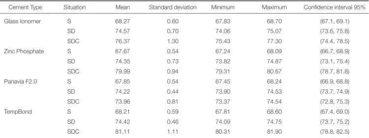
The means and standard deviations of the L* values for the four groups in three situations of S, SD, and SDC were demonstrated (Table 1). Repeated Measures ANOVA was used to evaluate the effects of cement type, situation type, and their interaction on the L* values. The results of this test showed that the cement type (P < .0001), situation type (P < .0001), and their interaction (P < .0001) affected the L* values. Pairwise comparisons of the four groups in the SDC, using Tukey Post Hoc and Bonferroni tests, showed significant differences among all the cement groups (P <
.0001), except between Zinc Phosphate and Tempbond (P
= .145).
Table 1. Measures of central tendency and dispersion for the L* values of specimens in the four groups
Cement Type Situation Mean Standard deviation Minimum Maximum Confidence interval 95%
Glass Ionomer S 68.27 0.60 67.83 68.70 (67.1, 69.1)
SD 74.57 0.70 74.06 75.07 (73.6, 75.8)
SDC 76.37 1.30 75.43 77.30 (74.4, 78.5)
Zinc Phosphate S 67.67 0.54 67.24 68.09 (66.7, 68.9)
SD 74.35 0.73 73.82 74.87 (73.1, 75.4)
SDC 79.99 0.94 79.31 80.67 (78.7, 81.8)
Panavia F2.0 S 67.85 0.54 67.45 68.24 (66.9, 68.8)
SD 74.22 0.44 73.90 74.53 (73.7, 74.9)
SDC 73.96 0.81 73.37 74.54 (72.8, 75.3)
TempBond S 68.21 0.59 67.81 68.60 (67.4, 69.0)
SD 74.42 0.46 74.09 74.75 (73.7, 75.2)
SDC 81.11 1.11 80.31 81.90 (78.8, 82.5)
The means and standard deviations of the a* values for the four groups in three situations of S, SD, and SDC were demonstrated (Table 2). Repeated Measures ANOVA was used to evaluate the effects of cement type, situation type, and their interaction on the a* values. The results of this test showed that the cement type (P < .0001), situation type (P < .0001), and their interaction (P < .0001) affected the a*
values. Pairwise comparisons of the four groups in the SDC, using Tukey Post Hoc and Bonferroni tests, showed no significant differences between Zinc Phosphate and Glass Ionomer (P = 1), and between Panavia F2.0 and TempBond (P = .88). The differences between Glass Ionomer and Panavia F2.0 (P < .0001), between Glass Ionomer and Tempbond (P < .0001), between Zinc Phosphate and Panavia F2.0 (P < .0001), and between Zinc
Phosphate and Tempbond (P = .043) were statistically sig- nificant.
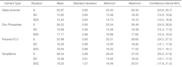
The means and standard deviations of the b* values for the four groups in three situations of S, SD, and SDC were demonstrated (Table 3). Repeated Measures ANOVA was used to evaluate the effects of cement type, situation type, and their interaction on the b* values. The results of this test showed that the cement type (P < .0001), situation type (P < .0001), and their interaction (P < .0001) affected the b*
values. Pairwise comparisons of the four groups in the SDC, using Tukey Post Hoc and Bonferroni tests, showed no significant difference between Zinc Phosphate and Panavia F2.0 (P = 1). The differences among the other cement groups were statistically significant (P < .0001).
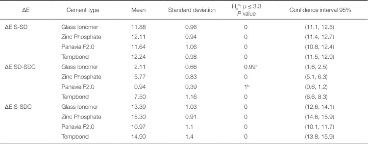
The means and standard deviations of the ΔES-SD, ΔESD-
Table 2. Measures of central tendency and dispersion for the a* values of specimens in the four groups
Cement Type Situation Mean Standard deviation Minimum Maximum Confidence interval 95%
Glass Ionomer S 3.02 0.09 2.95 3.08 (2.9, 3.1)
SD 1.65 0.10 1.57 1.72 (1.5, 1.8)
SDC 1.15 0.16 1.03 1.26 (0.9, 1.4)
Zinc Phosphate S 3.10 0.16 2.98 3.21 (2.8, 3.3)
SD 1.76 0.27 1.56 1.95 (1.5, 2.2)
SDC 1.31 0.26 1.12 1.49 (0.9, 1.7)
Panavia F2.0 S 3.09 0.13 2.99 3.18 (2.9, 3.3)
SD 1.84 0.30 1.62 2.05 (1.5, 2.3)
SDC 2.07 0.28 1.86 2.27 (1.6, 2.5)
TempBond S 3.09 0.13 2.99 3.18 (2.8, 3.3)
SD 1.63 0.14 1.52 1.73 (1.5, 1.9)
SDC 1.81 0.66 1.33 2.28 (0.9, 2.9)
Table 3. Measures of central tendency and dispersion for the b* values of specimens in the four groups
Cement Type Situation Mean Standard deviation Minimum Maximum Confidence interval 95%
Glass Ionomer S 25.87 0.65 25.40 26.33 (24.8, 26.7)
SD 15.92 0.60 15.48 16.35 (14.8, 16.8)
SDC 15.43 0.94 14.75 16.10 (13.5, 16.9)
Zinc Phosphate S 26.02 0.65 25.54 26.49 (24.9, 26.8)
SD 15.99 0.56 15.58 16.39 (15.3, 17.0)
SDC 17.17 0.66 16.69 17.64 (15.9, 18.0)
Panavia F2.0 S 25.98 0.93 25.31 26.65 (24.2, 27.2)
SD 16.29 0.89 15.65 16.92 (15.1, 17.9)
SDC 16.94 0.86 16.32 17.55 (15.7, 18.1)
TempBond S 26.54 0.68 26.04 27.03 (25.2, 27.6)
SD 16.08 0.61 15.63 16.52 (15.1, 17.0)
SDC 19.25 1.07 18.48 20.01 (17.8, 21.3)
SDC, and ΔES-SDC values for the four groups were demon- strated (Table 4) (Fig. 1). One-way ANOVA test showed no significant difference among the four groups in ΔES-SD val- ues(P = .55). Also, this test showed a significant difference among the four groups in ΔESD-SDC values (P < .0001).
Pairwise comparisons of the four groups, using Tukey Post Hoc test, represented significant differences among all the cement groups in ΔESD-SDC values (P < .0001). One-way ANOVA showed a significant difference among the four groups in ΔES-SDC values (P < .0001). Pairwise comparisons of the four groups, using Tukey Post Hoc test, represented significant differences among all the cement groups in ΔES-
SDC values(P < .0001), except between Zinc Phosphate and TempBond (P = .88).
In order to compare the means of the ΔES-SD, ΔESD-SDC, and ΔES-SDC values for the four groups with the predeter- mined perceptional threshold of ΔE = 3.3, One-sample t-test (one-sided) was employed. Considering the null hypothesis of µ ≤ 3.3, in ΔES-SD and ΔES-SDC, it was rejected for the four groups (P < .0001). In ΔESD-SCD the null hypothesis was accepted for Glass Ionomer (ΔE = 2.11) (P
= .99) and Panavia F2.0 (ΔE = .94) (P = 1) and rejected for Zinc Phosphate (ΔE = 5.77) (P < .0001) and TempBond (ΔE = 7.50) (P < .0001).
DISCUSSION
The present study evaluated the L*, a*, b*, and ΔE values for zirconia disk specimens before and after cementation using four different cements of Glass Ionomer, Panavia F2.0, Zinc Phosphate, and TempBond. Statistical analysis indicated significant differences among the four cement groups in the L*, a*, b*, and ΔE values, comparing before (SD) and after cementation (SDC). The results showed that the cement type affected the color attributes of zirconia ceramic. Hence, the null hypothesis of the study was reject- ed. The results of this study can be interpreted based on the color attributes of L*, a*, b*, and ΔE as follows:
According to Table 1, comparison of the L* values for the cement groups before and after cementation (SD and SDC) showed that all the tested cements increased the L*
values except Panavia F2.0. This may be due to the optical characteristics of Glass Ionomer, Zinc Phosphate, and TempBond. It seems that the higher L* values of these three cements compared to Panavia F2.0 are responsible for increasing the L* values in SDC. The three mentioned cements acted like bright backgrounds under zirconia ceramic. However, this effect of Glass Ionomer was less
Table 4. Characteristics of the studied cement groups according to their ΔE values
ΔE Cement type Mean Standard deviation H0*: µ ≤ 3.3
P value Confidence interval 95%
ΔE S-SD Glass Ionomer 11.88 0.96 0 (11.1, 12.5)
Zinc Phosphate 12.11 0.94 0 (11.4, 12.7)
Panavia F2.0 11.64 1.06 0 (10.8, 12.4)
Tempbond 12.24 0.98 0 (11.5, 12.9)
ΔE SD-SDC Glass Ionomer 2.11 0.66 0.99a (1.6, 2.5)
Zinc Phosphate 5.77 0.83 0 (5.1, 6.3)
Panavia F2.0 0.94 0.39 1b (0.6, 1.2)
Tempbond 7.50 1.16 0 (6.6, 8.3)
ΔE S-SDC Glass Ionomer 13.39 1.03 0 (12.6, 14.1)
Zinc Phosphate 15.30 0.91 0 (14.6, 15.9)
Panavia F2.0 10.97 1.1 0 (10.1, 11.7)
Tempbond 14.90 1.4 0 (13.8, 15.9)
* Null hypothesis of ΔE ≤ 3.3
a, b No statistically significant differences.
Fig. 1. The means and standard deviations of the ΔE values in the four groups.
than those of Zinc Phosphate and TempBond.
As indicated in Table 2, comparison of the a* values for the cement groups before and after cementation (SD and SDC) showed that Panavia F2.0 and TempBond increased the a* values, while Glass Ionomer and Zinc Phosphate decreased the a* values. This result may be related to the natural a* values of Panavia F2.0 and TempBond. The a*
value for TempBond at situation SDC showed a higher standard deviation compared to the other color attributes of the same group and also compared to the other cement groups. This may be due to a color difference between the accelerator and base pastes of the Tempbond cement, which affects the red color tendency of this cement. However, this had no significant influence on the standard deviation of the ΔE color change of this cement.
Based on the data in Table 3, comparison of the b* val- ues for the cement groups before and after cementation (SD and SDC) demonstrated that all the tested cements except Glass Ionomer increased the b* value. This result may be caused by the yellow shade tendency of Panavia F2.0, Zinc Phosphate, and Tempbond, which does not exist in Glass Ionomer.
According to Table 4 and Fig. 1, comparison of the ΔESD-SDC values for the cement groups before and after cementation (SD and SDC) demonstrated that the color dif- ferences induced by Glass Ionomer and Panavia F2.0 cements were less than the predetermined perceptional threshold (ΔE < 3.3). Accordingly, Panavia F2.0 and Glass Ionomer induced acceptable color changes. The color dif- ferences induced by Zinc Phosphate and Tempbond were more than the predetermined perceptional threshold (ΔE >
3.3). Consequently, Zinc Phosphate and Tempbond cements caused perceptible color changes. In other words, overall changes of the L*, a*, and b* values created by Zinc Phosphate and TempBond led to the increase of ΔE values beyond the perceptible limit. This color shift may be due to the opaque optical properties of Zinc Phosphate and Tempbond. No significant differences were found among the tested groups in the ΔES-SD values (P = .55), which can be a reason for accurate and precise sample matching of the groups. The ΔES-SCD values represented significant differ- ences among the groups (P < .0001), except between Zinc Phosphate and Tempbond (P = .88) due to their similar optical characteristics. The ΔES-SDC values displayed that the cement and zirconia created impressive color changes on the composite substrate (ΔE > 3.3).
Tracing the changes of L*, a*, and b* values (Table 1, Table 2, Table 3) indicated that the most amount of differ- ences belonged to the L* attribute. Accordingly, the cement types had the highest impact on the L* attribute. Therefore, the ΔE changes have mainly been derived from this attri- bute (Table 4).
Fazi et al.11 reported that among the four tested cements of Fuji Plus resin modified glass-ionomer, Zinc-Phosphate, Rely X Unicem resin cement, and Maxcem resin cement, only Zinc-Phosphate caused perceptible color changes (ΔE
> 3.3). This result on Zinc Phosphate cement was con-
firmed by the present study. Although the resin and glass ionomer cements used by Fazi et al.11 were different com- pared to the current study, both studies showed similar results on the cements, which created acceptable color changes (ΔE < 3.3). In the present study, the zirconia speci- mens were cemented to the substrates. However, in the study of Fazi et al.,11 the zirconia specimens were placed onto the cement disks without an interface. Obviously, the cementation procedure in the current research was more similar to a clinical setting. Additionally, the current study assessed TempBond cement, a temporary cement, which was not evaluated by Fazi et al.11
Chang et al.19 investigated the optical properties of resin- based composite cements and their effects on the color of Empress crowns and Katana zirconia crowns and conclud- ed that the tested composite cements created perceptible color differences in the cervical and body regions with par- ticular combinations of die material, cement, and ceramic crown. In their study, the color of luting cements could make color changes in the cervical thirds of Katana zirconia crowns. Although Chang et al.19 used pre-colored full zirco- nia crowns and luting cements different from the ones used in the current investigation, both studies revealed the possi- bility of cement-induced color changes for zirconia ceram- ics. A monolithic zirconia has a more translucency than a zirconia core in an equal thickness, however a zirconia core has a less thickness than a monolithic zirconia. This may be a reason for the similarity of the results.
Choi and Razzoog20 evaluated the masking ability of zir- conia ceramic with and without porcelain veneer on four different substrates and reported that the zirconia without veneer had a degree of masking ability. On the other hand, the current study surveyed the effect of four cements on the color attributes of a zirconia ceramic, and according to its results, this degree of masking ability of the zirconia without veneer mentioned by Choi and Razzoog20 seems inadequate to prevent a cement-induced color mismatch.
Based on the results of this study, Zinc Phosphate (a permanent cement) and TempBond (a temporary cement) can change the color of zirconia core beyond the predeter- mined perceptional threshold. The color change due to TempBond cement in its short term use may lead clinicians to an incorrect clinical judgment like concerning the other affecting factors like zirconia core, veneer porcelain, and staining, while a proper choice of permanent cement can subsequently correct the color mismatch. Thus, in zirconia- based restorations, these cements should not be applied, or their negative effects should be reduced by sufficient porce- lain veneers if possible. Moreover, Zinc Phosphate cement can be rationally substituted by the other cements like Panavia F2.0 and Glass Ionomer, especially in cases with limited restorative space in esthetic zones.
Considering the semi-translucent optical properties of zirconia ceramic and the thicknesses of zirconia cores (approximately 0.5 - 0.6 mm in a normal case), light trans- mission through the zirconia structure can be expected in zirconia-based restorations. Also, the underlying materials
such as cements may affect the color. Thus, clinicians should realize color behaviors of the cements used for zir- conia-based restorations to decrease the risk of a color mis- match. The color behaviors of four luting agents were eval- uated in this study, and more investigations on various lut- ing agents are recommended in zirconia-based restorations.
On the other hand, the zirconia core overlaying materials such as porcelain veneers may influence the final color of the zirconia-based restorations, which were not evaluated in this study. As several effective factors play a role in the final color of zirconia-based restorations, the impact of each fac- tor should be analyzed separately to determine its contribu- tion to the final result and, ultimately, the cumulative effect of the factors should be determined. Therefore, the authors should aim to evaluate the other factors in future studies.
This study had some limitations such as using a specific brand of zirconia ceramic, a specific shade of composite substrate, one shade of coloring liquid, and four types of cement. More studies on the above mentioned subjects are suggested.
CONCLUSION
Within the limitations of this study, it was concluded that Zinc Phosphate and Tempbond cements affected the color attributes of the tested zirconia ceramic beyond the percep- tional threshold. However, Glass Ionomer and Panavia F2.0 cements affected the color attributes of the tested zirconia ceramic within the perceptional threshold.
ORCID
Farhad Tabatabaian http://orcid.org/0000-0003-2394-6643 Maliheh Habib Khodaei http://orcid.org/0000-0001-5267-1322 REFERENCES
1. Vichi A, Louca C, Corciolani G, Ferrari M. Color related to ceramic and zirconia restorations: a review. Dent Mater 2011;
27:97-108.
2. Raigrodski AJ. Contemporary materials and technologies for all-ceramic fixed partial dentures: a review of the literature. J Prosthet Dent 2004;92:557-62.
3. Luthardt RG, Holzhüter M, Sandkuhl O, Herold V, Schnapp JD, Kuhlisch E, Walter M. Reliability and properties of ground Y-TZP-zirconia ceramics. J Dent Res 2002;81:487-91.
4. Manicone PF, Rossi Iommetti P, Raffaelli L. An overview of zirconia ceramics: basic properties and clinical applications. J Dent 2007;35:819-26.
5. Eschbach S, Wolfart S, Bohlsen F, Kern M. Clinical evaluation of all-ceramic posterior three-unit FDPs made of In-Ceram Zirconia. Int J Prosthodont 2009;22:490-2.
6. Schmitt J, Holst S, Wichmann M, Reich S, Gollner M, Hamel J. Zirconia posterior fixed partial dentures: a prospective clini- cal 3-year follow-up. Int J Prosthodont 2009;22:597-603.
7. Ozkurt Z, Kazazoğlu E. Clinical success of zirconia in dental applications. J Prosthodont 2010;19:64-8.
8. Della Bona A, Kelly JR. The clinical success of all-ceramic restorations. J Am Dent Assoc 2008;139:8S-13S.
9. Christensen GJ. Choosing an all-ceramic restorative material:
porcelain-fused-to-metal or zirconia-based? J Am Dent Assoc 2007;138:662-5.
10. Suputtamongkol K, Tulapornchai C, Mamani J, Kamchatphai W, Thongpun N. Effect of the shades of background sub- structures on the overall color of zirconia-based all-ceramic crowns. J Adv Prosthodont 2013;5:319-25.
11. Fazi G, Vichi A, Ferrari M. Influence of four different ce- ments on the colour of zirconia structures of varying ceramic thickness. Int Dent SA 2006;9:54-61.
12. Wang F, Takahashi H, Iwasaki N. Translucency of dental ce- ramics with different thicknesses. J Prosthet Dent 2013;110:
14-20.
13. Son HJ, Kim WC, Jun SH, Kim YS, Ju SW, Ahn JS. Influence of dentin porcelain thickness on layered all-ceramic restora- tion color. J Dent 2010;38:e71-7.
14. Bachhav VC, Aras MA. The effect of ceramic thickness and number of firings on the color of a zirconium oxide based all ceramic system fabricated using CAD/CAM technology. J Adv Prosthodont 2011;3:57-62.
15. Sinmazisik G, Demirbas B, Tarcin B. Influence of dentin and core porcelain thickness on the color of fully sintered zirco- nia ceramic restorations. J Prosthet Dent 2014;111:142-9.
16. Turgut S, Bagis B. Effect of resin cement and ceramic thick- ness on final color of laminate veneers: an in vitro study. J Prosthet Dent 2013;109:179-86.
17. de Azevedo Cubas GB, Camacho GB, Demarco FF, Pereira- Cenci T. The Effect of Luting Agents and Ceramic Thickness on the Color Variation of Different Ceramics against a Chromatic Background. Eur J Dent 2011;5:245-52.
18. Chaiyabutr Y, Kois JC, Lebeau D, Nunokawa G. Effect of abutment tooth color, cement color, and ceramic thickness on the resulting optical color of a CAD/CAM glass-ceramic lith- ium disilicate-reinforced crown. J Prosthet Dent 2011;105:83- 90.
19. Chang J, Da Silva JD, Sakai M, Kristiansen J, Ishikawa-Nagai S. The optical effect of composite luting cement on all ce- ramic crowns. J Dent 2009;37:937-43.
20. Choi YJ, Razzoog ME. Masking ability of zirconia with and without veneering porcelain. J Prosthodont 2013;22:98-104.
21. Swain VL, Pesun IJ, Hodges JS. The effect of metal ceramic restoration framework design on tooth color. J Prosthet Dent 2008;99:468-76.
22. Seghi RR, Hewlett ER, Kim J. Visual and instrumental colori- metric assessments of small color differences on translucent dental porcelain. J Dent Res 1989;68:1760-4.
23. Heffernan MJ, Aquilino SA, Diaz-Arnold AM, Haselton DR, Stanford CM, Vargas MA. Relative translucency of six all-ce- ramic systems. Part II: core and veneer materials. J Prosthet Dent 2002;88:10-5.
24. Sim CP, Yap AU, Teo J. Color perception among different dental personnel. Oper Dent 2001;26:435-9.
25. Douglas RD, Steinhauer TJ, Wee AG. Intraoral determination of the tolerance of dentists for perceptibility and acceptabili- ty of shade mismatch. J Prosthet Dent 2007;97:200-8.
26. Johnston WM, Kao EC. Assessment of appearance match by visual observation and clinical colorimetry. J Dent Res 1989;
68:819-22.
27. Douglas RD, Brewer JD. Acceptability of shade differences in metal ceramic crowns. J Prosthet Dent 1998;79:254-60.
28. Rosenstiel SF, Land MF, Fujimoto J. Contemporary fixed prosthodontics. 5th ed. St. Louis; Elsevier; 2016. p. 774.
29. Rödiger M, Rinke S, Ehret-Kleinau F, Pohlmeyer F, Lange K, Bürgers R, Gersdorff N. Evaluation of removal forces of im- plant-supported zirconia copings depending on abutment ge- ometry, luting agent and cleaning method during re-cementa- tion. J Adv Prosthodont 2014;6:233-40.
30. Denry I, Kelly JR. State of the art of zirconia for dental ap- plications. Dent Mater 2008;24:299-307.
31. Aboushelib MN, Dozic A, Liem JK. Influence of framework color and layering technique on the final color of zirconia ve- neered restorations. Quintessence Int 2010;41:e84-9.
32. Llena C, Lozano E, Amengual J, Forner L. Reliability of two color selection devices in matching and measuring tooth col- or. J Contemp Dent Pract 2011;12:19-23.