• 검색 결과가 없습니다.

Anterior interosseous nerve (AIN) syndrome is cha­

N/A
N/A
Protected

Academic year: 2021

Share "Anterior interosseous nerve (AIN) syndrome is cha­"

Copied!
4
0
0

로드 중.... (전체 텍스트 보기)

전체 글

(1)

Introduction

Anterior interosseous nerve (AIN) syndrome is cha­

racterized by palsy of the flexor pollcis longus, the flexor digitorum profundus of the index and middle fingers, and the pronator quadratus muscle, and is also

referred to as Kiloh­Nevin syndrome.

1,2

The incidence of AIN syndrome is low and accounts for less than 1% of all compression syndromes in the upper limb.

3

The exact cause and pathophysiology remain unclear, but the suspected cause of spontaneous AIN palsy has been reported as neuralgic amyotrophy (NA), isolated neuritis, and entrapment neuropathy.

2,4,5

Although a few cases of NA presenting as AIN palsy have been reported, predominant AIN paresis was very rare (9/246 cases, 3.6%).

6

Recently, we encountered a patient diagnosed with isolated AIN palsy after video­assisted thoracoscopic surgery (VATS). Here we report a case and review the

비디오흉강경수술 후 전방골간신경증후군 형태로 나타난 신경통성근위축 - 증례 보고 -

김철기

1

, 구정회

2

, 송선홍

2

1대구 명성요양병원, 2울산대학교 의과대학 강릉아산병원 재활의학교실

Neuralgic Amyotrophy Presenting as Anterior Interosseous Nerve Syndrome After Video-Assisted Thoracoscopic Surgery - Case Report -

Cheol Ki Kim

1

, Jung Hoi Koo

2

, Sun Hong Song

2

1

Daegu Myungsung Medical Center, Daegu,

2

Department of Rehabilitation Medicine, Gangneung Asan Hospital, University of Ulsan College of Medicine, Gangneung, Korea

Received February 7, 2017

Revised (1st) April 11, 2017, (2nd) May 11, 2017 Accepted May 12, 2017

Corresponding Author: Jung Hoi Koo

Department of Rehabilitation Medicine, Gangneung Asan Hospital, University of Ulsan College of Medicine, 38 Bangdong-gil, Sacheon- myeon, Gangneung 25440, Korea

Tel: 82-33-610-4951, Fax: 82-33-610-4960, E-mail: [email protected]

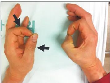
Anterior interosseous nerve (AIN) syndrome is a pure motor syndrome resulting in characteristic weakness of pinch between the thumb and index finger. Different etiologies such as spontaneous, traumatic, compression are suggested, but remain controversial. To our knowledge, a case of neuralgic amyotrophy (NA) presenting as predominant AIN palsy after video-assisted thoracoscopic surgery have not been reported yet. Hence, we report the case of a 34-year-old man who presented with thumb weakness and was diagnosed with AIN palsy due to NA.

Key Words: syndrome, neuralgic amyotrophy, palsy

Copyright © by Korean Association of EMG Electrodiagnostic Medicine

This is an Open Ac cess article distributed under the terms of the Creative Commons Attribution Non-Commercial License (http://creativecommons.org/licenses/by-nc/4.0) which permits unrestricted non-commercial use, distribution, and reproduction in any medium, provided the original work is properly cited.

CASE REPORT

ISSN 1229-6066 https://doi.org/10.18214/jkaem.2017.19.1.1 J Korean Assoc EMG Electrodiagn Med 19(1):1-4, 2017

J Korean Assoc

Electrodiagn Med EMG

(2)

2

J Korean Assoc EMG Electrodiagn Med Vol. 19, No. 1, Jun. 2017

literature.

Case Report

A 34­year­old man visited the outpatient rehabili­

tation clinic due to weakness of left thumb. Nine days ago, he underwent left VATS under general anesthesia with a right lateral decubitus position after being hospitalized in the department of thoracic surgery due to left pneumothorax. There were no specific problems associated with surgery or anesthesia. After surgery, he felt severe pain in the left arm, but he thought that it was a usual symptom related to the surgery. About seven days after the operation, the pain disappeared, but he noticed the weakness of left thumb. So he was referred to rehabilitation department by thoracic surgeon for further evaluation and treatment.

He had not had any other surgery, previous trauma, or medical history related to the problem. On physical examination, surgical site was clean and there were no external wounds in the left upper extremity. Muscle strength of flexor pollicis longus (FPL) was grade 0. The strength of left flexor digitorum profundus of index finger (FDP1) and middle finger (FDP2) was grade 2.

And the muscle power of left pronator was grade 3.

Other muscles showed normal strength. Sensory testing was normal, and the deep tendon reflex was normally induced. Also, the patient was unable to make an “OK”

sign (Fig. 1) with the thumb and index finger.

The electrodiagnostic study was performed on the 22

nd

day after the operation, and there were no definite abnormalities in the sensory nerve conduction study (NCS). A motor NCS was also normal except that the amplitude of compound muscle action potential of the left axillary and musculocutaneous were decreased compared to the right side (Table 1). On needle electro­

myography (EMG), positive sharp waves and fibrilla tions were observed in the left FPL, FDP1, FDP2, and pronator quadratus muscles, and there were no abnormal findings in other muscles (Table 2).

According to EMG findings and characteristic history

of the patient, we concluded that his symptoms were caused by neuralgic amyotrophy, predominantly invol­

ving anterior interosseous nerve.

Two months later, the muscle power was slightly improved, but there was no definite interval change in the follow­up EMG study. Since then the patient did not come to hospital for further follow­up. However, we identified that he was able to make an “OK” sign by 8 months after the onset of symptoms, through a telephone interview.

Discussion

A number of etiologies, such as spontaneous (idio­

pathic), trauma, iatrogenic injuries, have been impli­

cated in AIN syndrome. But the exact cause and patho­

physiology remain unclear, and the incidence is low.

3

In 1948, Parsonage and Turner reported several cases of AIN palsy caused by NA.

4

The etiology of NA remains unknown but appears to be immune mediated.

Possible triggers may include viral illness, immunizations, perioperative and peripartum periods, and strenuous exercise.

7,8

In our case, the patient underwent thoracic surgery. The classic symptoms begin with a sudden onset of severe pain in the shoulder and upper extremity. As the pain subsides, weakness occurs. Weakness may occur

Fig. 1. Typical “OK” sign of the case patient. The patient was unable

to flex left thumb and index fingers at the interphalangeal and the

distal interphalangeal joint, respectively (arrows).

(3)

3

Cheol Ki Kim, et al. AIN Syndrome After VATS or AINS After VATS

in one of several patterns: muscles innervated by one peripheral nerve, by multiple peripheral nerves, or by a combination of peripheral nerves and brachial plexus

trunks. The most commonly affected nerves are axillary, suprascapular, long thoracic, and musculocutaneous.

8

AIN may also rarely be involved. Alfen et al.

6

investigated

Table 1. Results of Nerve Conduction Study

Nerve Recording Stimulation site Lat (ms) Amp (mV) Dist (cm) CV (m/s)

Motor

Rt median (APB)

Wrist 3.05 11.4

Elbow 7.45 11.4 25 56.8

Lt median (APB)

Wrist 3.25 13.3

Elbow 7.90 13.0 25 53.8

Rt ulnar (ADM)

Wrist 2.30 19.0

Elbow 6.15 19.9 24 62.3

Lt ulnar (ADM)

Wrist 2.35 22.9

Elbow 6.35 22.1 25 62.5

Rt radial (EIP) 1.55 15.9

Lt radial (EIP) 2.05 14.1

Rt axiallry (Erb’s point) 2.35 30.2

Lt axiallry (Erb’s point) 3.45 17.4

Rt MC (Erb’s point) 4.05 29.8

Lt MC (Erb’s point) 4.10 17.8

Sensory Lat (ms) Amp (μV)

Rt median (3rd finger) 3.00 56.5

Lt median (3rd finger) 3.10 58.2

Rt ulnar (5th finger) 2.70 43.3

Lt ulnar (5th finger) 3.10 40.8

Rt supf radial (thumb) 2.55 49.5

Lt supf radial (thumb) 2.45 41.4

Lat: latency, Amp: amplitude, Dist: distance, CV: conduction velocity, Rt: right, Lt: left, APB: abductor pollicis brevis, ADM: abductor digiti minimi, EIP:

Extensor indicis pollicis, MC: musculocutaneous, supf: superficial

Table 2. Results of Needle Electromyography

ASA MUAP

Recruitment

IA Fib PSW Amp Dur Poly

Lt C7-T1 paraspinal N None None N N -

Lt BB N None None N N - N

Lt Deltoid N None None N N - N

Lt Infraspinatus N None None N N - N

Lt PT N None None N N - N

Lt FCR N None None N N - N

Lt FPL N 1+ 1+ No motor unit

Lt FCU N None None N N - N

Lt FDP (I&II) N None 1+ N N + Reduced

Lt APB N None None N N - N

Lt FDI N None None N N - N

Lt EI N None None N N - N

Lt PQ N 1+ 2+ N N + Reduced

ASA: abnormal spontaneous activity, MUAP: motor unit action potential, IA: insertional activity, Fib: fibrillation, PSW: positive sharp wave, Lt: left, BB: biceps brachii, PT: pronator teres, FCR: flexor carpi radialis, FPL: flexor pollicis longus, FCU: flexor carpi ulnaris, FDP: flexor digitorum profundus, APB: abductor pollicis brevis, FDI:

first dorsal interosseous, EI: extensor indicis, PQ: pronator quadratus, N: normal, Amp: amplitude, Dur: duration, Poly: polyphasic motor units

(4)

4

J Korean Assoc EMG Electrodiagn Med Vol. 19, No. 1, Jun. 2017

246 cases of NA. Among them, predominant AIN involvement in males was very rare (1.2%). Wong et al.

9

reported a brachial neuritis case that was presenting as AIN compression. In their case, EMG study revealed abnormalities in the FPL, triceps, and biceps, consistent with patchy brachial neuritis. On the other hand, our case showed abnormal EMG findings only in the muscles innervated by AIN.

The literature regarding AIN syndrome can be divided into compressive neuropathy and peripheral nerve manifestation of NA. Important points to distinguish NA from local compression are pain in the arm, elbow, and/or forearm often preceding motor symptoms, and absence of trauma.

9

However, AIN palsy is usually a transient idiopathic nerve dysfunction similar to NA rather than a compressive neuropathy.

10

The differential diagnosis of AIN syndrome includes tendon rupture, proximal sites of nerve compression and pronator syndrome and so on.

7

Among them, FPL tendon rupture can mimic a complete FPL palsy in a patient with AIN syndrome. So it should not be overlooked, and could be evaluated by the tenodesis effect. Electrodiagnostic studies are essential to identify the site and determine the severity of the lesion.

Schollen et al.

1

defined patients with brachial neuritis by EMG findings of AIN denervation and additional diffuse proximal abnormalities along with a history of shoulder and upper arm pain followed by the AIN motor deficit.

Because the natural history of AIN syndrome has not yet been fully determined, controversy exists regarding its management.

7

Because of high probability of spontaneous resolution after even 1 year of symptoms, conservative treatment is recommended. There is

a study that the recovery of paralysis was faster in patients with steroids.

6

Although we did not use it our patient was fully recovered. Surgical treatment should be considered in patients with a known compressing lesion or in those who have failed about 12 months of nonsurgical treatment.

7

In summary, we should keep in mind that AIN syndrome may be occurred due to NA. Then unnecessary study or surgery may be avoided.

References

1. Schollen W, Degreef I, De Smet L: Kiloh­Nevin syndrome:

a compression neuropathy or brachial plexus neuritis? Acta Orthop Belg 2007: 73: 315­318

2. Kiloh LG, Nevin S: Isolated neuritis of the anterior interosseous nerve. Br Med J 1952: 1: 850­851

3. Ulrich D, Piatkowski A, Pallua N: Anterior interosseous nerve syndrome: retrospective analysis of 14 patients. Arch Orthop Trauma Surg 2011: 131: 1561­1565

4. Parsonage MJ, Turner JW: Neuralgic amyotrophy; the shoulder girdle syndrome. Lancet 1948: 1: 973­978

5. Fearn C, Goodfellow J: Anterior interosseous nerve palsy. J Bone Joint Surg Br 1965: 47: 91­93

6. van Alfen N, van Engelen BG: The clinical spectrum of neuralgic amyotrophy in 246 cases. Brain 2006: 129: 438­

450

7. Rodner CM, Tinsley BA, O’malley MP: Pronator syndrome and anterior interosseous nerve syndrome. J Am Acad Orthop Surg 2013: 21: 268­275

8. McCarty EC, Tsairis P, Warren RF: Brachial neuritis. Clin Orthop Relat Res 1999: 368: 37­43

9. Wong L, Dellon AL: Brachial neuritis presenting as anterior interosseous nerve compression­Implications for diagnosis and treatment: A case report. J Hand Surg 1997: 22: 536­539 10. Chi Y, Harness NG: Anterior interosseous nerve syndrome. J

Hand Surg Am 2010: 35: 2078­2080

수치

Fig. 1.	Typical	“OK”	sign	of	the	case	patient.	The	patient	was	unable	 to	flex	left	thumb	and	index	fingers	at	the	interphalangeal	and	the	 distal	interphalangeal	joint,	respectively	(arrows).
Table 1. Results of Nerve Conduction Study

참조

관련 문서

Hwa Jung Kim, Bon-Cheol Ku, Jae Kwan Lee “Use of Pyrolyzed Amino Acids as Versatile Dopants for Synthesis of Heteroatoms-doped

전경부(앞목부위, anterior cervical region): 좌·우

 The PL bundle is hidden behind the AM bundle and becomes visible during flexion as the femoral insertion moves

상완동맥(위팔동맥)과 같이 위팔의 안쪽을 따라 하행하여 주와(팔오금)에 이른 후, 전완의 천지굴근(얕은손가락굽힘근)의 밑을 지나.. 손바닥으로 나오는데, 피지는

1) 신경전도 속도검사 (Nerve conduction velocity test).. 162. Electrodiagnostic

Smed-prep , encoding a TALE class homeodomain, is described as the first gene necessary for correct anterior fate and patterning during

신장의 구조와 생리.. 피질과 수질로 구성.. 1) 신증후군, 신증..

Chest X-ray: Posterio-anterior (PA) View - Left Anterior Oblique