INTRODUCTION
Treatment options using implants can be mainly divided into fixed types and removable types in edentulous patients.1The appropriate type should be determined by considering the radi- ographic findings, the intra-oral examination, the diagnostic cast, and the patient's demand in the diagnostic stage, and by assessing the amount of residual ridge resorption and the biomechanical condition.2The removable type is classified into implant-supported overdentures that achieve support from multiple implants, and implant-tissue-supported overden- tures that achieve support from both tissues and implants by having distal extension areas.3
The attachments used for implant overdentures are mainly divided into the bar type and the solitary type, and into the resilient type and the rigid type, depending on the move-
ment allowance. The attachment selection is affected by the implant number, distribution and alignment, bone quality, arch shape, retention, and denture design.4
A locator can be used in implant overdentures with a bar for retention improvement. Three methods-the gold bar casting method, the laser welding method, and the drill and tapping method-are used to manufacture locators bar system.5If the reten- tion in the plastic male part of the locator is reduced due to fric- tion, the locator can be replaced, as can other attachments. If the retention in the metal female part of the locator is reduced due to wear, it is difficult to restore.6Using the drill and tap- ping technique, however, the female part can be replaced, there- by easily recovering the retention.
In these cases, locator bar overdentures using the drill and tap technique were presented.
Implant overdenture using a locator bar system by drill and tapping technique in a mandible edentulous
patient: a case report
Min-Su Kim, DDS, MSD, Mi-Jung Yoon, DDS, MSD, Jung-Bo Huh, DDS, MSD, PhD, Young-Chan Jeon, DDS, MSD, PhD, Chang-Mo Jeong*, DDS, MSD, PhD
Department of Prosthodontics, School of Dentistry, Pusan National University, Yangsan, Korea
Various options have been introduced for the selection of implant overdenture attachments. Attachment wear due to the repeated insertion and removal of dentures has caused problems such as decreased retention and the requirement for suprastructure remanufacturing. In these cas- es, a Locator bar system was applied using the drill and tapping technique to achieve total retrievability. In a 55-year-old female patient who showed three degrees of mobility in most of her teeth due to severe alveolar bone loss, a complete denture in the maxilla and an implant sup- ported type overdenture in the mandible were planned after extracting all the remaining teeth. Six implants were placed from canine region to the distal molar region, and the locator was connected to the milled bar using the drill and tapping technique. For a 61-year-old female eden- tulous patient who complained of poor retention with old denture, a complete denture in the maxilla and an implant-tissue supported type over- denture in the mandible were planned. Four implants were placed in front of mental foramen, and the Locator was also connected to the Hader bar using the drill and tapping technique. With this technique, female parts can be easily replaced, and retention can be continuously maintained.
[J Adv Prosthodont 2012;4:116-20]
KEY WORDS: Implant overdenture; Locator bar system; Drill & tapping technique; Total retrievability
Corresponding author: Chang-Mo Jeong
Department of Prosthodontics, School of Dentistry, Pusan National University, Beom-eo li, Mul-geum eup, Yangsan, 626-770, Korea
Tel. 82 55 360 5130: e-mail, [email protected]
Received August 24, 2011 / Last Revison November 18, 2011 / Accepted November 21, 2011
ⓒ 2012 The Korean Academy of Prosthodontics
This is an Open Access article distributed under the terms of the Creative Commons Attribution Non-Commercial License (http://creativecommons.org/licenses/by- nc/3.0) which permits unrestricted non-commercial use, distribution, and reproduction in any medium, provided the original work is properly cited.
CASE REPORT
In the first case, a 55-year-old healthy woman visited the authors' hospital for re-treatment of her maxillary fixed par- tial denture and mandibular removable partial denture, which had been used for more than 10 years. All the remaining teeth were floating. After the extraction of all the remaining teeth, a complete denture in the maxilla and an implant over- denture in the mandible were planned. No medical history of implant installation concern was found.
One week after the extraction of all the remaining teeth, an impression was obtained using alginate impression materials (Tokuso A-1, Tokuyama Dental Corp., Tokyo, Japan) for the manufacture of a temporary denture. A temporary denture was prepared and the mandibular temporary denture was duplicated to prepare a stent for the diagnosis and operation of implants, followed by the installation of the implants at 3 months after the extraction (Osstem US II, Osstem Implant Co., Busan, Korea) at sites #33, 36, 37, 43, 46, and 47 (Fig. 1). A rigid type prosthesis which was supported by multiple implants was planned, so milled bar and Locator were assembled.
Two weeks after the implantation, relining was performed using a tissue conditioner (Coe comfort, GC America Inc., Alsip IL, USA). After 6 month healing time for osseointegration, a final impression for a complete denture in the maxilla and a pre- liminary impression in the mandible at the fixture level were obtained using vinyl polysiloxane impression materials
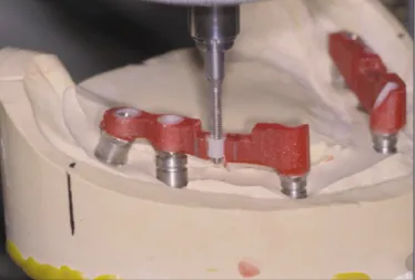
(Imprint II, 3M ESPE, St. Paul, MN, USA) by pick-up impression technique. To accurately transfer the position of the implants to the master cast, the impression coping (Osstem Implant Co., Busan, Korea) was splinted in the master cast using autopolymerized acrylic resin (GC Pattern Resin, GC Corp., Tokyo, Japan). After the impression coping was separated into individual copings, fastening and splinting were performed in the oral cavity, after which a final impression was made using vinyl polysiloxane impression materials (Imprint II, 3M ESPE, St. Paul, MN, USA) at the fixture level. The mas- ter cast was prepared using improved dental stone (GC Fujirock, GC Europe, Interleuvenlaan, Leuven, Belgium), and the recording base was prepared using autopolymerized acrylic resin (Orthodontic resin, Dentsply International, Milford, DE, USA). The lip support was checked using a sil- icone medium (Fit checker, GC Corp., Tokyo, Japan), and the interocclusal record was obtained using recording wax (Aluwax, Aluwax Dental Products Co., Allendale, MI, USA), followed by mounting. After preliminary artificial teeth arrangement was performed, the lip support and facial appear- ance were again assessed in the oral cavity. The interoc- clusal space was also assessed. A silicone index (Express STD, 3E ESPE, St. Paul, MN, USA) was prepared using the arranged teeth, after which it was used as a reference for setting the bar location. In this case study, a large dead space was expected to be formed in the anterior region of the mandible if the splinting was performed with a cross-arch. Based on the maxillary complete denture and the implantation in the mandible with good bone quality, the milled bar was bilater- ally prepared. Drill and tapping technique was presented by schematic diagram (Fig. 2).The plastic sleeve was placed for the Locator (Zest Anchors, Inc., Escondido, CA, USA) (Fig.
3), after which the bar was cast. Subsequently, after the loca- tor was checked with a tapping tool, the Locator female part
Fig. 2. Schematic diagram of 'drill and tapping'.
Wax-up Thread-tapping Insertion of locator
Fig. 3. Location of the plastic sleeve for the drill and tap.
Fig. 1. Post-operative panoramic radiograph.
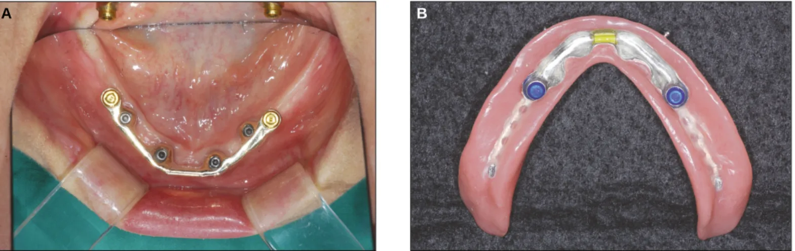
was fastened to the bar with a force of 20 Nm (Fig. 4). The bar fitness was checked in oral cavity, as was the sufficiency of the interocclusal space. The metal framework and the Locator met- al housing were connected using autopolymerized acrylic resin (ADFA, Shofu, Inc., Kyoto, Japan) (Fig. 5). In oral cavity, milled bar and the metal framework were checked, and the lip support and facial appearance with the wax denture were finally checked after the teeth arrangement. After denture curing and lab remounting, selective grinding was performed to form a fully balanced occlusion. The final denture was placed in the oral cavity. The stability, retention, and support of the lip support, esthetic, and denture were shown to be appropriately secured. The blue plastic male part (Zest Anchors, Inc., Escondido, CA, USA) of the Locator was used for reten- tion (Fig. 6. A, B).
In the second case, a 61-year-old healthy woman visited the
authors' hospital for re-treatment of her unsatisfying complete denture. According to the intraoral findings from her first exam- ination, in the mandible, severe residual bone loss was observed. A complete denture in the maxilla and an implant overdenture in the mandible were planned. No medical history of implant installation concern was found.
The mandibular denture was relined by a tissue cocndi- tioner (Coe comfort, GC America Inc., Alsip, IL, USA) and was duplicated, a stent was prepared for the diagnosis and oper- ation of implant, followed by the installation of the 4 implants (Osstem US II, Osstem Implant Co., Busan, Korea) in front of mental foramen area because of alveolar bone deficiency in the molar region (Fig. 7). Overall procedure was simlliar to the first case. But in this case, resilient type prosthesis which allows move- ment in the distal edentulous area was planned, so Hader bar, clip, and locator were assembled. (Fig. 8. A, B).
Fig. 4. Tightening of the female part by 20 Ncm torque. Fig. 5. Connecting metal framework and metal housing by autopolymerized acrylic resin (ADFA, Shofu, Inc., Kyoto, Japan).
Fig. 6. A: Final prosthesis (Milled bar and Locator), B: Final prosthesis (tissue aspect, milled bar).
A B
DISCUSSION
Overdenture treatment using implants can secure the appro- priate stability, support, and retention of the denture, and allows functional recoveries such as lip support, esthetic, and phonation via a denture flange.7Celik and Uludag8report- ed that more stress was observed in the solitary type than in the bar splinting type when the photoelastic stress distribution was assessed in overdentures with three mandibular implants according to the retention mechanism. When a bar attachment is planned after three or more implants are installed for stress distribution, the inter-arch space must be assessed. Pasciuta et al.9 reported that at least 14 mm interocclusal space was required when considering teeth size , denture base thickness, bar thickness for the rigidity, the space from the mucosa to the bar for hygiene, and the soft tissue thickness. If the interocclusal space is insufficient, the bar manufacturing may cause a prosthesis overcontour, denture fracture, or poor oral hygiene.10 When considering attachments for retention with bar splint- ing, ERA attachments for the resilient type, and a multiple clip
with different directions, friction pin, and swivel latchet for the rigid type have been previously used.4Such attachments had the disadvantage of retention loss due to the wear caused by the repeated insertion and removal of the denture.11 In case of wear, plastic male parts can be replaced. Metal female parts, however, require remanufacturing of the prosthesis.
To solve this problem, a Locator can be used. It is very durable and allows 0.2 mm vertical movement. It can be used with a bar as it has the lowest vertical height6 and enables the metal female part to be replaced using the drill and tapping technique, unlike with other methods. The biggest advan- tage of a drill and tapping technique is that it can achieve total retrievability by just replacing new metal female part.
In these cases, one case was a rigid-type prosthesis that was supported by the implants, the other was a resilient-type prosthesis that was supported by the tissue and implants.
Using the drill and tapping technique to connect the bar and the locator, female part wear can be easily managed during reg- ular management.
Fig. 8. A: Final prosthesis (Hader bar and Locator), B: Final prosthesis (Denture, tissue aspect).
A B
Fig. 7. Post-operative panoramic radiograph.
CONCLUSION
Overdenture treatment using implants can provide stability, support, and retention to a denture for fully edentulous patients. Via the Locator bar system using the drill and tapping technique, retention loss due to the wear of the attachments can be resolved and total retievability can be achieved by easily replacing the female parts.
REFERENCES
1. DeBoer J. Edentulous implants: overdenture versus fixed. J Prosthet Dent 1993;69:386-90.
2. Sadowsky SJ. The implant-supported prosthesis for the edentulous arch: design considerations. J Prosthet Dent 1997;78:28-33.
3. Adell R, Lekholm U, Rockler B, Bra�nemark PI. A 15-year study of osseointegrated implants in the treatment of the eden- tulous jaw. Int J Oral Surg 1981;10:387-416.
4. Trakas T, Michalakis K, Kang K, Hirayama H. Attachment sys- tems for implant retained overdentures: a literature review.
Implant Dent 2006;15:24-34.
5. Kurtzman GM. Lab techniques for use of the locator attachment in bar-overdenture applications. TeamWork 2008;1:72-8.
6. Schneider AL, Kurtzman GM. Bar overdentures utilizing the Locator attachment. Gen Dent 2001;49:210-4.
7. Naert I, De Clercq M, Theuniers G, Schepers E. Overdentures supported by osseointegrated fixtures for the edentulous mandible: a 2.5-year report. Int J Oral Maxillofac Implants 1988;3:191-6.
8. Celik G, Uludag B. Photoelastic stress analysis of various retention mechanisms on 3-implant-retained mandibular overdentures. J Prosthet Dent 2007;97:229-35.
9. Pasciuta M, Grossmann Y, Finger IM. A prosthetic solution to restoring the edentulous mandible with limited interarch space using an implant-tissue-supported overdenture: a clinical report.
J Prosthet Dent 2005;93:116-20.
10. Lee CK, Agar JR. Surgical and prosthetic planning for a two-im- plant-retained mandibular overdenture: a clinical report. J Prosthet Dent 2006;95:102-5.
11. Chung KH, Chung CY, Cagna DR, Cronin RJ Jr. Retention char- acteristics of attachment systems for implant overdentures. J Prosthodont 2004;13:221-6.