Received August 28, 2018, Revised May 20, 2019, Accepted for publication July 1, 2019
Corresponding author: Fatma Pelin Cengiz, Department of Dermatove- neorology, Bezmialem Vakif University, 34710, Fatih, Istanbul, Turkey. Tel:
90-2124531700, Fax: 90-45317701, E-mail: fpelinozgen@hotmail.com ORCID: https://orcid.org/0000-0003-0669-6232
This is an Open Access article distributed under the terms of the Creative Commons Attribution Non-Commercial License (http://creativecommons.
org/licenses/by-nc/4.0) which permits unrestricted non-commercial use, distribution, and reproduction in any medium, provided the original work is properly cited.
Copyright © The Korean Dermatological Association and The Korean Society for Investigative Dermatology
Ann Dermatol Vol. 31, No. 5, 2019 https://doi.org/10.5021/ad.2019.31.5.518
ORIGINAL ARTICLE
Dermoscopic Evolution of Pediatric Nevi
Fatma Pelin Cengiz, Yaren Yılmaz1, Nazan Emiroglu, Nahide Onsun
Department of Dermatoveneorology, Bezmialem Vakif University, 1Bezmialem Vakif University Faculty of Medicine, Istanbul, Turkey
Background: The incidence of pediatric melanoma is very rare. Dermoscopic features help to distinguish pediatric mel- anoma and common nevi. Objective: To study the evolution of dermoscopic findings in benign nevi in childhood through serial observation and photography. Methods: We examined 504 melanocytic lesions in 100 patients. From each partic- ipant, dermoscopic images of the nevi from 4-year dermo- scopic follow-up were obtained, including randomly se- lected nevi. Results: The most common dermoscopic pat- terns were homogeneous (193 nevi; 38.3%), globular (92 ne- vi; 18.3%), and reticular (86 nevi; 17.1%). Dermoscopic pat- tern changes were detected in 27% of patients aged 2∼10 years and in 20% of patients aged 11∼16 years. The main pattern changes consisted of the transition from homoge- neous to globular-homogeneous (16%), from homogeneous to reticular-homogeneous (12%) and from globular to glob- ular-homogeneous (10%). Although 257 of the 504 nevi (51.0%) have stable duration without size changes, 169 of the 504 nevi (33.5%) were enlarged, and 78 of the 504 nevi (15.5%) had become smaller. Conclusion: These results con- trast with the prevailing view that dermoscopic patterns in pediatric nevi are usually characterized by globular patterns and that melanocytic nevi generally undergo a characteristic transition from a globular pattern to a reticular pattern. Fifty one percent of patients did not exhibit a size change. While 33% of patients had symmetrical enlargement, 15% of pa-
tients had involution. Therefore, enlargement is a common dermoscopic change in pediatric nevi, and is not a specific sign of pediatric melanoma. (Ann Dermatol 31(5) 518∼524, 2019)
-Keywords-
Child, Dermoscopy, Nevus
INTRODUCTION
‘This nevus has grown’ or ‘My child has a new mole’ are frequent concerns raised by parents. Childhood is a dy- namic period, where many new nevi will appear and grow. Dermoscopy is a pivotal tool for examining children and has been shown to improve the sensitivity for detect- ing melanomas while, at the same time, improving specificity. This can help determine which lesions actually require excision. The primary end points of our study in- cluded changes in nevus size and dermoscopic features in pediatric melanocytic lesions, with a focus on nonpro- blematic lesions that may occur in childhood.
MATERIALS AND METHODS
The dermoscopic images evaluated in this study were col- lected in the database of the Department of Dermatove- neorology of the Bezmialem Vakif University. This study was approved by the Institutional Review Board of the Bezmialem Vakif University (IRB no. 54022451-050.05.04).
The written consent from the patient to report and publish the identifiable patient images was obtained from the patients. In total, images of 504 melanocytic nevi were ob- tained from patients (47 females and 53 males). All pa- tients had skin types II, III, or IV according to Fitzpatrick’s classification1. Dermoscopic evaluations at each visit were performed using a computerized polarized-light video der- moscopy system (FotoFinder dermoscope; Teach Screen
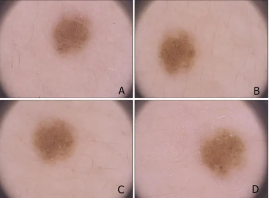
Fig. 1. A 9-year-old boy, nevus on the arm, dermoscopic change of homogeneous pattern to homoge- neous-globular pattern (A: baseline, B: 12 months after, C: 24 months after, D: 48 months after).
Table 1. Dermoscopic patterns of groups younger and older than 10 years old per nevus count Homogeneous Globular Reticular Reticulo-
homogeneous
Globular-
homogeneous Cobblestone Reticuloglobular
≤10 years (n=159) 67 (42.1) 30 (18.9) 34 (21.4) 12 (7.5) 12 (7.5) 3 (1.9) 1 (0.6)
>10 years (n=345) 126 (36.5) 62 (18.0) 52 (15.1) 36 (10.4) 66 (19.1) 3 (0.9) 0 Total (n=504) 193 (38.3) 92 (18.3) 86 (17.1) 48 (9.5) 78 (15.5) 6 (1.2) 1 (0.2) Values are presented as number (%).
Software, Bad Birnbach, Germany) with an alcohol inter- face solution. Dermoscopic images of follow-up visits, which were performed at baseline, 12 months, 24 months, and 48 months were evaluated. Dermoscopic changes in lesions were evaluated by the observation of 2 groups of changes in subsequent visits, changes in size and changes in color, to determine their dermoscopy scores. Patterns were defined according to the 2003 consensus confer- ence2. Congenital types of nevi were excluded from the study, and only nevi acquired after the first two years of life were included in this study. Dermoscopic images of up to five nevi were obtained.
Statistical analysis
Descriptive analysis of the sample was performed, includ- ing percentages for categorical variables and the mean, minimum, and maximum values and standard deviations for continuous variables. All statistical analyses were per- formed using the SPSS Statistical software program ver.
15.0 (SPSS Inc., Chicago, IL, USA). The results were con-
sidered to be statistically significant when the p-value was less than 0.05.
RESULTS
Five hundred and four nevi from one hundred patients were enrolled in the study: 53 male (53%) and 47 female (47%). The mean age of the patients was 12.01±3.38 (minimum: 3, maximum: 16). Only nevi located on the trunk were included in the study. Nine patients had Fitzpatrick skin type II, 52 had Fitzpatrick skin type III, and 39 had Fitzpatrick skin type IV. The most common dermo- scopic patterns were homogeneous (193 nevi; 38.3%), globular (92 nevi; 18.3%), reticular (86 nevi; 17.1%), globular-homogeneous (78 nevi; 15.5%), reticulo-homo- geneous (48 nevi; 9.5%), reticuloglobular (1 nevus; 0.2%), and cobblestone (6 nevi; 1.2%) (Table 1). There were 11 halo nevi at baseline; at the end of the 4 years, all of these nevi had faded.
The mean diameter of the nevi with complex patterns was
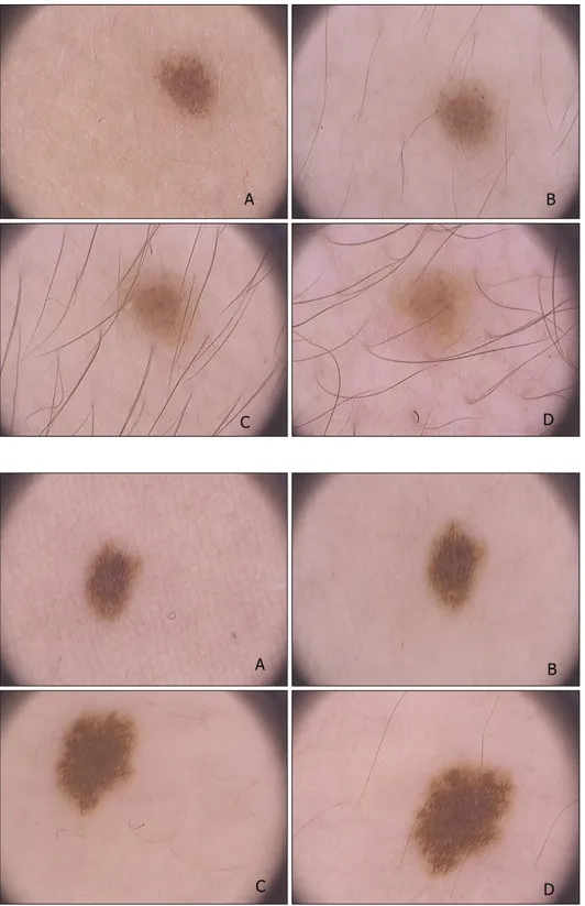
Fig. 3. A 10-year-old girl, nevus on the back, dermoscopic change of homogeneous pattern to reticular pattern, enlargement of nevus (A:
baseline, B: 12 months after, C: 24 months after, D: 48 months after).
Fig. 2. A 14-year-old boy, nevus on the leg, dermoscopic change of globular pattern to homogeneous pattern (A: baseline, B: 12 months after, C: 24 months after, D: 48 months after).
9.06±1.42 mm, that of nevi with only one pattern (reticular, globular, and cobblestone) was 7.22±0.74 mm, and that of nevi with homogenous patterns was 6.83±
0.92 mm. There were statistically significant differences between the the groups related to the diameter of the nevi (p=0.041).
The transformation from a homogeneous to a glob-
ular-homogeneous pattern was the most frequent pattern change (19 of 114; 16.7%) (Fig. 1). The transformation from a homogeneous pattern to a reticular-homogeneous pattern was the second most common pattern change (14 of 114; 12.2%), and globular to globular-homogeneous was the third most common pattern change (12 of 114;
10.5%) (Fig. 2).
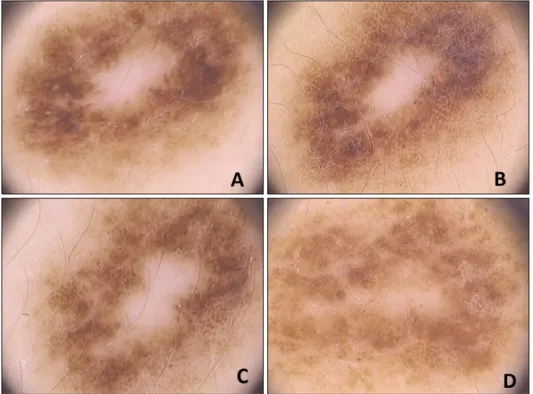
Fig. 4. A 12-year-old girl, nevus on the back, dermoscopic change of homogeneous-globular pattern to homogeneous-reticular pattern (A:
baseline, B: 12 months after, C: 24 months after, D: 48 months after).
Table 2. Differences of groups younger and older than 10 years old per nevus count Stabile Size
enlargement
Decreasing in size
Dermoscopic pattern transformation
Common transformation type of dermoscopic pattern
≤10 years (n=159) 77 (48.4) 65 (40.9) 17 (10.7) 44 (27.7) Globular-globulohomogeneous
>10 years (n=345) 180 (52.2) 104 (30.1) 61 (17.7) 70 (20.3) Homogeneous-globulohomogeneous
Total 257 169 78 114
Values are presented as number (%) and number only.
Although 257 of the 504 nevi (51.0%) have shown a sta- ble duration without size changes, 169 of the 504 nevi (33.5%) had enlarged (Fig. 3, 4), and 78 of the 504 nevi (15.5%) had become smaller during our 4-year experience of dermoscopic monitoring. Of the 169 nevi, 81 had ho- mogeneous pattern (47.9%), 37 had globular pattern (21.9%), 21 had reticular pattern (12.4%), 14 had glob- ular-homogeneous (8.3%), 13 had reticular-homogeneous pattern (7.7%), 3 had cobblestone pattern (1.8%). There was statistical difference between the groups related to en- largement (p=0.041). All of the halo nevi had disappeared at this time. One hundred and fourteen nevi had pattern transformation (22.6%).
Thirty-three of 61 patients (54.1%) who had pattern changes were male, and 28 of 61 patients (45.9%) were female. No significant difference between genders related to pattern changes was observed (p=0.839). There were 30 children younger than 10 years old (159 nevi) and 70 children older than 10 years old (345 nevi). There was no statistical association between age and pattern changes
(p=0.372) (Table 2).
Color changes were observed in 70 of 504 nevi (13.9%).
Fifty-one nevi were fading in color (Fig. 5). Nineteen of the nevi darkened in color. There was no significant differ- ence between genders and age-related groups (p=0.243, p=0.751). Peripheral dots and clods were found in 49 of 100 patients at baseline (198 nevi). At the end of 4 years, 128 of 198 nevi (64.6%), lost these structures.
DISCUSSION
Commonly acquired nevi are defined as small, flat, brown lesions that can appear anywhere on the surface of the body after birth. Childhood is a dynamic period with re- gard to the development of nevi. Many new nevi will ap- pear, enlarge, and finally begin to disappear. To make an accurate diagnosis of melanoma in childhood and ado- lescence without performing unnecessary biopsies and ex- cisions, dermoscopic patterns, size changes, and color changes must be well understood.
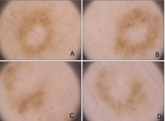
Fig. 5. A 8-year-old boy, nevus on the back, dermoscopy of homo- geneous-reticular pattern, fading in color (A: baseline, B: 12 months after, C: 24 months after, D: 48 months after).
Table 3. Comparison of studies about dermoscopy of pediatric nevi
Zalaudek et al.6 Scope et al.7 Our study
Nevus count 1,268 1,181 504
Mean age at enrollment 13.3 10.7 12.0
Anatomic sites Lesions located on the soles, palms, face, head and mucosal or
subungual sites were excluded
Back Back
Skin types No information Fair 278 (62.7%),
light olive 33 (7.4%), dark (dark olive, brown, and black)
131 (29.6%)
Fitzpatrick II 9 (9.0%), Fitzpatrick III 52 (52.0%), Fitzpatrick IV 39 (39.0%)
Prevalent dermoscopic pattern Globular Homogeneous Homogeneous
Prevalent transformation type - - Homogeneous to
globular-homogeneous
Peripheral dots and clods are usually found in common nevi of children, and during follow-up examinations, these structures disappear while the sizes of the lesions enlarge.
This finding is consistent with observations described in the literature3,4.
All halo nevi have a central globular pattern at first, and during follow-up, they begin to fade.
Finally, all of the nevi disappeared with only a gray-brown homogeneous pigmentation. It has been proposed that the central part of the nevus has a progressive involution by T lymphocytes5. Due to the disappearance of all halo nevi by four years, we suggest that symmetrical halo nevi should not be excised, and the preferential management for these nevi with this pattern is simply monitoring.
It has been thought that the globular pattern is the most common dermoscopic pattern in childhood, and nevi in children evolve from globular to reticular through the years6. Zalaudek et al.6 found that the globular pattern is the most prevalent pattern (60%), followed by complex (21%), reticular (12%), and homogeneous (5%) patterns.
There are significant differences in the distribution of nevi by dermoscopic classification between studies present in the literature: Zalaudek et al.6 classified 8 of 151 nevi (5%) as homogeneous, Scope et al.7 classified 468 of 936 nevi (50%) as homogenous, and we classified 193 of 504 nevi (38%) as homogenous. The mean age of the children was 13.3 years in the study of Zalaudek et al.6, 10.7 years in the study of Scope et al.7, and 12.01 years in our study.
Zalaudek et al.6 studied patients presenting nevi >5 mm.
For comparison, if we analyze larger nevi in our study, the frequency of homogeneous patterns decreases, similar to the findings of Scope et al.7. We also enrolled the smallest nevi as well as the largest nevi. In addition, Zalaudek et al.6 studied older children, with a mean age of 13.3 years (10.7 in Scope et al.7 vs. 12 in our study). The SONIC study demonstrated that the frequency of the homoge- neous pattern decreased from 38% at baseline (mean age 12 years) to 28% after 1 year (mean age 13 years) (Table 3)8. Most of our patients had Fitzpatrick skin type III and IV. The phenotypic features of our population were similar to those examined by Scope et al.7. The proportion of re- ticular pattern was lower among children under the age of 10 years, and our results support this observation.
In this study, we aimed to identify dermoscopic trans- formations in our country. We found the homogeneous pattern to be the prevalent dermoscopic pattern (38%), and the transformation from a homogeneous pattern to a globular-homogeneous pattern (16%) was the prevalent transformation type. Although the reticular nevus pattern on dermoscopy correlates with junctional nevi histopatho- logically, the globular nevus pattern correlates with com- pound or dermal nevi9,10.
However, it is more difficult to histopathologically predict the location of homogeneous pattern nevus. Worret and Burgdorf11 reported that in their series, none of the chil- dren under the age of 10 years showed completely junc- tional nevi, and their nevi were often characterized by compound and dermal nevi.
From a histological perspective, nevi with a globular pat- tern are dermal proliferations of melanocytes, while nevi with a reticular pattern are epidermal proliferations of mel- anocytes12.
Although dermoscopy correlates with the histopatho- logical pattern of melanocytic neoplasms, there are limi- tations to the dermoscopic- pathological correlation. Using dermoscopic-reflectance confocal microscopy (RCM)-his- topathological correlation, Pellacani et al.13 showed that nevi with a globular dermoscopic pattern correlated either with a predominantly junctional or a predominantly der- mal nested proliferation of melanocytes. Furthermore, ne- vi with a reticular dermoscopic pattern correlated with ei- ther predominantly lentiginous or predominantly nested junctional proliferations of melanocytes. Using RCM for longitudinal tracking of changes in nevi, the authors showed that while some nevi changed dermoscopically from the globular pattern to reticular pattern, they did not demon- strate a change in their overall RCM pattern. These nevi showed a meshwork pattern on RCM throughout their evolution, consistent with a nested junctional proliferation
of melanocytes13.
Thus, we posited that, while such growing nevi can show a dermoscopic change from ahomogeneous to globular pattern, melanocytic proliferation occurs within the same anatomical compartment.
Fifty two percent of nevi did not demonstrate a change in size. While 33% of the nevi had symmetrical enlarge- ment, 15% of nevi had involution. Cordoro et al.14 inves- tigated whether conventional asymmetry, border, color, diameter, evolution (ABCDE) melanoma detection criteria adequately define pediatric melanoma and analyzed clin- icopathological and outcome differences between young- er and older children. They suggested that conventional ABCDE criteria and additional amelanotic; bleeding, bump; color uniformity; de novo, any diameter detection criteria may be helpful for earlier recognition and treat- ment of melanoma in children when these criteria were used together14. Therefore, size changes, either enlarge- ment or involution are common in pediatric population, and we recommend that parents not be concerned all en- largements of nevi. Even though, 15% of nevi had in- volution, 33% of nevi had enlargement in our study. As well as enlargement may be related with the growth of children, homogeneous pattern is more common in pedia- tric population in our study, and the most frequent pattern in the enlargement group was homogeneous pattern, therefore these nevi are more prone to enlarge in terms of diameter. These results showed that dermoscopic changes associated with suspicion for development of melanoma are common in children. Therefore, excision is not a good option when the clinician observes these findings in a pe- diatric population.
Our limitation was the lack of dermoscopic and patho- logical correlation. Since we did not excise the lesions, our study was based on a dermoscopic follow-up. There- fore, we observed the predominance of the homogeneous pattern, but we could not determine the actual histology of these nevi. Thus, further studies investigating dermo- scopic patterns of children from different races are needed to better understand the anatomic correlates and clinical significance of each pattern.
CONFLICTS OF INTEREST
The authors have nothing to disclose.
ORCID
Fatma Pelin Cengiz, https://orcid.org/0000-0003-0669-6232 Yaren Yılmaz, https://orcid.org/0000-0003-1585-4087 Nazan Emiroglu, https://orcid.org/0000-0002-5575-6484
Nahide Onsun, https://orcid.org/0000-0001-6259-0219
REFERENCES
1. Fitzpatrick TB. The validity and practicality of sun-reactive skin types I through VI. Arch Dermatol 1988;124:869-871.
2. Argenziano G, Soyer HP, Chimenti S, Talamini R, Corona R, Sera F, et al. Dermoscopy of pigmented skin lesions:
results of a consensus meeting via the Internet. J Am Acad Dermatol 2003;48:679-693.
3. Kittler H, Pehamberger H, Wolff K, Binder M. Follow-up of melanocytic skin lesions with digital epiluminescence microscopy: patterns of modifications observed in early melanoma, atypical nevi, and common nevi. J Am Acad Dermatol 2000;43:467-476.
4. Kittler H, Seltenheim M, Dawid M, Pehamberger H, Wolff K, Binder M. Frequency and characteristics of enlarging common melanocytic nevi. Arch Dermatol 2000;136:316- 320.
5. Akasu R, From L, Kahn HJ. Characterization of the mononuclear infiltrate involved in regression of halo nevi. J Cutan Pathol 1994;21:302-311.
6. Zalaudek I, Grinschgl S, Argenziano G, Marghoob AA, Blum A, Richtig E, et al. Age-related prevalence of dermo- scopy patterns in acquired melanocytic naevi. Br J Dermatol 2006;154:299-304.
7. Scope A, Marghoob AA, Dusza SW, Satagopan JM, Agero AL, Benvenuto-Andrade C, et al. Dermoscopic patterns of
naevi in fifth grade children of the Framingham school system. Br J Dermatol 2008;158:1041-1049.
8. Oliveria SA, Geller AC, Dusza SW, Marghoob AA, Sachs D, Weinstock MA, et al. The Framingham school nevus study:
a pilot study. Arch Dermatol 2004;140:545-551.
9. Bauer J, Blum A. Dermoscopy features of common melano- cytic nevi of the junctional, compound and dermal type. In:
Marghoob AA, Braun RP, Kopf AW, editors. Atlas of dermoscopy. London: Taylor&Francis, 2006:181-187.
10. Argenziano G, Zalaudek I, Ferrara G, Hofmann-Wellenhof R, Soyer HP. Proposal of a new classification system for melanocytic naevi. Br J Dermatol 2007;157:217-227.
11. Worret WI, Burgdorf WH. Which direction do nevus cells move? Abtropfung reexamined. Am J Dermatopathol 1998;
20:135-139.
12. Zalaudek I, Hofmann-Wellenhof R, Kittler H, Argenziano G, Ferrara G, Petrillo L, et al. A dual concept of nevogenesis:
theoretical considerations based on dermoscopic features of melanocytic nevi. J Dtsch Dermatol Ges 2007;5:985-992.
13. Pellacani G, Scope A, Ferrari B, Pupelli G, Bassoli S, Longo C, et al. New insights into nevogenesis: in vivo charac- terization and follow-up of melanocytic nevi by reflectance confocal microscopy. J Am Acad Dermatol 2009;61:1001- 1013.
14. Cordoro KM, Gupta D, Frieden IJ, McCalmont T, Kashani- Sabet M. Pediatric melanoma: results of a large cohort study and proposal for modified ABCD detection criteria for children. J Am Acad Dermatol 2013;68:913-925.