CASE REPORT
Copyright © 2011, the Korean Surgical Society J Korean Surg Soc 2011;80:S47-50
DOI: 10.4174/jkss.2011.80.Suppl 1.S47
JKSS
Journal of the Korean Surgical Society pISSN 2233-7903ㆍeISSN 2093-0488
Received May 28, 2010, Accepted July 6, 2010 Correspondence to: Jae-Won Joh
Department of Surgery, Samsung Medical Center, Sungkyunkwan University School of Medicine, 50 Irwon-dong, Gangnam-gu, Seoul 135-710, Korea
Tel: +82-2-3410-3466, Fax: +82-2-3410-0040, E-mail: [email protected]
cc Journal of the Korean Surgical Society is an Open Access Journal. All articles are distributed under the terms of the Creative Commons Attribution Non-Commercial License (http://creativecommons.org/licenses/by-nc/3.0/) which permits unrestricted non-commercial use, distribution, and reproduction in any medium, provided the original work is properly cited.
Pneumatosis intestinalis after adult liver transplantation
Jong Man Kim, Yulri Park
1, Jae-Won Joh, Choon Hyuck David Kwon, Sung Joo Kim, Seung Heui Hong
2, Suk-Koo Lee
Departments of Surgery and 1Radiology, 2Organ Transplant Center, Samsung Medical Center, Sungkyunkwan University School of Medicine, Seoul, Korea
Pneumatosis intestinalis is an uncommon disorder characterized by an accumulation of gas in the bowel wall. We described three cases undertaking liver transplantation. The patients developed diarrhea in three cases and high fever in two. An ab- dominal X-ray and computed tomography scan demonstrated extensive pneumatosis intestinalis in the colon with pneumo- peritoneum mimicking hollow organ perforation. However, the patients had no abdominal symptoms and there was no evi- dence of peritonitis. The infection work-up was negative except one case with cytomegalovirus antigenemia. After one week of conservative management including bowel rest and antibiotic therapy, their pneumoperitoneum resolved spontaneously without any complication. Pneumatosis intestinalis should be considered as a differential diagnosis after adult liver trans- plantation with patients suffering from watery diarrhea and fever. Pneumoperitoneum, air-density in mesentery and retro- peritoneum in patients with pneumatosis intestinalis without signs of peritonitis improved with conservative management, which included bowel rest and antibiotic therapy.
Key Words: Liver transplantation, Pneumatosis intestinalis, Watery diarrhea
INTRODUCTION
Pneumatosis intestinalis (PI) is a rare and unexpected disorder defined as accumulation of gas in the submucosa or subserosa of the large or small intestine. It is associated with a variety of diseases, and its pathogenesis is still obscure. PI is an infrequent complication of liver trans- plantation (LT). Until now, it has been reported in 12 cases after pediatric orthotopic LT [1-5], although nearly none has been reported in adult liver recipients.
We report three cases of adult liver transplant recipients
in whom the diagnosis of PI was made by computer to- mography (CT) and simple X-ray (Table 1).
CASE REPORT
The patients in case 1 and 2 were unremarkable during post-operative period, and they were discharged home.
The patient in case 1 experienced high fever and watery di- arrhea after two months and the patient in case 2 com- plained of watery diarrhea after four months. The patient
Jong Man Kim, et al.
S48 thesurgery.or.kr
Table 1. Pneumatosis intestinalis: epidemiology, immunosuppression, and outcome
Case Sex/Age Diagnosis CTP MELD OP Immunosuppression Drug level
(FK506/MMF)
Resolution (wk)
1 F/55 LC-NBNC C 20 LDLT FK506 (2 mg/day) 4.6 ng/mL 2
MPD (8 mg/day)
MMF (1,500 mg/day) 0.51 mg/L
2 M/62 HBV B 9 LDLT FK506 (2 mg/day) 4.0 ng/mL 1
HCC MPD (4 mg/day)
MMF (1,500 mg/day) 1.2 mg/L
3 M/63 HBV C 45 DDLT FK506 (1 mg/day) 0.5 ng/mL 1
HCC MMF (500 mg/day) 0.52 mg/L
CTP, child-turchotte-pugh score; MELD, model for end-stage liver disease; OP, operation; LC-NBNC, liver cirrhosis-non B, non C; HBV, hepatitis B virus; HCC, hepatocellular carcinoma; LDLT, living donor liver transplantation; DDLT, deceased donor liver transplantation;
MPD, methylprednisolone; MMF, mycophenolate mofetil.
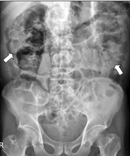
Fig. 1. Plain film of abdomen shows diffuse linear intramural air (arrows) along cecum to proximal descending colon.
in case 3 experienced high fever and watery diarrhea one month after the transplantation without discharge. At ad- mission, all patients were stable in blood pressure, pulse rate, and respiratory rate, but in case 1 and 3, patients had high body temperature (38.8oC and 38.5oC).
The physical examination showed severely distended, non-tender abdomen, with hypoactive bowel sound and tympanic percussion in case 1. Other patients showed no specific findings. The laboratory findings of all cases did not reveal abnormal findings except mildly elevated C-re- active protein, ranging 0.38 to 1.59 mg/dL (normal range,
<0.3 mg/dL). Serial blood tests of case 1 were positive twice for cytomegalovirus (CMV) antigenemia, but other cases showed all negative results. The peak titer of CMV antigenemia was 10/400,000 and colonoscopic finding did not show abnormal finding in case 1. The cytotoxicity as- say of the all patient’s stool was negative for Clostridium difficile toxin. Stool cultures and special stains for micro- organisms were all negative in all patients. The work-up failed to demonstrate an infectious etiology for the gastro- intestinal symptom.
Abdomen simple X-rays and CT in abdomen revealed diffuse pneumatosis intestinalis of the colon, ileus, and free air under diaphragm, but no air in the portal vein in case 1 and 2 (Fig. 1). An abdominal CT of case 3 demon- strated pneumopericardium, diffuse linear pneumatosis intestinalis of the colon and extraluminal air within retro- peritoneum and mesentery but no free intraperitoneal air (Figs. 2, 3).
All patients were conservatively managed with bowel rest, parenteral nutritional support, intravenous anti- biotics (cefotaxime, ampicillin/sulbactam, and metronida- zole) because of no signs of peritonitis. The patients in case 1 was treated with additional antiviral agent (ganciclovir) for positive CMV antigenemia and nasogastric tube de- compression for free air, but other patients did not receive antiviral agent and did not have nasogastric tube decompression. The patients in case 1 and 3 did not receive immunosuppressive agent for 1 week, but the patient in case 2 received reduced immunosuppressive treatment with FK506 (2 mg daily), methylprednisolone (2 mg daily),
Pneumatosis intestinalis after LT
thesurgery.or.kr S49
Fig. 3. Computed tomography scan of abdomen shows air density in the retroperitoneum (arrow) and mesentery (arrowhead).
Fig. 2. Computed tomography scan of abdomen shows pneumo- pericardium (arrow) and pneumoperitoneum (arrowhead).
and mycophenolate mofetil (500 mg daily). He remained in excellent general condition from the admission and had a sense of well-being during hospitalization.
The abdominal distension of case 1 resolved and diar- rhea improved within a week and antibiotics were discontinued. All patients were given a diet, and were dis- charged home after the PI resolved clinically and radiographically.
DISCUSSION
The common factor linking PI to organ transplant recip- ients is steroid treatment [6]. In this report, one patient did not receive steroid treatment and the other two had steroid treatment but the amount was only 4 mg/day. Drug con- centrations of FK506 and mycophenolate mofetil (MMF) in all patients have low levels as well. It might be possible that immunosuppression including FK506, steroid, and MMF did not cause of PI in our cases.
Symptoms that have been attributed to PI are in de- creasing order of frequency: diarrhea, bloody stool, ab- dominal pain, abdominal distension, constipation, weight loss, and tenesmus [7]. In this report, three recipients had watery diarrhea, and two had high fever.
The physical examination of recipients did not show
specific findings except abdominal distension in one case.
The patients did not have abdominal pain, tenderness, re- bound tenderness, and rigidity of abdominal wall. There was no clear evidence of peritonitis despite of pneumo- peritoneum and air density in mesentery and retro- peritoneum in simple X-rays and CT scan of abdomen. We assumed that there was no direct communication between lumen and peritoneum, therefore conservative treatment should be performed rather than surgical exploration. PI improved spontaneously after nearly one week of bowel rest and antibiotic therapy.
However, patients with white blood cells of more than 12,000/mm3 with or without the symptoms of clinical ob- struction such as emesis, vomiting, and pain, age over 60 years were considered to be candidates for surgical inter- vention [8].
Based on this report, PI should be considered as the dif- ferential diagnosis of adult after LT who suffers from wa- tery diarrhea and fever. Pneumoperitoneum, air-density in mesentery and retroperitoneum in patients with pneu- matosis intestinalis without signs of peritonitis improved with conservative management.
Jong Man Kim, et al.
S50 thesurgery.or.kr
CONFLICTS OF INTEREST
No potential conflict of interest relevant to this article was reported.
REFERENCES
1. Burress GC, Ben-Ami T, Whitington PF. Pneumatosis in- testinalis in infants after orthotopic liver transplantation. J Pediatr Gastroenterol Nutr 1996;23:577-82.
2. Janssen DA, Kalayoglu M, Sollinger HW. Pneumatosis cys- toides intestinalis following lactulose and steroid treatment in a liver transplant patient with an intermittently enlarged scrotum. Transplant Proc 1987;19:2949-52.
3. King S, Shuckett B. Sonographic diagnosis of portal venous
gas in two pediatric liver transplant patients with benign pneumatosis intestinalis: case reports and literature review.
Pediatr Radiol 1992;22:577-8.
4. Sachse RE, Burke GW 3rd, Jonas M, Milgrom M, Miller J.
Benign pneumatosis intestinalis with subcutaneous emphy- sema in a liver transplant recipient. Am J Gastroenterol 1990;85:876-9.
5. Koep LJ, Peters TG, Starzl TE. Major colonic complications of hepatic transplantation. Dis Colon Rectum 1979;22:218- 20.
6. Andorsky RI. Pneumatosis cystoides intestinalis after organ transplantation. Am J Gastroenterol 1990;85:189-94.
7. Jamart J. Pneumatosis cystoides intestinalis: a statistical study of 919 cases. Acta Hepatogastroenterol (Stuttg) 1979;26:419-22.
8. Greenstein AJ, Nguyen SQ, Berlin A, Corona J, Lee J, Wong E, et al. Pneumatosis intestinalis in adults: management, surgical indications, and risk factors for mortality. J Gastrointest Surg 2007;11:1268-74.