Received March 21, 2016, Revised July 13, 2016, Accepted for publication August 10, 2016
Corresponding author: Jeong Hee Kim, Department of Beauty Design, Wonkwang University, 460 Iksan-daero, Iksan 54538, Korea. Tel:
82-63-850-6898, Fax: 82-63-850-7301, E-mail: jh@wku.ac.kr
This is an Open Access article distributed under the terms of the Creative Commons Attribution Non-Commercial License (http://creativecommons.
org/licenses/by-nc/4.0) which permits unrestricted non-commercial use, distribution, and reproduction in any medium, provided the original work is properly cited.
Copyright © The Korean Dermatological Association and The Korean Society for Investigative Dermatology
Ann Dermatol Vol. 28, No. 5, 2016 http://dx.doi.org/10.5021/ad.2016.28.5.615
ORIGINAL ARTICLE
Values of a Patient and Observer Scar Assessment Scale to Evaluate the Facial Skin Graft Scar
Jin Kyung Chae, Jeong Hee Kim1, Eun Jung Kim, Kun Park
Department of Dermatology, Wonkwang University Hospital, Wonkwang University School of Medicine, 1Department of Beauty Design, Wonkwang University, Iksan, Korea
Background: The patient and observer scar assessment scale (POSAS) recently emerged as a promising method, reflecting both observer’s and patient’s opinions in evaluating scar.
This tool was shown to be consistent and reliable in burn scar assessment, but it has not been tested in the setting of skin graft scar in skin cancer patients. Objective: To evaluate fa- cial skin graft scar applied to POSAS and to compare with ob- jective scar assessment tools. Methods: Twenty three pa- tients, who diagnosed with facial cutaneous malignancy and transplanted skin after Mohs micrographic surgery, were recruited. Observer assessment was performed by three in- dependent rates using the observer component of the POSAS and Vancouver scar scale (VSS). Patient self-assessment was performed using the patient component of the POSAS. To quantify scar color and scar thickness more objectively, spectrophotometer and ultrasonography was applied.
Results: Inter-observer reliability was substantial with both VSS and the observer component of the POSAS (average measure intraclass coefficient correlation, 0.76 and 0.80, re- spectively). The observer component consistently showed significant correlations with patients’ ratings for the parame- ters of the POSAS (all p-values<0.05). The correlation be- tween subjective assessment using POSAS and objective as- sessment using spectrophotometer and ultrasonography
showed low relationship. Conclusion: In facial skin graft scar assessment in skin cancer patients, the POSAS showed ac- ceptable inter-observer reliability. This tool was more com- prehensive and had higher correlation with patient’s opinion. (Ann Dermatol 28(5) 615∼623, 2016)
-Keywords-
Cicatrix, Patient and observer scar assessment scale, Skin graft
INTRODUCTION
Mohs micrographic surgery is preferred treatment option for facial malignant tumor due to assurance tumor re- moval and minimal loss of surrounding normal tissue1. Skin grafting is simple and better reconstruction procedure according to anatomic site, but it can lead to poor aes- thetic results due to mismatch of thickness, texture and scar contraction2. The development of a skin graft scar is inevitable. In particularly, facial skin graft scar might be associated with adverse physical and psychological dis- turbances in patients undergoing treatment for cutaneous malignancy.
The ideal scar assessment tool should contain the follow- ing parameters: noninvasiveness, painlessness, easiness of work and reliability. The objective measurement parame- ters to evaluate the scar include color, thickness, surface texture, suppleness, and surface area3. The objective measurement apparatus like computerized image capture systems and digital color analysis methods require com- plex equipment and experienced operators, which may limit its use in a busy clinical setting. Hence, although ob- jective measurements for scar evaluation are essential, there is a need for subjective assessment of scars.
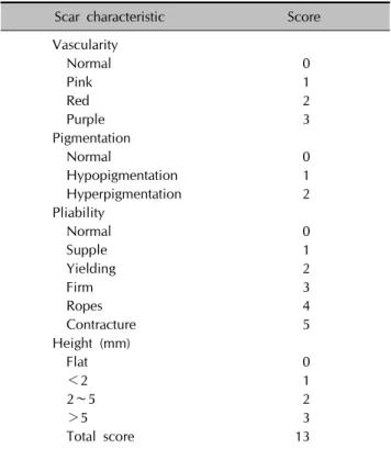
Table 1. The Vancouver scar scale
Scar characteristic Score
Vascularity
Normal 0
Pink 1
Red 2
Purple 3
Pigmentation
Normal 0
Hypopigmentation 1
Hyperpigmentation 2
Pliability
Normal 0
Supple 1
Yielding 2
Firm 3
Ropes 4
Contracture 5
Height (mm)
Flat 0
<2 1
2∼5 2
>5 3
Total score 13
The patient and observer scar assessment scale (POSAS) was designed to evaluate various types of scar sub- jectively4,5. This tool provides both the observers’ and the patient’s insights and it is easy to use, proving to be more advantageous than other tools. It was used to evaluate burn scars4 and linear surgical scars5, which showed reli- able and valid results for scar evaluation.
Therefore, the aim of this study was to evaluate the useful- ness of POSAS in facial skin graft scars in skin cancer pa- tients and to compare POSAS with objective scar assess- ment tools like spectrophotometer and ultrasonography.
MATERIALS AND METHODS
Study design
This was a prospective, single-center study conducted from June 2015 to October 2015 at the Department of Dermatology, Wonkwang University Hospital, Republic of Korea. Informed consent was obtained from all partic- ipants after providing them with written and oral in- formation about the study. The study protocol was ap- proved by the ethics committee of Wonkwang University Hospital (IRB no. WKUH 201506-HR-043).
Patients
Twenty-three patients who underwent Mohs micrographic surgery from October 2011 to June 2015 for facial cuta- neous malignancy were assessed for inclusion. The criteria for patient selection were as follows: patients with cuta- neous malignancy on the face who had undergone Mohs micrographic surgery at least 2 months after the surgery;
patients must be able to provide written informed consent;
and, patients must not have any severe dermatologic, mental, or physical illness.
Measures
1) The Vancouver scar scale and the patient and observer scar assessment scale
Three observers (two dermatologists and one outpatient nurse) from the department of dermatology independently assessed all facial skin graft scar, using the Vancouver scar scale (VSS) and POSAS on the same day. The VSS, which was designed by Sullivan et al.6 in 1990, rated the scars according to four parameters: vascularity, pigmentation, pliability, and height. Each parameter contained ranked subscales that may be summed to obtain a total score ranging from 0 (representing normal skin) to 13 (represen- ting worst scar imaginable) (Table 1). In POSAS, which was developed by Draaijers et al.4 in 2004, the observer component was composed of six parameters of scars: vas-
cularity, pigmentation, thickness, relief, pliability, and sur- face area. Each parameter consisted of several categories.
The degree of vascularity might be difficult to measure vis- ually when there is pigmentation of the wound. A trans- parent plate can be used to compress the blood vessels7 and the amount of blood return after blanching can be scored numerically3. Also, when measuring the degree of pigmentation, the plate can be used to eliminate the effect of vascularity. According to Draaijers et al.4 a Plexiglas tool was used to evaluate vascularity and pigmentation in their study. However, this study used slide glass as a substitute. The patients, who were blinded from observers’
scores, rated their own scars using the patient component of the POSAS during the same day. The patient compo- nent consisted of six parameters: scar-related pain, itchi- ness, color, stiffness, thickness, and irregularity. Each pa- rameter used a 10-point scoring system, with 1 represent- ing normal skin and 60 representing the worst scar imagi- nable (Table 2, 3).
2) Spectrophotometer
To quantify the scar color objectively, Minolta Spectro- photometerⓇ CM-700d (Minolta Camera Co., Osaka, Japan) was used with identical room lighting. The meas- urement of scar color was compared to normal skin color in the same cosmetic units. The face could be divided into six cosmetic units, (i.e., forehead, eyes and periorbital
Table 2. The patient and observer scar assessment scale
Observer component* Normal skin Worst scar imaginable
1 2 3 4 5 6 7 8 9 10
Vascularity ○ ○ ○ ○ ○ ○ ○ ○ ○ ○
Pigmentation ○ ○ ○ ○ ○ ○ ○ ○ ○ ○
Thickness ○ ○ ○ ○ ○ ○ ○ ○ ○ ○
Relief ○ ○ ○ ○ ○ ○ ○ ○ ○ ○
Pliability ○ ○ ○ ○ ○ ○ ○ ○ ○ ○
Surface area ○ ○ ○ ○ ○ ○ ○ ○ ○ ○
Overall opinion ○ ○ ○ ○ ○ ○ ○ ○ ○ ○
Patient component No Yes
1 2 3 4 5 6 7 8 9 10
Is the scar painful? ○ ○ ○ ○ ○ ○ ○ ○ ○ ○
Is the scar itching? ○ ○ ○ ○ ○ ○ ○ ○ ○ ○
Is the color of the scar different? ○ ○ ○ ○ ○ ○ ○ ○ ○ ○
Is the scar more stiff? ○ ○ ○ ○ ○ ○ ○ ○ ○ ○
Is the thickness of the scar different? ○ ○ ○ ○ ○ ○ ○ ○ ○ ○
Is the scar irregular? ○ ○ ○ ○ ○ ○ ○ ○ ○ ○
Overall opinion ○ ○ ○ ○ ○ ○ ○ ○ ○ ○
*In observer component, all parameters consisted of additional category: Vascularity: pale, pink, red, purple or mix; Pigmentation:
hypopigmentaion, hyperpigmentaion or mix; Thickness: thicker or thinner; Relief: more, less or mix; Pliability: supple, stiff or mix;
Surface area: expansion, contraction or mix.
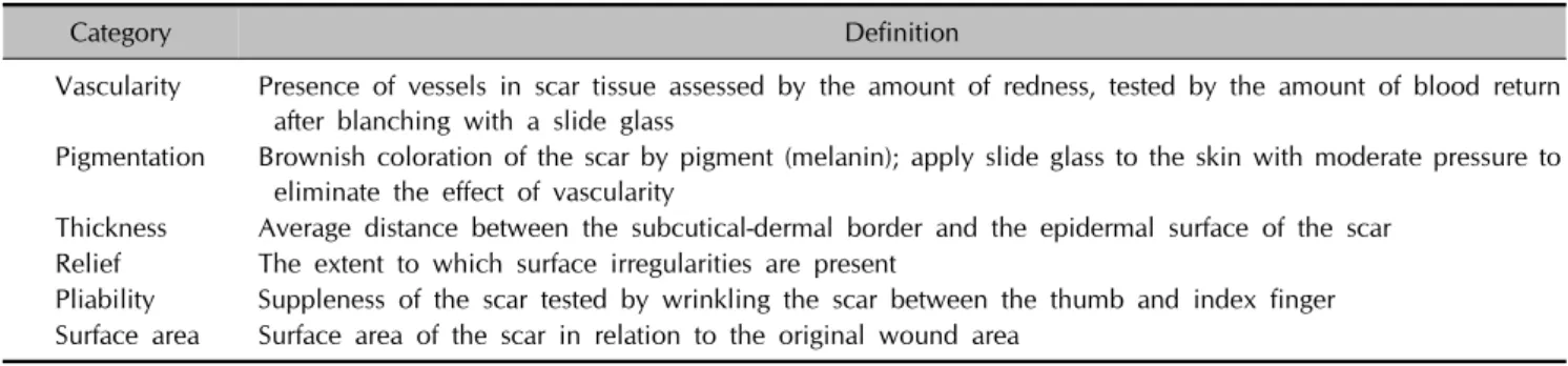
Table 3. The definitions of terms used in the patient and observer scar assessment scale
Category Definition
Vascularity Presence of vessels in scar tissue assessed by the amount of redness, tested by the amount of blood return after blanching with a slide glass
Pigmentation Brownish coloration of the scar by pigment (melanin); apply slide glass to the skin with moderate pressure to eliminate the effect of vascularity
Thickness Average distance between the subcutical-dermal border and the epidermal surface of the scar Relief The extent to which surface irregularities are present
Pliability Suppleness of the scar tested by wrinkling the scar between the thumb and index finger Surface area Surface area of the scar in relation to the original wound area
area, cheeks, nose, lips and perioral area, and chin) ac- cording to skin texture, color, and contour8. The Minolta SpectrophotometerⓇ is a tri-stimulus colorimeter that con- tains a Xenon lamp as a light source. The light that is re- flected perpendicular to the skin is collected by photo- detectors with color filter for a tristimulus color analysis at 450, 560, and 600 nm. This equipment uses the L*a*b*
system, where L* signifies brightness (scored from 0 for white to 100 for black), a* indicates color values from green to red (negative values indicated green and positive values indicated red), and b* indicates color values from blue to yellow (negative values indicated blue and pos- itive values indicated yellow)9. The a* parameter used to evaluate scar vascularity, while the L* and b* parameters were used to evaluate scar pigmentation10.
3) Ultrasonography
When evaluating scar thickness objectively, ultrasono- graphy provided reliable and accurate quantitative in- formation11. The average distance between the subcuti- cal-dermal border and epidermal surface of the scar was measured with LOGIQⓇ 9 (General Electric Company, Niskayuna, NY, USA) ultrasonography system. Scar thick- ness was compared to normal skin using in the same cos- metic units.
4) Statistical analysis
Internal consistency was defined as “the homogeneity of a set of categories and the degree to which they all share the same characteristics.” It was assessed by using Cronbach’s
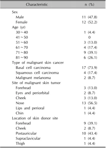
Table 4. Characteristics of 23 patients with facial skin graft scar
Characteristic n (%)
Sex
Male 11 (47.8)
Female 12 (52.2)
Age (yr)
30∼40 1 (4.4)
41∼50 0
51∼60 3 (13.0)
61∼70 4 (17.4)
71∼80 9 (39.1)
81∼90 6 (26.1)
Type of malignant skin cancer
Basal cell carcinoma 17 (73.9) Squamous cell carcinoma 4 (17.4)
Malignant melanoma 2 (8.7)
Site of malignant skin tumor
Forehead 3 (13.0)
Eyes and periorbital 2 (8.7)
Cheek 3 (13.0)
Nose 13 (56.5)
Lips and perioral 1 (4.4)
Chin 1 (4.4)
Location of skin donor site
Forehead 9 (39.1)
Cheek 2 (8.7)
Postauricular 10 (43.4)
Supraclavicular 1 (4.4)
Thigh 1 (4.4)
alpha statistics which considered the values greater than or equal to 0.70 to be acceptable12. Interobserver reli- ability was defined as “the extent of agreement between three observers” and was assessed by computing the intra- class correlation coefficient (ICC) using a two-way mixed model with measures of consistency. An ICC within the range of 0 to 0.20 was considered as “slight”, 0.21 to 0.40 as “fair”, 0.41 to 0.60 as “moderate”, 0.61 to 0.80 as
“substantial”, and 0.81 to 1.0 as “almost perfect”13. Convergent validity, which refers to the correlation among independently gathered rating, was evaluated using Pearson’s correlation statistics. Simple linear regression analysis was used to identify variables that significantly in- fluenced the patients’ overall opinions of their scars. The differences in skin color and thickness between the skin graft scar and normal skin, which were measured with spectrophotometer and ultrasonography, were analyzed statistically by using t-test. All statistical analyses were per- formed by using SPSS ver. 12.0 (SPSS Inc., Chicago, IL, USA) and p-values of <0.05 were considered significant.
RESULTS
Patient characteristics
Twenty-three patients (11 men, 12 women) were recruited for the study (Table 4). The median age was 70.9 years (range, 33∼86 years). The mean time that had passed from Mohs micrographic surgery to the study was 23.9 months (range, 2∼48 months). The most common type of malignant cutaneous neoplasm was basal cell carcinoma (73.9%) and the most common skin graft recipient site was the nose (56.5%).
Scar assessment using VSS and POSAS
1) Overall opinion of the VSS and POSAS
The mean total score using the VSS for facial skin graft scar was 3.4±1.8. The mean total score using the ob- server component of POSAS for facial skin graft scar was 15.8±7.0 and that using the patient component of POSAS was 18.4±10.3 (p=0.09) (Fig. 1).
2) Internal consistency
The internal consistency (Table 5) was acceptable for the VSS and observer and patient components of the POSAS, with Cronbach’s alpha values of 0.76, 0.84, and 0.88, respectively.
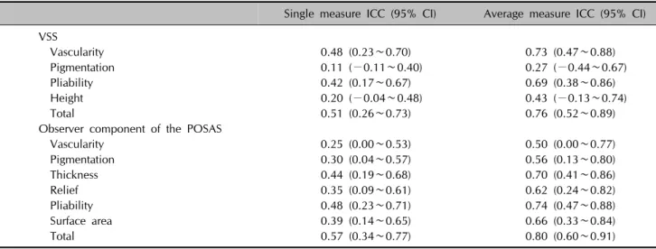
3) Interobserver reliability
The interobserver reliability (Table 6) was substantial for both the VSS and the observer component of the POSAS
in terms of total score (the average measures of ICC were, 0.76 and 0.80, respectively). For the individual VSS cate- gories, the interobserver reliability was substantial for vas- cularity (0.73) and, pliability (0.69), moderate for height (0.43), and fair for pigmentation (0.27). For the individual observer component of the POSAS, interobserver reli- ability was substantial for pliability, thickness, surface area, and relief (0.74, 0.70, 0.66, and 0.62, respectively), and moderate for pigmentation and vascularity (0.56 and 0.50, respectively).
4) Convergent validity
The correlations between the observer ratings of VSS and the observer component of POSAS were found to be sig- nificant (all p-values<0.05; Table 7). The observer com- ponent consistently showed significant correlations with the patients’ ratings for the individual categories (all p-val- ues<0.05; Table 8). In VSS, pliability, height, and total score correlated significantly with the patient components of stiffness, thickness, and total scores.
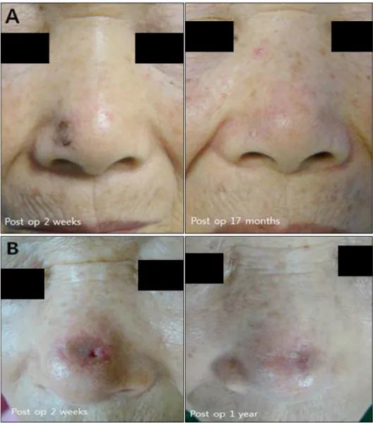
Fig. 1. Clinical images of skin graft scar. (A) A 73-year-old woman who underwent skin graft because of basal cell carcinoma on the ala of nose. Evaluating skin graft scar using patient and observer scar assessment scale (POSAS) 17 months later, patient scar score was 13 and the mean observer scar score was 8.6. The patient score of POSAS:
pain (2), itchiness (3), color (2), stiffness (2), thickness (2), irregularity (2). The mean observer score of POSAS: vascularity (1.3), pigmen- tation (1.3), thickness (2.0), relief (1.0), pliability (1.3), surface area (1.3). (B) The patient score of skin graft scar who had Moh’s micro- graphic surgery on dorsum of nose 1 year ago was 22. The observer score was 15.3. The patient score of POSAS: pain (1), itchiness (1), color (5), stiffness (5), thickness (5), irregularity (2). The mean observer score of POSAS: vascularity (1.7), pigmentation (3.0), thickness (3.0), relief (2.3), pliability (3.3), surface area (2.0) (presented with permi- ssion patients). Preop: preoperative, postop: postoperative.
Table 5. The reliability of the Vancouver scar scale and patient and observer scar assessment scale (POSAS)
Items Cronbach’s
alpha
Number of items
Vancouver scar scale 0.76 4
Observer component of POSAS 0.84 6
Patient component of POSAS 0.88 6
5) Patient self-assessment and linear regression analysis On simple linear regression analysis, the patients’ overall opinion regarding their own scars was significantly influ- enced by scar-related itchiness, color, stiffness, thickness, and irregularity (p<0.05; Table 9).
Scar evaluation using spectrophotometer and ultrasonography
1) Scar color
The interobserver reliability of the observer component of POSAS was moderate in terms of vascularity and pigmen- tation (0.50 and 0.56, respectively). When measuring scar color using spectrophotometer, there was no significant difference between the scar and normal skin. The value of L* in the facial skin graft scar was 54.4±6.1 and that of the normal facial skin was 55.7±4.4 (p=0.44). The value of a* in the facial skin graft scar was 14.0±3.1 and that of the normal skin was 13.9±2.3 (p=0.87). The value of b*
in the facial skin graft scar was 17.1±2.9 and that of the normal skin was 18.2±2.7 (p=0.21). The correlation be- tween the subjective assessment using the observer com- ponent of POSAS and the objective assessment using spectrophotometer showed moderate but significant rela-
Table 7. Correlations between the VSS and the observer component of the patient and observer scar assessment scale
Pearson’s correlation coefficient p-value
VSS vascularity score vs. OSAS vascularity score 0.56 0.005
VSS pigmentation score vs. OSAS pigmentation score 0.57 0.004
VSS pliability score vs. OSAS pliability score 0.83 <0.001
VSS height score vs. OSAS thickness score 0.82 <0.001
VSS total score vs. OSAS total score 0.80 <0.001
VSS: Vancouver scar scale, OSAS: observer scar assessment scale.
Table 6. Interobserver reliability of the Vancouver scar scale (VSS) and the observer component of the patient and observer scar assessment scale (POSAS)
Single measure ICC (95% CI) Average measure ICC (95% CI) VSS
Vascularity 0.48 (0.23∼0.70) 0.73 (0.47∼0.88)
Pigmentation 0.11 (−0.11∼0.40) 0.27 (−0.44∼0.67)
Pliability 0.42 (0.17∼0.67) 0.69 (0.38∼0.86)
Height 0.20 (−0.04∼0.48) 0.43 (−0.13∼0.74)
Total 0.51 (0.26∼0.73) 0.76 (0.52∼0.89)
Observer component of the POSAS
Vascularity 0.25 (0.00∼0.53) 0.50 (0.00∼0.77)
Pigmentation 0.30 (0.04∼0.57) 0.56 (0.13∼0.80)
Thickness 0.44 (0.19∼0.68) 0.70 (0.41∼0.86)
Relief 0.35 (0.09∼0.61) 0.62 (0.24∼0.82)
Pliability 0.48 (0.23∼0.71) 0.74 (0.47∼0.88)
Surface area 0.39 (0.14∼0.65) 0.66 (0.33∼0.84)
Total 0.57 (0.34∼0.77) 0.80 (0.60∼0.91)
CI: confidence interval, single measure ICC: intraclass correlation coeficient for a single observer, average measure ICC: intraclass correlation coefficient for the group of three observer.
Table 8. Correlation between observer scores using the VSS and the observer component of the patient and observer scar assessment scale (POSAS) and patient scores using the POSAS
Pearson’s correlation coefficient p-value
VSS vascularity score vs. PSAS color score 0.28 0.181
VSS pigmentation score vs. PSAS color score 0.35 0.097
VSS pliability score vs. PSAS stiffness score 0.65 0.001
VSS height score vs. PSAS thickness score 0.62 0.001
VSS total score vs. PSAS total score 0.61 0.002
OSAS vascularity score vs. PSAS color score 0.47 0.022
OSAS pigmentation score vs. PSAS color score 0.47 0.024
OSAS pliability score vs. PSAS stiffness score 0.56 0.005
OSAS thickness score vs. PSAS thickness score 0.63 0.001
OSAS relief score vs. PSAS irregularity score 0.59 0.003
OSAS total score vs. PSAS total score 0.49 0.015
VSS: Vancouver scar scale, PSAS: patient scar assessment scale, OSAS: observer scar assessment scale.
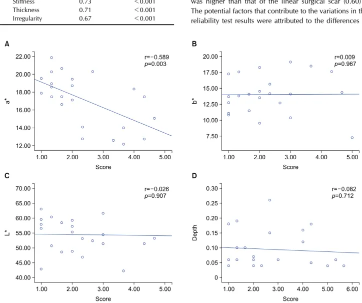
tionship in the vascularity subscale. However, the degree of correlation about pigmentation showed insignificant re- sults between two different scar evaluation tools (Fig. 2).
2) Scar thickness
The interobserver reliability of the observer component of POSAS was substantial in the thickness category (0.70).
Fig. 2. The correlation between patient and observer scar assessment scale (POSAS) to objective scar assessment tool. (A) a* indicated color values from green to red and it was applied to scar vascularity. The correlation between POSAS and spectrometer was significant but moderate relationship. (B, C) L* expressed brightness and it was used for the evaluation of scar pigmentation. b* designated values from blue to yellow and it was used for the measurement of scar pigmentation along with L*. The degree of correlation about pigmentation showed insignificant results between two different scar evaluation tool. (D) The correlation between POSAS and objective assessment using ultrasonography showed low relationship.
Table 9. Simple linear regression analysis of variables associated with patient scar assessment scale scores
Items Slope coefficient p-value
Pain 0.79 0.096
Itchness 0.74 0.006
Color 0.73 <0.001
Stiffness 0.73 <0.001
Thickness 0.71 <0.001
Irregularity 0.67 <0.001
When measuring scar thickness using ultrasonography, a significant difference between the scar and normal control group was observed. The mean depth of the facial skin graft scar was 0.10±0.01 cm, while that of the normal fa- cial skin was 0.09±0.06 cm (p<0.001). The correlation between the subjective assessment using the observer
component of POSAS and the objective assessment using ultrasonography showed low relationship (Fig. 2).
DISCUSSION
In evaluating the facial skin graft scars by using subjective methods, both VSS and POSAS had acceptable internal consistency and interobserver reliability. This indicated that both VSS and POSAS had good feasibility as a scar as- sessment tool for clinical follow-up and research purposes in skin graft scar. The reliability of the VSS for facial skin graft scar (0.76) was similar to linear scars as previously reported (0.78)14. The score of the interclass correlation coefficient of the observer component of POSAS (0.80) was higher than that of the linear surgical scar (0.60)14. The potential factors that contribute to the variations in the reliability test results were attributed to the differences in
the type of scars and observer’s training.
In the analysis of the individual components of the VSS and POSAS, the results of our study suggested that the ob- server component of POSAS had an advantage over the VSS. The observer component of the POSAS subscale ex- hibited significant correlations with the patient component of POSAS for color, stiffness, thickness, and irregularity.
However, the VSS subscale showed significant correla- tions with the patient component of POSAS for stiffness and thickness only. Also, the POSAS reflected the patients’
subjective scar-related symptoms. This study showed that color, stiffness, irregularity, and itchiness significantly af- fected the patients’ overall opinion. This suggests that not does the visibility of scars as an important factor, but also the scar symptoms, which needed to be monitored more closely in scar evaluation.
Scars distorted the physical appearance, especially when found on the head and neck, which could cause a neg- ative impact on quality of life15,16. Nose is the most com- mon site of skin cancer in the head and neck, which is re- lated to sun exposure and ultraviolet damage17. Because of its complex subunit, with intersecting concavities and convexities, the nose is complicated aesthetic units to operate. Full-thickness skin graft takes large portion of na- sal reconstruction along with nasolabial flap, especially in nasal sidewall and nasal dorsum skin defect18. In our study, nose is the most common site of operation (56.5%).
In nasal subunit, the ala of nose (46.1%), dorsum (30.7%) and tip (23.0%) are the common recipient site of oper- ation and forehead is the most common donor site (61.5%). The mean total score using the observer compo- nent of POSAS for nasal skin graft scar was 15.5±7.5 and that using the patient component of POSAS was 17.4±9.6 (p=0.19). In observer component of POSAS, the relief, pliability and thickness had a high score. The irregularity, thickness and color were a high proportion than sub- jective symptom like pain and itchiness in patient compo- nent of POSAS. These results showed that scar irregularity was a major considering factor in nasal skin graft scar. It is very important that the operating surgeon should be con- sidered postoperative care for patients undergoing nasal reconstruction. In this regard, the POSAS is easy and effi- cient evaluation tool in scar assessment.
The color of the skin was affected by the distribution of the blood vessels and the pigmentation of the skin. The correlation between subjective assessment using POSAS and objective assessment using Spectrophotometer showed significant and moderate relationships in terms of vascu- larity and low relationships in pigmentation. POSAS has a limitation in that it may assess hypopigmented scar as high as a hyperpigmented scar because of pigmentation was
compared to normal skin. In burn scar evaluation, the cor- relation between the vascularity score in POSAS and ob- jective assessments in chromameter was moderate. On the other hand, the correlation coefficient in pigmentation was varied enormous per observer9. It is difficult to rate the pigmentation of the scar reliably. Wei et al.19 sug- gested the use of dermoscopy in assessment of vascularity and pigmentation of scar. The dermoscopy is a non- invasive method that allows the visualization of dilated ca- pillaries and pigments in the dermal and epidermal layers of scar. It expresses great advantage in assessing the vascu- larity and pigmentation accurately. Also, he applied trans- formed VSS pigmentation score when correlating with ob- jective scar assessment tool. The negative value repre- sented hyperpigmentation and positive value implied hypopigmentation. This method showed a significant cor- relation between scar evaluation in objective tools and the subjective assessment.
To evaluate scar thickness objectively, ultrasound techni- ques can be used. It can produce images that are reflected from the interfaces of different tissues7. The interobserver reliability of the POSAS among three observers was sub- stantial in scar thickness. The correlation between sub- jective assessment using POSAS and objective assessment using ultrasonography showed poor relationships. When we evaluate scar thickness by POSAS, there is no differ- ence in scores between hypertrophic scar and hypo- trophic scar.
Over the years, a lot of efforts have tried in exploring more objective and accurate assessment tools for scar measurement with the development in electronics and in- formation technology. Objective tools, such as Laser Doppler can be used to measure the blood perfusion of scars which is also an important indicator of vascularity20. However, the measurements are difficult in the border of scar, and pigmentation characteristics cannot be measured with Laser Doppler. A non-contact 3D digitiser (Kino- ca-Minolta Vivid 900Ⓡ;Konica Minolta Co., Tokyo, Japan) can measure the volume of a scar21. This tool used initially in industry to scan a 3D image of an object. When evalu- ating a scar, this method showed a significant positive cor- relation between the volume of scar and the clinical se- verity according to scar scale. An optical system that pro- duces three-dimensional measurement of the skin surface, the PRIMOSⓇ system, can be used to evaluate the scar surface22. It is available to measure skin surface level dif- ference up to 10 mm with a resolution over 0.004 mm.
However, these kinds of instruments are usually extremely expensive and difficult to be carried around, thus it is not easily applicable tools to be widely used for clinical practice.
The POSAS is a standardized, validated, and compre- hensive scar assessment tool in clinical care. To our knowledge, this is the first attempt to evaluate facial skin graft scar by POSAS. The POSAS had more advantage as its observer component showed better correlation to the patient’s rating. Additionally, the POSAS reflected the pa- tient’s perspective about scar-related symptoms like pain and itchiness, which were not considered in previous scar assessment tool4,5. These findings support the use of POSAS as a reliable, valid, and comprehensive tool to as- sess facial skin graft scar. Even though there are many ad- vantages of POSAS as scar evaluation tool, the correlation between the POSAS and objective scar evaluation tools shows low relationships. To compensate these limitations, there are several things to complement. First, it is in- sufficient to evaluate scar color with naked eye, so addi- tional tools like dermoscopy could increase the exactitude of scar evaluation. Second, the POSAS lacks the score sys- tem in the category of parameters. It cannot make dis- tinction between hyperpigmentation and hypopigmentation because of pigmentation was compared to normal skin.
The transformed POSAS scoring system which included positive and negative value will enable accurate scar evaluation. Our study has some limitation that we in- cluded a limited number of patients in single center.
Future study should contain large sample size and explore the improvement of facial skin graft scar through the man- agement of scar.
ACKNOWLEDGMENT
This work was supported by Wonkwang University in 2014.
REFERENCES
1. Viola KV, Jhaveri MB, Soulos PR, Turner RB, Tolpinrud WL, Doshi D, et al. Mohs micrographic surgery and surgical excision for nonmelanoma skin cancer treatment in the Medicare population. Arch Dermatol 2012;148:473-477.
2. Sheehan JM, Kingsley M, Rohrer TE. Excisional surgery and repair, flaps, and graft. In: Goldsmith LA, Katz SI, Gilchrest BA, Paller AS, Leffell DJ, Wolff K, editors. Fitzpatrick’s dermatology in general medicine. 8th ed. New York:
McGraw-Hill, 2012:2944-2949.
3. Idriss N, Maibach HI. Scar assessment scales: a derma- tologic overview. Skin Res Technol 2009;15:1-5.
4. Draaijers LJ, Tempelman FR, Botman YA, Tuinebreijer WE, Middelkoop E, Kreis RW, et al. The patient and observer scar assessment scale: a reliable and feasible tool for scar evaluation. Plast Reconstr Surg 2004;113:1960-1965.
5. van de Kar AL, Corion LU, Smeulders MJ, Draaijers LJ, van
der Horst CM, van Zuijlen PP. Reliable and feasible evalu- ation of linear scars by the Patient and Observer Scar Assessment Scale. Plast Reconstr Surg 2005;116:514-522.
6. Sullivan T, Smith J, Kermode J, McIver E, Courtemanche DJ.
Rating the burn scar. J Burn Care Rehabil 1990;11:256-260.
7. Roques C, Teot L. A critical analysis of measurements used to assess and manage scars. Int J Low Extrem Wounds 2007;6:249-253.
8. Robinson JK, Anderson ER. Skin structure and surgical anatomy. In: Robinson JK, Hnke CW, Sengelmann RD, Siegel DM, editors Surgery of the skin. Philadelphia: Elsevier Mosby, 2005:6-10.
9. Draaijers LJ, Tempelman FR, Botman YA, Kreis RW, Middelkoop E, van Zuijlen PP. Colour evaluation in scars:
tristimulus colorimeter, narrow-band simple reflectance meter or subjective evaluation? Burns 2004;30:103-107.
10. Takiwaki H. Measurement of skin color: practical appli- cation and theoretical considerations. J Med Invest 1998;
44:121-126.
11. Beausang E, Floyd H, Dunn KW, Orton CI, Ferguson MW.
A new quantitative scale for clinical scar assessment. Plast Reconstr Surg 1998;102:1954-1061.
12. Bland JM, Altman DG. Cronbach's alpha. BMJ 1997;
314:572.
13. Landis JR, Koch GG. The measurement of observer agreement for categorical data. Biometrics 1977;33:159-174.
14. Truong PT, Lee JC, Soer B, Gaul CA, Olivotto IA. Reliability and validity testing of the Patient and Observer Scar Assessment Scale in evaluating linear scars after breast cancer surgery. Plast Reconstr Surg 2007;119:487-494.
15. Sobanko JF, Sarwer DB, Zvargulis Z, Miller CJ. Importance of physical appearance in patients with skin cancer.
Dermatol Surg 2015;41:183-188.
16. Choi Y, Lee JH, Kim YH, Lee YS, Chang HS, Park CS, et al.
Impact of postthyroidectomy scar on the quality of life of thyroid cancer patients. Ann Dermatol 2014;26:693-699.
17. Evans GR, Williams JZ, Ainslie NB. Cutaneous nasal malignancies: is primary reconstruction safe? Head Neck 1997;19:182-187.
18. Weathers WM, Bhadkamkar M, Wolfswinkel EM, Thornton JF. Full-thickness skin grafting in nasal reconstruction. Semin Plast Surg 2013;27:90-95.
19. Wei Y, Li-Tsang CW, Luk DC, Tan T, Zhang W, Chiu TW. A validation study of scar vascularity and pigmentation assess- ment using dermoscopy. Burns 2015;41:1717-1723.
20. Bray R, Forrester K, Leonard C, McArthur R, Tulip J, Lindsay R. Laser Doppler imaging of burn scars: a comparison of wavelength and scanning methods. Burns 2003;29:199- 206.
21. Taylor B, McGrouther DA, Bayat A. Use of a non-contact 3D digitiser to measure the volume of keloid scars: a useful tool for scar assessment. J Plast Reconstr Aesthet Surg 2007;60:87-94.
22. Roques C, Téot L, Frasson N, Meaume S. PRIMOS: an optical system that produces three-dimensional measurements of skin surfaces. J Wound Care 2003;12:362-364.