Platelet-rich Plasma in Arthroscopic Rotator Cuff Repair
Do Hoon Kim, Sae Hoon Kim
Department of Orthopedic Surgery, Seoul National University Hospital, Seoul National University College of Medicine, Seoul, Korea
Rotator cuff tear is a common reason for shoulder pain. Although the surgical technique of rotator cuff repair is developing, high retear rate requires additional supplementary methods. Among these supplementary methods, as a kind of biologic augmentation, platelet-rich plasma (PRP) has been spotlighted and has recently been studied by many researchers. PRP, a concentrate of platelet extract obtained from whole blood, contains numerous growth factors. As this is known to play an important role in the tissue recovery process, it had been used for research in a variety of fields including orthopedics. Use of PRP has been attempted in surgical treatments of rotator cuff tear for better results; however, only a few large-scale research studies on the effect of PRP have been reported. Clinical results of each study are also variable. Therefore research using large-scale randomized, double-blind trials should be conducted in order to prove the application range, safety, and clinical effects of PRP.
(Clin Shoulder Elbow 2015;18(2):113-118)
Key Words: Shoulder; Rotator cuff; Platelet-rich plasma
Clinics in Shoulder and Elbow
Copyright © 2015 Korean Shoulder and Elbow Society. All Rights Reserved. pISSN 2383-8337
Clinics in Shoulder and Elbow Vol. 18, No. 2, June, 2015 http://dx.doi.org/10.5397/cise.2015.18.2.113
Received December 11, 2014. Revised January 23, 2015. Accepted January 28, 2015.
Correspondence to: Sae Hoon Kim
Department of Orthopedic Surgery, Seoul National University Hospital, 103 Daehak-ro, Jongno-gu, Seoul 110-799, Korea Tel: +82-2-2072-3930, Fax: +82-2-764-2718, E-mail: [email protected]
Financial support: None. Conflict of interests: None.
Introduction
Rotator cuff lesion, which was reported from 14% to 50% of adults over the age of 60 years and 80% of adults over the age of 80 years, is the main reason for shoulder pain and causes much discomfort in daily life by causing decreased range of motion or muscular weakness of shoulder.1,2) Numerous cases of rota- tor cuff lesion require surgical treatment. Surgical techniques for rotator cuff tear have been continuously developed for decades, from open repair with transosseous suture to all-arthroscopic single row technique or double row suture bridge technique.3) Despite development of surgical techniques, the reported retear rate is still high. Single row technique, the most commonly used technique, showed retear rates of 30% to 94%.4,5) According to a recent meta-analysis, double row suture bridge technique showed a lower retear rate when compared to single row tech- nique, yet 27.3% of patients in whom double row suture bridge technique was performed also suffered from retear.3,6) Because chronic rotator cuff tear makes attachment of the terminal part of the tendon to the bone difficult and prolongs the process, ad-
ditional biologic augmentation other than suture methods are needed to help the tissues recover.7)
Usage of platelet-rich plasma (PRP) is a form of biologic aug- mentation. PRP could be directly injected inside the joint, or may be applied by suturing organized PRP directly to the rup- tured site during surgery.8) Because numerous kinds of growth factors secreted from platelets have been regarded as having a positive effect on tendon recovery, studies researching the us- age of PRP on rotator cuff repair have been conducted by many researchers for several years. In this study, the role of PRP on rotator cuff tear will be discussed and the effect of PRP will be analyzed by investigating existing literature.
Platelet-rich Plasma
Platelet is a component of blood plasma that takes part in hemostasis. Its life expectancy is 7 to 10 days and it contains intracellular structures including glycogen, lysosome, and two types of granules. Among these, alpha granule secretes growth factors involved in tissue repair. Activation of platelets in the
resting state by thrombin leads to secretion of more than 1,500 kinds of materials, such as growth factors including transforming growth factor-β, platelet-derived growth factor, fibroblast growth factor, and vascular endothelial growth factor, and proteins that take part in hemostasis.9,10) Once platelets are activated, 70%
of restored growth factors are secreted within 10 minutes, and all restored growth factors are released within an hour. More growth factors are synthesized and secreted for the next 7 to 10 days until platelets die.11)
PRP is a concentrate of platelets extracted from autologous blood. Whole blood is primarily centrifuged to separate red blood cells from plasma, and then undergoes secondary cen- trifugation to separate leukocytes and platelets together with a few red blood cells from platelet-poor plasma finely.11) Average number of platelets within whole blood is 200,000 cells/ml. Opti- mal concentration of PRP that is effective for vascularization and tissue regeneration of 1.5 to 3 million cells/ml has been reported, which is around 7 to 10 times greater than the normal amount.12)
First researched by Ferrari et al.13) in 1987, the effect of PRP has been extensively researched in various fields for more than
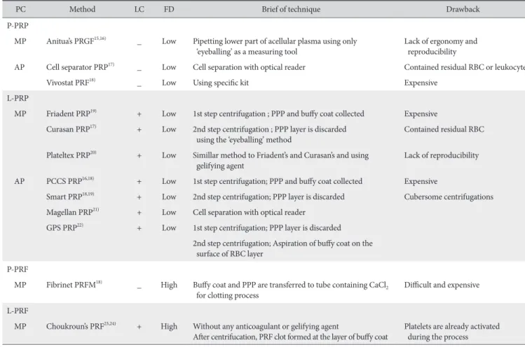
20 years. Due to various kinds of platelet concentration meth- ods and components, questions regarding the validity of corre- lation among research data have arisen. In order to standardize the result, Dohan Ehrenfest et al.14) classified PRP according to four categories, depending on the presence of white blood cells and fibrin. The characteristics of classes and each protocol are shown in Table 1.14-24) Although the effect of leukocytes in PRP has not yet been clearly determined, no effects that negatively affect PRP were found. In addition, when acromioplasty was performed in patients who suffered from subacromial impinge- ment, the group that used leukocyte-rich PRP showed improve- ment of pain and inflammation.25) Further research is necessary to study the effect of leukocyte-rich PRP, leukocyte-rich platelet- rich fibrin, pure PRP, and pure platelet-rich fibrin.
The Effect of Applying Platelet-rich Plasma in Rotator Cuff Repair; Review of Literature
Laboratory Study
Jo et al.,26) who harvested human tenocytes during degenera-
Table 1. Classification of the Platelet Concentrates Protocols
PC Method LC FD Brief of technique Drawback
P-PRP
MP Anitua’s PRGF15,16) _ Low Pipetting lower part of acellular plasma using only
‘eyeballing’ as a measuring tool Lack of ergonomy and reproducibility
AP Cell separator PRP17) _ Low Cell separation with optical reader Contained residual RBC or leukocyte
Vivostat PRF18) _ Low Using specific kit Expensive
L-PRP
MP Friadent PRP19) + Low 1st step centrifugation ; PPP and buffy coat collected Expensive Curasan PRP17) + Low 2nd step centrifugation ; PPP layer is discarded
using the ‘eyeballing’ method Contained residual RBC
Plateltex PRP20) + Low Simillar method to Friadent’s and Curasan’s and using
gelifying agent Lack of reproducibility
AP PCCS PRP16,18) + Low 1st step centrifugation; PPP and buffy coat collected Expensive
Smart PRP18,19) + Low 2nd step centrifugation; PPP layer is discarded Cubersome centrifugations Magellan PRP21) + Low Cell separation with optical reader
GPS PRP22) + Low 1st step centrifugation; PPP layer is discarded 2nd step centrifugation; Aspiration of buffy coat on the
surface of RBC layer P-PRF
MP Fibrinet PRFM18) _ High Buffy coat and PPP are transferred to tube containing CaCl2
for clotting process Difficult and expensive
L-PRF
MP Choukroun’s PRF23,24) + High Without any anticoagulant or gelifying agent
After centrifucation, PRF clot formed at the layer of buffy coat Platelets are already activated during the process
PC: platelet concentrates, LC: leukocyte collection, FD: fibrin density, P-PRP: pure platelet-rich plasma, L-PRP: leukocyte rich platelet-rich plasma, P-PRF: pure platelet-rich fibrin, L-PRF: leukocyte-rich platelet-rich fibrin, MP: manual protocol, AP: automatized protocol, PRGF: preparation rich in growth factors, PCCS:
platelet concentrate collection system, GPS: gravitational platelet separation system, PPP: platelet-poor plasma, PRFM: platelet-rich plasma fibrin matrix, RBC:
red blood cell.
Table 2. Controlled Clinical Studies Dealing with the Surgical Use of PRP in Rotator Cuff Tears Author (year)Evidence level and study designSample size (persons) (PRP/control)Tear sizeMaterialSurgical techniqueMean F/U (mo)Clinical outcomeImaging outcome Barber et al. (2011)32)Level 3 Case-control20/20All size
PRFM SuenupRP gte in Pr raeaetwer rLodounces froerannifico sigN31owgle rSint diff eabletur upe slatIso43/45atinicini et al 1veLeus l. spstrCara 33)TRC(2011)tear
PRFM Suendces fenert diffannifico sigNdounces founerowDt diffble rou16No significan bleeatur t diffannifico sigutN20e urenert diffces foundNo significanerces foundble row/senouel19/23All sizePRP gl 2veLeDl. Jo et a 34)ctProspe(2011)ivbre coeaturbleidgeSurtho antial difference, but no significr rat Lower reteate for smaller tear Iniow24ARandelli et agle rLevel 126/27l. ll sizePRSinP 35) RPweblta(2011)ith Pjecce aIndiffenert final F/UTRC l 3edio ml talSm16/21umveLel. on et agesBer 36) Cohort(2012)
PRFM SueruproRP gte in Peaether rigHdounces fenr rat diffle rangle or doubSinownificNo sig12 bleeatur enert diffo sigannificounNces fte in PdLower retear raRP groupow13RFMgle rl 1Sinumina et al. LeveG39/37LargeL-P 37) eaTbleSutur(2012)RC LeA40/39l 2vel. deo et aRoll size 38) (2012)Prospective RCT
PRFM Suo sigerences foundNnificananerences foundt difft diffnificow/o sigSinr double rgle o12N sutureabletureridge b ounow24No significant differences fces fdNo significant differenoundSingle rivuñl 2Lel. 14/14Massa et aePRFntAve 39) ta(2013)jecPrpective RCTIneblos anenert diffe nifico sigN12urces fwer roundLoetear rate in PRP groupow/sutrgeble rsiveJo et al. vel 120/18La to masLePRP gDelou 40) eaeSutur(2013)blebridgRCT Do sigouble row12Nces fnificant differenounNandLet diffl 1Ruiz-Moneo et dnificveounerces f32/31All sizePRPeno sig 41) jecetaInTRCal. (2013)bl LeA30/30l 1vel. er et aebWll size 42) (2013)RCT
PRFM Suces founces fenert diffannifico sigNdounden12Sinerowgle rNo significant diff bleeatur enert diffanble rnifico sigN24owounces fetdNo differences of rear rate ; rear DouetCh toLearou35/35Large ml 3assiveL-PRPl. et et assve 43) blsize waaller in L-ProupeRP gs sm(2014)tase-controlInjecCa ert diffan24nifico sigNces fowennificoundNo sigant differences foundSingle ro malalLevel 1ta et aol27/27Sml tl. ediumPRPavM 44) ebl(2014)TInjectaRC ounNo significant differences feeo sigdNnificant differences foundksll size12 WAowstein et al. Level 110/10ZumL-PDRFble rou 45) turable(2014)SuRCT er6 WeeksNo significant differences found Not pformedSingle rowveLe12/13Small to mediumPRPl. ak et aHl 2 46)e(2015)blTInjecRCta brich plasmich pelet-rlatcyte-rkoRFM: leu-Prix, La fi-Pin matrix, PRF: platelet-rich fibrin, LRP: in matlasmbrndoPRP: pa fielet-rich pa, F/U: follow-up, RCT: ralatmizeRFM: pd colasmelet-rlatich pl, Priad tlleront RFbrich fielet-rlatich pe-rcytko: leuich p-Pa, Llasmich pelet-rlate rcytkoleuin.
tive rotator cuff repair, and cultivated them for 2 weeks using platelet-poor plasma and PRP with various concentrations, re- ported that the PRP applied group showed better cell prolifera- tion, gene expression, and synthesis of tendon matrix.
Beck et al.27) researched PRP application in rotator cuff tear using a rat model. Supraspinatus of rat model was detached and repaired using PRP. Follow-ups for 7, 14, and 21 days showed that while failure load showed no significant difference, the group that used PRP showed high stiffness of tendons and more organized collagen fiber. Hapa et al.,28) who used PRP for rotator cuff tear on a rat model and followed for 2 weeks, reported that the group that used PRP showed less inflammation, and better vascularization and mechanical strength. Dolkart et al.,29) who used PRP for rotator cuff repair on a rat model and observed for 3 weeks, reported that the group that used PRP showed significantly higher maximal load, stiffness and collagen birefri- ence. However they reported that no significant difference was shown in tendon organization and vascularization. Ersen et al.30) also conducted research using a rat model and reported that even though maximal load and stiffness of the group that used PRP was significantly superior, no histological differences were shown.
Chung et al.31) used a rabbit model to perform repair surgery 6 weeks after incising supraspinatus to imitate chronic rotator cuff tear and used PRP for rotator cuff repair and observed the results at 4 and 8 weeks after the surgery. They reported that the group that used PRP showed better tendon status in continuity and orientation of collagen, and higher maximal load of ten- dons.
In cases of cell level or animal experiments, results are mostly positive. However, these results came from a controlled situ- ation and may not represent the status of the human body. In addition, current animal experiments could not reflect human’s status of chronic rotator cuff tear or recovery ability. Therefore, application of the results of animal experiments to clinical situa- tions is still limited.
Clinical Study
Clinically, PRP has been applied to tissues of repaired site during rotator cuff repair. The results of research that used PRP during surgeries are described in Table 2.32-46)
Conclusion could not be made easily since the consistency of study design and PRP formula for each study has not been for- mulated. According to the study so far, clinical results of the us- age of PRP for rotator cuff tear showed no significant difference, but some studies reported a low retear rate,1,32,35,37) while the study conducted by Bergeson et al.36) reported a higher retear rate.
A systemic review conducted by Chahal et al.47) in 2012 which analyzed 5 studies32-36) reported that PRP usage is not ef- fective, and meta-analysis conducted by Zhao et al.48) and Li et
al.49) on randomized controlled trials33,35,37-42) in 2014 reported that PRP usage does not help clinical results or decrease rotator cuff retear rate. In meta-analysis conducted by Zhang et al.50) in 2013, it was reported that PRP usage did not help clinical re- sults, but retear rate showed a significant decrease in small and middle sized tears.
In response to these inconsistent results, some researchers who support PRP stated that ‘All PRP is not created equal.’ In other words, they suggested that the differences result from dif- ferent methods of PRP manufacture, activation, and application.
Therefore, more standardized criteria for manufacture, main- tenance, dosage, and application of PRP will be required, and adequate additional large-scale randomized study should be conducted. Analysis of the effects of PRP is also necessary, con- sidering the size and the condition of torn rotator cuff tendon.
Conclusion
Although surgical techniques for rotator cuff tear are be- ing developed, retear rate is significantly high. For this reason, biologic augmentation that could enhance the recovery has become prominent. PRP, a concentration of platelets, could be useful for recovery of rotator cuff tear through secretion of nu- merous growth factors that could help in the recovery of tissues.
Research has been conducted in order to prove the effectiveness of PRP. However, the effect of PRP has not been significantly proven. For the usage of PRP during rotator cuff repair surgery, it should be additionally necessary that large-scale research studies use the standardization of manufacture, dosage, and method for application of PRP.
References
1. Milgrom C, Schaffler M, Gilbert S, van Holsbeeck M. Rotator- cuff changes in asymptomatic adults. The effect of age, hand dominance and gender. J Bone Joint Surg Br. 1995;77(2):296- 8.
2. Tempelhof S, Rupp S, Seil R. Age-related prevalence of rotator cuff tears in asymptomatic shoulders. J Shoulder Elbow Surg.
1999;8(4):296-9.
3. Slabaugh MA, Nho SJ, Grumet RC, et al. Does the literature confirm superior clinical results in radiographically healed rota- tor cuffs after rotator cuff repair? Arthroscopy. 2010;26(3):393- 403.
4. Galatz LM, Rothermich SY, Zaegel M, Silva MJ, Havlioglu N, Thomopoulos S. Delayed repair of tendon to bone injuries leads to decreased biomechanical properties and bone loss. J Orthop Res. 2005;23(6):1441-7.
5. Boileau P, Brassart N, Watkinson DJ, Carles M, Hatzidakis AM, Krishnan SG. Arthroscopic repair of full-thickness tears of the supraspinatus: does the tendon really heal? J Bone Joint Surg
Am. 2005;87(6):1229-40.
6. DeHaan AM, Axelrad TW, Kaye E, Silvestri L, Puskas B, Foster TE. Does double-row rotator cuff repair improve functional outcome of patients compared with single-row technique? A systematic review. Am J Sports Med. 2012;40(5):1176-85.
7. Jo CH, Yoon KS, Lee JH, et al. The effect of multiple channel- ing on the structural integrity of repaired rotator cuff. Knee Surg Sports Traumatol Arthrosc. 2011;19(12):2098-107.
8. Hall MP, Band PA, Meislin RJ, Jazrawi LM, Cardone DA. Plate- let-rich plasma: current concepts and application in sports medicine. J Am Acad Orthop Surg. 2009;17(10):602-8.
9. Anitua E, Andia I, Ardanza B, Nurden P, Nurden AT. Autologous platelets as a source of proteins for healing and tissue regenera- tion. Thromb Haemost. 2004;91(1):4-15.
10. Lopez-Vidriero E, Goulding KA, Simon DA, Sanchez M, John- son DH. The use of platelet-rich plasma in arthroscopy and sports medicine: optimizing the healing environment. Arthros- copy. 2010;26(2):269-78.
11. Marx RE. Platelet-rich plasma (PRP): what is PRP and what is not PRP? Implant Dent. 2001;10(4):225-8.
12. Giusti I, Rughetti A, D’Ascenzo S, et al. Identification of an op- timal concentration of platelet gel for promoting angiogenesis in human endothelial cells. Transfusion. 2009;49(4):771-8.
13. Ferrari M, Zia S, Valbonesi M, et al. A new technique for he- modilution, preparation of autologous platelet-rich plasma and intraoperative blood salvage in cardiac surgery. Int J Artif Organs. 1987;10(1):47-50.
14. Dohan Ehrenfest DM, Rasmusson L, Albrektsson T. Classification of platelet concentrates: from pure platelet-rich plasma (P-PRP) to leucocyte- and platelet-rich fibrin (L-PRF). Trends Biotechnol.
2009;27(3):158-67.
15. Anitua E, Aguirre JJ, Algorta J, et al. Effectiveness of autologous preparation rich in growth factors for the treatment of chronic cutaneous ulcers. J Biomed Mater Res B Appl Biomater.
2008;84(2):415-21.
16. Weibrich G, Kleis WK, Hitzler WE, Hafner G. Comparison of the platelet concentrate collection system with the plasma-rich- in-growth-factors kit to produce platelet-rich plasma: a techni- cal report. Int J Oral Maxillofac Implants. 2005;20(1):118-23.
17. Weibrich G, Kleis WK, Hafner G, Hitzler WE, Wagner W.
Comparison of platelet, leukocyte, and growth factor levels in point-of-care platelet-enriched plasma, prepared using a modified Curasan kit, with preparations received from a local blood bank. Clin Oral Implants Res. 2003;14(3):357-62.
18. Leitner GC, Gruber R, Neumüller J, et al. Platelet content and growth factor release in platelet-rich plasma: a comparison of four different systems. Vox Sang. 2006;91(2):135-9.
19. Weibrich G, Kleis WK, Buch R, Hitzler WE, Hafner G.
The Harvest Smart PRePTM system versus the Friadent- Schütze platelet-rich plasma kit. Clin Oral Implants Res.
2003;14(2):233-9.
20. Mazzucco L, Balbo V, Cattana E, Borzini P. Platelet-rich plasma and platelet gel preparation using Plateltex. Vox Sang. 2008;
94(3):202-8.
21. Christensen K, Vang S, Brady C, et al. Autologous platelet gel:
an in vitro analysis of platelet-rich plasma using multiple cycles.
J Extra Corpor Technol. 2006;38(3):249-53.
22. Marlovits S, Mousavi M, Gäbler C, Erdös J, Vécsei V. A new simplified technique for producing platelet-rich plasma: a short technical note. Eur Spine J. 2004;13 Suppl 1:S102-6.
23. Dohan DM, Choukroun J, Diss A, et al. Platelet-rich fibrin (PRF):
a second-generation platelet concentrate. Part I: technological concepts and evolution. Oral Surg Oral Med Oral Pathol Oral Radiol Endod. 2006;101(3):e37-44.
24. Choukroun JI, Braccini F, Diss A, Giordano G, Doglioli P, Do- han DM. Influence of platelet rich fibrin (PRF) on proliferation of human preadipocytes and tympanic keratinocytes: a new opportunity in facial lipostructure (Coleman’s technique) and tympanoplasty? Rev Laryngol Otol Rhinol (Bord). 2007;128(1- 2):27-32.
25. Everts PA, Devilee RJ, Brown Mahoney C, et al. Exogenous application of platelet-leukocyte gel during open subacromial decompression contributes to improved patient outcome. A prospective randomized double-blind study. Eur Surg Res.
2008;40(2):203-10.
26. Jo CH, Kim JE, Yoon KS, Shin S. Platelet-rich plasma stimulates cell proliferation and enhances matrix gene expression and synthesis in tenocytes from human rotator cuff tendons with degenerative tears. Am J Sports Med. 2012;40(5):1035-45.
27. Beck J, Evans D, Tonino PM, Yong S, Callaci JJ. The biome- chanical and histologic effects of platelet-rich plasma on rat rotator cuff repairs. Am J Sports Med. 2012;40(9):2037-44.
28. Hapa O, Cakıcı H, Kükner A, Aygün H, Sarkalan N, Baysal G.
Effect of platelet-rich plasma on tendon-to-bone healing after rotator cuff repair in rats: an in vivo experimental study. Acta Orthop Traumatol Turc. 2012;46(4):301-7.
29. Dolkart O, Chechik O, Zarfati Y, Brosh T, Alhajajra F, Maman E.
A single dose of platelet-rich plasma improves the organization and strength of a surgically repaired rotator cuff tendon in rats.
Arch Orthop Trauma Surg. 2014;134(9):1271-7.
30. Ersen A, Demirhan M, Atalar AC, Kapicioğlu M, Baysal G.
Platelet-rich plasma for enhancing surgical rotator cuff repair:
evaluation and comparison of two application methods in a rat model. Arch Orthop Trauma Surg. 2014;134(3):405-11.
31. Chung SW, Song BW, Kim YH, Park KU, Oh JH. Effect of plate- let-rich plasma and porcine dermal collagen graft augmenta- tion for rotator cuff healing in a rabbit model. Am J Sports Med. 2013;41(12):2909-18.
32. Barber FA, Hrnack SA, Snyder SJ, Hapa O. Rotator cuff repair healing influenced by platelet-rich plasma construct augmen- tation. Arthroscopy. 2011;27(8):1029-35.
33. Castricini R, Longo UG, De Benedetto M, et al. Platelet-rich
plasma augmentation for arthroscopic rotator cuff repair: a ran- domized controlled trial. Am J Sports Med. 2011;39(2):258- 65.
34. Jo CH, Kim JE, Yoon KS, et al. Does platelet-rich plasma ac- celerate recovery after rotator cuff repair? A prospective cohort study. Am J Sports Med. 2011;39(10):2082-90.
35. Randelli P, Arrigoni P, Ragone V, Aliprandi A, Cabitza P. Platelet rich plasma in arthroscopic rotator cuff repair: a prospec- tive RCT study, 2-year follow-up. J Shoulder Elbow Surg.
2011;20(4):518-28.
36. Bergeson AG, Tashjian RZ, Greis PE, Crim J, Stoddard GJ, Burks RT. Effects of platelet-rich fibrin matrix on repair integrity of at-risk rotator cuff tears. Am J Sports Med. 2012;40(2):286- 93.
37. Gumina S, Campagna V, Ferrazza G, et al. Use of platelet- leukocyte membrane in arthroscopic repair of large rotator cuff tears: a prospective randomized study. J Bone Joint Surg Am. 2012;94(15):1345-52.
38. Rodeo SA, Delos D, Williams RJ, Adler RS, Pearle A, Warren RF. The effect of platelet-rich fibrin matrix on rotator cuff ten- don healing: a prospective, randomized clinical study. Am J Sports Med. 2012;40(6):1234-41.
39. Antuña S, Barco R, Martínez Diez JM, Sánchez Márquez JM.
Platelet-rich fibrin in arthroscopic repair of massive rotator cuff tears: a prospective randomized pilot clinical trial. Acta Orthop Belg. 2013;79(1):25-30.
40. Jo CH, Shin JS, Lee YG, et al. Platelet-rich plasma for ar- throscopic repair of large to massive rotator cuff tears: a ran- domized, single-blind, parallel-group trial. Am J Sports Med.
2013;41(10):2240-8.
41. Ruiz-Moneo P, Molano-Muñoz J, Prieto E, Algorta J. Plasma rich in growth factors in arthroscopic rotator cuff repair: a ran- domized, double-blind, controlled clinical trial. Arthroscopy.
2013;29(1):2-9.
42. Weber SC, Kauffman JI, Parise C, Weber SJ, Katz SD. Platelet- rich fibrin matrix in the management of arthroscopic repair of
the rotator cuff: a prospective, randomized, double-blinded study. Am J Sports Med. 2013;41(2):263-70.
43. Charousset C, Zaoui A, Bellaïche L, Piterman M. Does autolo- gous leukocyte-platelet-rich plasma improve tendon healing in arthroscopic repair of large or massive rotator cuff tears?
Arthroscopy. 2014;30(4):428-35.
44. Malavolta EA, Gracitelli ME, Ferreira Neto AA, Assunção JH, Bordalo-Rodrigues M, de Camargo OP. Platelet-rich plasma in rotator cuff repair: a prospective randomized study. Am J Sports Med. 2014;42(10):2446-54.
45. Zumstein MA, Rumian A, Lesbats V, Schaer M, Boileau P.
Increased vascularization during early healing after biologic augmentation in repair of chronic rotator cuff tears using au- tologous leukocyte- and platelet-rich fibrin (L-PRF): a prospec- tive randomized controlled pilot trial. J Shoulder Elbow Surg.
2014;23(1):3-12.
46. Hak A, Rajaratnam K, Ayeni OR, et al. A double-blinded pla- cebo randomized controlled trial evaluating short-term efficacy of platelet-rich plasma in reducing postoperative pain after arthroscopic rotator cuff repair: a pilot study. Sports Health.
2015;7(1):58-66.
47. Chahal J, Van Thiel GS, Mall N, et al. The role of platelet-rich plasma in arthroscopic rotator cuff repair: a systematic review with quantitative synthesis. Arthroscopy. 2012;28(11):1718- 27.
48. Zhao JG, Zhao L, Jiang YX, Wang ZL, Wang J, Zhang P. Platelet- rich plasma in arthroscopic rotator cuff repair: a meta-analysis of randomized controlled trials. Arthroscopy. 2015;31(1):125- 35.
49. Li X, Xu CP, Hou YL, Song JQ, Cui Z, Yu B. Are platelet con- centrates an ideal biomaterial for arthroscopic rotator cuff re- pair? A meta-analysis of randomized controlled trials. Arthros- copy. 2014;30(11):1483-90.
50. Zhang Q, Ge H, Zhou J, Cheng B. Are platelet-rich products necessary during the arthroscopic repair of full-thickness rota- tor cuff tears: a meta-analysis. PLoS One. 2013;8(7):e69731.