It is well known that portal hypertension opens the anastomotic channels between the portal and systemic circulatory systems. The major such channel is the pa- raumbilical vein. Hemoperitoneum due to intraperi- toneal variceal bleeding is an uncommon complication in cirrhotic patients with portal hypertension. Our re- view of the literature brought to light 21 cases of in- traperitoneal ruptured variceal bleeding (1, 2), though no such case has been reported in Korea. We describe a case in which this unusual intra-abdominal bleeding from a ruptured paraumbilical vein was treated by means of coil embolization.
Case Report
A 40-year old woman with known alcoholic cirrhosis was admitted to our hospital with abdominal pain, ab- dominal distension, and jaundice, symptoms which had developed three days earlier. Physical examination re- vealed icteric sclera, anemic conjuntiva, and a palpable mass, about 10 cm in size, at the mid lower abdomen.
Laboratory examination showed a hemoglobin level of 5 . 05 gm/dl, WBC 14,900/mm3, and total bilirubin 1 1 .6 mg/dl. At presentation, the patient was hypoten- sive, with systolic blood pressure of 90mmHg. Emer- gency ultrasound revealed a heterogeneous hyperechoic mass in the mid abdominal wall and another hypere- choic mass in the mid lower abdominal cavity (Fig. 1A).
In addition, a dilated paraumbilical vein extending from the left portal vein to the umbilicus was seen in the right paramedial abdominal wall (Fig. 1B). Computed tomog- raphy following intravenous contrast administration re-
H e m o p e r i toneum due to Ru p t u red Pa raumbilical Vein in a Cirrhotic Patient with Portal Hypertension:
Treatment by means of Coil Embolization
1Jong Myeong Lee, M.D., Hyung Lyul Kim, M.D. Young Hwan Lee, M.D., So Hyun Lee, M.D., Jong Kun Kim, M.D.
The paraumbilical vein is one of the anastomotic channels between the portal and systemic circulatory systems, and rupture of the intra-abdominal varix is an unusual complication of portal hypertension that can lead to life-threatening hemoperitoneum.
We experienced a case of hemoperitoneum due to a ruptured paraumbilical vein re- vealed by ultrasonography (US), computed tomography (CT) and percutaneous tran- shepatic portography. The last mentioned demonstrated a dilated paraumbilical ve i n draining two branches of the left portal vein into the right external iliac vein, and we performed coil embolization at the site at which the presumed site of paraumbilical vein was presumed to cause hemoperitoneum. We describe this unusual case of hemo- peritoneum due to a ruptured paraumbilical vein in a known liver cirrhosis patient in whom portal hypertension was treated by means of coil embolization.
Index words :Peritoneum, hemorrhage Po r t o g r a p hy
Hypertension, portal
1Department of Radiology, Taejon Sun General Hospital Received September 8, 1999 ; Accepted March 7, 2000
Address reprint requests to : Jong Myeong Lee, M.D., Department of Radio- logy, Taejon Sun General Hospital
10-7, Mok-dong, Jung-Ku, Taejon 301-070, Korea.
Tel. 82-42-220-8945 Fax. 82-42-226-8945
vealed a high density hematoma in the left paramedial abdominal wall and mid lower abdominal cavity, and an enhanced dilated paraumbilical vein extending from the left portal vein to the abdominal wall hematoma (Fig. 2). A GI bleeding 9 9 mTc-RBC scan demonstrated a linear vessel like structure on the right paramedial side of the abdomen and tracer uptake in the mid lower ab- domen (Fig. 3), and this was taken to be a dilated pa- raumbilical vein bleeding intra-abdominally. After con- servative treatment, we first attempted direct percuta- neous puncture of this vein under ultrasonic guidance, and performed coil embolization. The attempt failed, however, so we considered percutaneous transhepatic portography and coil embolization via the portal vein.
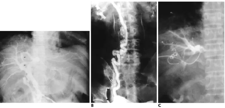
We first punctured the right portal vein under ultrasonic guidance and inserted a 4-F gliding catheter (Cobra type, Terumo) in the main portal vein via a guide wire. Direct percutaneous transhepatic portography revealed a dilat- ed paraumbilical vein originating from two branches of the left portal vein, heading toward the umbilicus, and communicating with the external iliac vein (Figs. 4A, 4B). Coil embolization of this dilated vein which origi- nated from the superior and inferior branches of the left portal vein, involved the placement of spring coils (Cook, Bloomington, U. S .A.) at the umbilical recess l e v- el, and nearly complete occlusion was achieved (Fig. 4C).
The coils used were one 3 c m×8 mm, one 3 c m×5 m m , and two 5 c m×5 mm in the superior branch of the left portal vein; and one 5 c m×8 mm, two 3 c m×5 mm, and one 5 c m×5 mm in the inferior branch of this vein.
After the procedure, the patient’s blood pressure was stabilized at 120/80 mmHg, her hemoglobin level re- mained above 10 gm/dl, and no more bleeding occurred.
Follow up CT scans obtained one month later showed a markedly decreased resolving hematoma in the ab- domen (Fig. 5).
D i s c u s s i o n
Spontaneous bleeding from an intraperitoneal varix is a rare condition, and the first such case was reported by Ellis et al in 1958 (3).
Among the anastomotic channels between the portal and systemic circulatory systems, the paraumbilical vein follows the path of the falciform ligament of the liv- er and anastomoses the anterior abdominal wall vein with the branches of the left portal vein. The umbilical vein normally collapses after birth, and in adults forms the ligamentum teres. With the development of portal hypertension, however, the umbilical vein may recanal- ize and serve as a porto-systemic collateral route. This condition is known as Cruveilhier-Baumgarten syn- drome, defined by Armstrong et al. as liver cirrhosis with portal hypertension, systemic collateral venous cir- culation including dilated umbilical or paraumbilical vein, caput medusa, venous hum, and splenomegaly (4).
The case we describe involves liver cirrhosis and in- cludes portal hypertension, splenomegaly and a dilated paraumbilical vein, and is thus considered to be Cru- veilhier-Baumgarten syndrome.
Fig. 1. A. Transverse sonogram in low- er abdomen shows heterogenous hy- perechoic mass of hematoma in lower abdominal cavity.
B . Longitudinal sonogram shows dilat- ed paraumbilical vein in abdominal w a l l .
There is controversy as to whether recanalization of the umbilical vein occurs in portal hypertension or whether this vein simply fails to be obliterated during development in patients in whom it is patent. Using per- cutaneous transhepatic portography, Aagaard et al re- ported a 26% incidence of umbilical vein patency in 107 patients with liver cirrhosis (5). Lafortune et al found no examples of portosystemic shunting via the umbilical vein in portal hypertension (6), but reported an increase in the number and caliber of the paraumbilical vein in 12% of their patients. The issue of recanalization versus congenital patency of the portal vein is thus unresolved,
and the fact remains that some portal hypertensives use the umbilical or paraumbilical vein as a porto-systemic collateral pathway.
A special characteristic of the paraumbilical vein is the absence of valves. Anson and McVay have claimed that in cases of portal hypertension, the diameter of the por- tal vein may be similar to that of the little finger, and that these veins are anastomosed with a vein from the gall bladder, the lesser omentum, and the lesser curve of the stomach (7). We therefore believe that rupture of a dilated paraumbilical vein could be the cause of this in- tra-abdominal bleeding. We have found 21 cases of vari-
A B
Fig. 2. Contrast enhanced pelvic CT.
A, B. Contrast enhanced CT obtained at iliac bone level (A) and more infeior level than A. (B) shows large hematoma in left abdom- inal wall (arrowhead) and intraperitoneal pelvic cavity(double arrowhead). Note prominent right inferior epigastric vein (arrow).
A B
Fig. 3. GI bleeding 9 9 mTc-RBC scan.
A, B. Early 15 minutes image (A) shows vertical oriented linear structure pre- sumed to be paraumbilical vein in right paramedian abdomen (arrows) and de- layed 4 hours image (B) also shows focal tracer uptake at mid-lower abdomen s u ggesting active state intraabdominal bleeding (double arrows)
ceal bleeding following intraperitoneal rupture, and six of these were shown to be due to rupture of a paraum- bilical vein involved in surgery.
variceal bleeding, early recognition and operative inter- vention, with ligation of the varix, are critical features of successful management (8). To identify the dilated pa- raumbilical vein and locate the focus of bleeding, we planned to perform percutaneous transhepatic portogra- phy, and to this end considered coil embolization rather than surgical intervention. To our knowledge, there are many reports of transhepatic portal venous emboliza- tion of varices (9), but no report has described coil em- bolization of the paraumbilical vein in hemoperitoneum due to ruptured intra-abdominal varix in a cirrhotic pa- tient with portal hypertension. Intuitively, it may ap- pear that occlusion of these large collateral umbilical veins would increase portal pressure and lead to in- creased variceal bleeding, but no association has been found between umbilical vein patency and portal pres- sure. A large patent paraumbilical vein does not, there- fore, effectively relieve portal hypertension, prevent
A B C
Fig. 4. Percutaneous transhepatic portography.
A, B. Percutaneous transhepatic portography shows dilated paraumbilical vein originated from two branches (arrows) of left portal vein heading toward umbilicus, and drained into the right external iliac vein (arrowhead).
C. After Coil embolization at proximal level of paraumbilical vein, no flow into dilated paraumbilical vein is noted.
Fig. 5. Follow-up CT scans after 1 month showed markedly de- creased size of the hematoma in abdomen.
R e f e r e n c e s
1 . Allan M. Goldstein, Neal Gorlick, David Gibbs, Carlos Fernandez- del Castillo. Hemoperitoneum due to spontaneous rupture of the umbilical vein. Am J Gastroenterol 1 9 9 5 ; 9 0 : 3 1 5 - 3 1 6
2 . Paizis B, Krespis E, Filiotou A, et al. Rupture of a peiumbilical vein causing hemoperitoneum in a cirrhotic patient. Mt Sinai J Med 1 9 8 6 : 5 3 : 1 2 3 - 1 2 5
3 . Ellis H, Griffiths PWW, Macintyre A. Haemoperitoneum-a record of 129 consecutive patients with notes on some unusual cases. Br J S u r g1 9 5 8 ; 4 5 : 6 0 6 - 6 1 0
4 . Armstrong EL, Adams WL. Tragerman LJ, et al. The Cruveilhier- Baumgarten syndrome: Review of the literature and report of two additional cases. Ann Intern Med 1 9 4 2 ; 1 6 : 1 1 3 - 1 5 1
5 . Aagaard J, Jensen LI, Sorensen TIA, Chritensen U, Burcharth F.
Recanalized umbilical vein in portal hypertension. AJR Am J Roentgenol 1 9 8 2 ; 1 3 9 : 1 1 0 7 - 1 1 0 9
6 . Lafortune M, Constantin A, Breton G, Legare AG, Lavoie P. The recanalized umbilical vein in portal hypertension: a myth. AJR Am J Roentgenol 1 9 8 5 ; 1 4 4 : 5 4 9 - 5 5 3
7 . Anson BJ, McVay CB. Surgical Anatomy, 5th ed. W.B. Saunders, 1971, vol. 1,582
8 . Sato H, Kamibayashi S, Tatsumura T, Yamamoto K. Intraabdomi- nal bleeding attributed to ruptured periumbilical varices. Jpa J Surg 1 9 8 7 ; 1 7 ; 3 3 - 3 6
9 . Sos TA. Transhepatic portal venous embolization of varices:pros and cons. Radiology 1 9 8 3 ; 1 4 8 : 5 6 9 - 5 7 0
1 0 . Burchell AR, Panke WF, Moreno AH, Rousselot LM. The patent umbilical vein in portal hypertension. Surg Gynecol Obstet 1 9 7 0 ; 1 3 0 : 7 7 - 8 6
대한방사선의학회지 2 0 00;42:7 51- 7 5 5
문맥고혈압을 가진 간경화 환자에서 부제대정맥 파열로 인한 복강내 혈종:
코일색전술 1예 보고11대전 선병원 방사선과
이종명・김형렬・이영환・이소현・김종건
부제대정맥은 문맥과 체순환과의 문합경로중의 하나로, 이런 복부정맥류의 파열은 문맥고혈압환자에서 보일 수 있는 드문 합병증중의 하나로 치명적인 복강내 혈종을 초래할 수 있다. 저자들은 초음파와 전산화 단층 촬영 술, 경피경간 문맥조영술로 확인한 부제대정맥의 파열로 인한 복강내 혈종예를 경험하였다. 문맥조영술상 확장 된 부제대정맥이 좌측 문맥에서 2개의 분지가 발생하여 우측 외장골 정맥으로 유입되는 것이 보였고, 복강내 혈 종의 발생부위로 생각되는 부제대정맥에 코일로 색전술을 시행하였다. 이에 문맥고혈압을 가진 간경화 환자에서 파열된 부제대정맥으로 인한 복강내 혈종과 이를 코일 색전술로 치료한 예를 보고한다.
2 0 0 0년도 제 1 1차 한일방사선의학회에 이어 제 2 2차 진단방사선과 전문의 연수강좌를 제주 K A L호텔에서 아래 일정과 같이 개최합니다. 여러 회원님들의 많은 협조와 참여를 바랍니다.
◈ 연 수 강 좌 ◈
대 회 일 시 : 2 0 0 0년 6월 1 0일(토)
대 회 장 소 : 제주 K A L호텔, 제주도 제주시 2도1동 1 6 9 1 - 9 연 락 처 : 전화: (064)724-2001, FAX: 064)720-6515
주 제 : Radiology in Emergency Medicine and Current Issues in Radiology 사 전등 록 비 : 2 0 , 0 0 0원(현장등록시 3 0 , 0 0 0원)
사전등록마감 : 2 0 0 0년 4월 2 9일까지(항공, 숙박, 가족관광, 친선체육대회 포함)
일 정 :
6월 1 0일(토) 13:00- 등 록
14:00-14:30 응급의학에서의 단순촬영: 근골격계 질환 정선관(원광의대) 14:30-15:00 응급의학에서의 자기공명영상: 뇌신경계 질환 김용선(경북의대) 15:00-15:30 응급의학에서의 CT: 흉부질환 최영희(단국의대) 15:30-16:00 응급의학에서의 초음파: 복부질환 차상훈(고려의대) 16:00-16:20 Coffee break
16:20-16:50 방사선과개원의의 향후 전망 한경민(개원의협의회)
16:50-17:20 상대가치의 의미와 방향 함창곡(한양의대)
17:20-17:50 최근 의료보험급여정책 방향 전병율(보건복지부)
17:50-18:20 간접촬영 필름 판독 임정기(서울의대)
19:00- 회원 친선의 밤
13:30-18:30 미니관광(동반가족에 한함)