INTRODUCTION
Fine needle aspiration (FNA) for thyroid nodule is a standard diagnostic procedure. One major limitation of FNA is a non-di- agnostic cytology result due to inadequate cytology specimen.
Although FNA generally yields sufficient cytology results, there have been questions with regard to aspiration pressure and nee- dle size. Previous studies evaluated the differences of sufficient sample rates between thick and thin needles and between the FNA with active suctions (FNAS) and FNA capillary techniques
(FNAC) and the result of the comparative studies were inconclu- sive (1-8). In this study, we compared the incidence of insufficient cytology results according to the aspiration techniques, especially focusing on the degree of suction pressure and needle size.
MATERIALS AND METHODS
From May 2009 to December 2010, three experienced radiolo- gists who are familiar to the ultrasound-guided aspiration, per- formed FNA studies consecutively in 1174 thyroid nodules from
J Korean Soc Radiol 2013;69(1):23-27 http://dx.doi.org/10.3348/jksr.2013.69.1.23
Received March 25, 2013; Accepted April 24, 2013 Corresponding author: Ji Kang Park, MD Department of Radiology, Jeju National University Hospital, Jeju National University School of Medicine, 15 Aran 13-gil, Jeju 690-767, Korea.
Tel. 82-64-717-1371 Fax. 82-64-757-8276 E-mail: [email protected]
This is an Open Access article distributed under the terms of the Creative Commons Attribution Non-Commercial License (http://creativecommons.org/licenses/by-nc/3.0) which permits unrestricted non-commercial use, distri- bution, and reproduction in any medium, provided the original work is properly cited.
This study was supported by a grant from the Jeju Natio- nal University Hospital Research Fund (2011).
Purpose: We compared the incidence of insufficient thyroid cytology due to blood- stained materials or low cellularity in terms of aspiration technique, especially fo- cusing on the degree of suction pressure and needle size.
Materials and Methods: Three experienced radiologists performed ultrasound-guid- ed aspiration for thyroid nodules in 1174 thyroid nodules consecutively. Three differ- ent techniques were used; (A) using a 25 gauge needle with mainly capillary tech- nique in 269 nodules; (B) using a 25 gauge needle with 3 cc syringe and minimal suction pressure in 303 nodules; (C) using a 22-23 gauge needle with 10 cc syringe and aspirator in 602 nodules. The differences of the incidence of the insufficient cy- tology among the three aspiration techniques and relationships of the incidence and needle size/degree of suction pressure was statistically analyzed using the Mann- Whitney U test and the chi-square test with linear-by-linear association.
Results: Overall, the difference in insufficient cytology was significant across the three aspiration technique (p = 0.004), and the incidence tended to increase signifi- cantly with increase of needle size and degree of suction pressure (p < 0.001). A pairwise comparison of aspiration techniques found significant differences (p = 0.003) between techniques (A) and (C), and no differences between technique (B) and (C) (p = 0.07) and between techniques (A) and (B) (p = 0.10).
Conclusion: The incidence of insufficient cytology was significantly low in the cap- illary technique, and it increased significantly with the increase of needle size and degree of suction pressure.
Index terms Thyroid Nodule Fine Needle Aspiration Insufficient Cytology
Evaluation of the Incidence on Insufficient Cytology Results
Comparing Different Ultrasound-Guided Aspiration Techniques for Thyroid Nodules
상이한 초음파 유도 흡인 기술에 따른 불충분 갑상선 세포검사 발생빈도의 비교 평가
Seong Hyung Kim, MD, Ju Hyun Park, MD, Ji Kang Park, MD
Department of Radiology, Jeju National University Hospital, Jeju National University School of Medicine, Jeju, Korea
between the incidence and invasiveness of the aspiration tech- nique among three technique groups was statistically analyzed using Pearson’s chi-square test with linear-by-linear association.
The pairwise incidence differences between each pair of groups was statistically analyzed using the Mann-Whitney U test and the relationships between the incidence of insufficient cytology results and invasiveness of aspiration technique among groups was analyzed by the Pearson’s chi-square test with linear-by-lin- ear association. Statistical analysis was performed using SPSS for Windows (SPSS Inc., an IBM Company, Chicago, IL, USA), and p-values of less than 0.05 were considered to represent statistical significance.
RESULTS
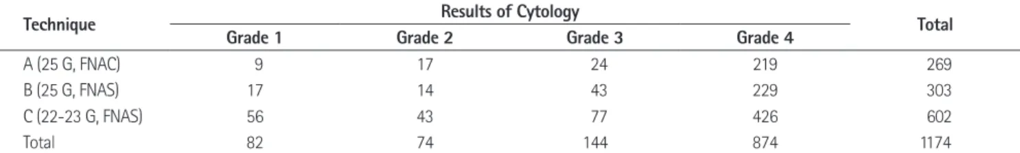
Malignant lesions were present in 15% (178/1174) of all nod- ules [14.9% (40/269) for group of technique (A), 15.5% (47/303) for group of technique (B), 15.1% (91/602) for group of technique (C)], and there was no significant differences in the incidence of malignant lesions between three groups. The insufficient cytology results occurred in 18.6% for the technique (A), 24.4% for the technique (B), and 29.2% for the technique (C), and detailed re- sults are summarized in Table 1. Overall, the difference in inci- dence of the insufficient cytology results was significant among the three aspiration techniques (p = 0.004), and the incidence of insufficient cytology results tended to increase significantly with the increase of needle size and degree of suction pressure (linear- by-linear association, p < 0.001). By comparing the two aspiration techniques, the difference was significant (p = 0.003) between technique (A) and (C), and not significant between technique (B) and (C) (p = 0.07) or between technique (A) and (B) (p = 0.10).
The proportional tendency between the incidence of insufficient results and needle size/degree of suction pressure was statistically 987 patients (322 men, 665 women; mean age 52.7 years) during
different time periods. The operators performed FNA studies over 5 to 17 months consecutively with or without overlapping time periods. The three radiologists used different aspiration techniques [we arbitrarily described as the technique (A), (B), and (C)], and they did not acknowledge that their FNA studies were conducted in different ways from one another. For tech- nique (A), 25 gauge needles were used mainly with capillary technique, and the radiologist had 2 years of experience in FNA.
For technique (B), 25 gauge needles with 3 cc syringe were used with intermittent minimal suction pressure, and the radiologist had 8 years of experience in FNA. For technique (C), 22-23 gauge needles with 10 cc syringe and aspirator were used for ac- tive aspiration, and the radiologist had 5 years of experience in FNA. The aspiration volume in technique (C) was usually to fill the needle hub. The aspiration volume in technique (A) and (B) was usually one third of the needle hub. Aspiration was usually conducted once or twice per nodule.
During the study period, the cytology was not reported ac- cording to the Bethesda system criteria. We retrospectively cate- gorized cytology results as four grades: grade 1 for blood only or mostly blood cells with scant cellularity, grade 2 for scanty follic- ular cells with hemorrhagic background, grade 3 for insufficient results due to scanty cellularity without mention of hemorrhagic background, and grade 4 for sufficient cytology specimen. We arbitrarily assumed invasiveness of three aspiration techniques according to the needle size and degree of suction pressure, and we assigned ordinal scales as followed; technique (A) as 1, tech- nique (B) as 2, and technique (C) as 3.
We evaluated the incidence of malignancy and analyzed whether the incidence of malignant lesions was different among the three groups by using the Kruskal-Wallis test. The difference in incidence of insufficient cytology results and the relationships
Table 1. Results of Cytology Specimen According to the Aspiration Technique
Technique Results of Cytology Total
Grade 1 Grade 2 Grade 3 Grade 4
A (25 G, FNAC) 9 17 24 219 269
B (25 G, FNAS) 17 14 43 229 303
C (22-23 G, FNAS) 56 43 77 426 602
Total 82 74 144 874 1174
Note.-Grade 1 for blood only or mostly blood cells with scant cellularity, Grade 2 for scanty follicular cells with hemorrhagic background, Grade 3 for in- sufficient results due to scanty cellularity without mention of hemorrhagic background, Grade 4 for sufficient cytology specimen.
FNAC = fine needle aspiration capillary technique, FNAS = fine needle aspiration with active suction
ume of aspiration. If we combined large needle sizes with active suction, we may have a large volume of aspiration rapidly, but the chance of blood-stained materials occupying the volume may be increased and the incidence of insufficient cytology may increase.
From the patient’s perspective, a thinner needle with capillary technique may cause the least discomfort such as pain and bleed- ing within the thyroid gland. In addition, although the chances are extremely low, the possibility of needle tract seeding during FNA may be smaller in finer needles and FNAC (10).
We have encountered some limitations in this study. First, we did not consider nodule characteristics such as vascularity, echo- genicity, and size of the nodules. The imaging characteristics such as echogenicity, vascularity, and size may affect the adequa- cy of cytology specimen. Heterogeneous echoic nodule, cystic portions > 50%, and sizes less than 5 mm or larger than 10 mm were all related to the inadequate cytology results (9, 11). In our study, the operator performed FNA studies consecutively and in- dependently, and the incidences of malignancy among three groups were not statistically different. Therefore, we think that the imaging characteristics of the nodules may not be significant- ly different between the three groups. Second, the cytology re- sults might not be consistent because specimens were read by several pathologists during the study, and cytology reports were not made by the standardized form such as the Bethesda system.
In conclusion, the incidence of insufficient cytology result was significantly different according to aspiration technique, and it was least in the capillary technique. The incidence of insufficient cytology result tended to increase significantly with increase of needle size and degree of suction pressure.
REFERENCES
1. Tublin ME, Martin JA, Rollin LJ, Pealer K, Kurs-Lasky M, significant between technique (A) and (C) (linear-by-linear asso-
ciation, p < 0.001), technique (B) and (C) (p = 0.02).
DISCUSSION
In our study, FNAC with a smaller fine needle yielded better results in terms of sufficient cytology specimen. Cytology ade- quacy is important for accurate diagnosis of thyroid nodule.
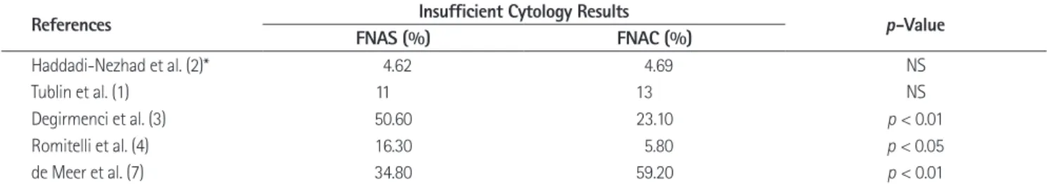
There have been controversial results about the needle sizes and aspiration techniques for adequate cytology specimen in thyroid nodules, and the previous results were summarized in Table 2.
Previous comparative studies have shown no significant differ- ences between FNAC and FNAS (1, 2). However, there have been one-sided results favoring FNAC (3, 4) or FNAS (7). Simi- larly in the aspiration technique, there have been no prominent differences in results on needle sizes (5, 6, 9), or one-sided re- sults favoring smaller needles (3, 7) or larger needles (8). In summary, although sufficient volume of aspiration may be ob- tained by using larger needles and active suction, it may not guarantee adequacy of cytology specimen and may be more eas- ily obscured by red blood cells (5, 6).
In our study, a mixed combination of the aspiration techniques and needle sizes could be compared fortuitously, and FNAC re- sulted in better cytology adequacy. The volume of aspiration in FNAC was relatively smaller than the volume of FNAS within our study. Although it may seem peculiar that the smaller volume of aspiration could result in better cytology adequacy, we interpret our results in terms of needle sizes and degrees of suction. Gener- ally, we can see several vessel structures in thyroid nodules on a color Doppler study. In our opinion, although the difference of needle size was about 0.2 mm in our study, large sized needles may be more likely to puncture or penetrate the vessel structure.
One of the criteria of needle withdrawal during FNA is the vol-
Table 2. Summary of the Comparative Studies between FNAS and FNAC
References Insufficient Cytology Results p-Value
FNAS (%) FNAC (%)
Haddadi-Nezhad et al. (2)* 4.62 4.69 NS
Tublin et al. (1) 11 13 NS
Degirmenci et al. (3) 50.60 23.10 p < 0.01
Romitelli et al. (4) 16.30 5.80 p < 0.05
de Meer et al. (7) 34.80 59.20 p < 0.01
Note.-*Haddadi-Nezhad (2003) compared the qualitative score of cytology specimen.
FNAC = fine needle aspiration with capillary technique, FNAS = fine needle aspiration with active suction
fine-needle aspiration biopsy of the thyroid but may not provide increased diagnostic accuracy. Thyroid 2001;11:
973-976
7. de Meer SG, Schreinemakers JM, Zelissen PM, Stapper G, Sie-Go DM, Rinkes IH, et al. Fine-needle aspiration of thy- roid tumors: identifying factors associated with adequacy rate in a large academic center in the Netherlands. Diagn Cytopathol 2012;40 Suppl 1:E21-E26
8. Hanbidge AE, Arenson AM, Shaw PA, Szalai JP, Hamilton PA, Leonhardt C. Needle size and sample adequacy in ul- trasound-guided biopsy of thyroid nodules. Can Assoc Ra- diol J 1995;46:199-201
9. Inci MF, Özkan F, Yüksel M, S¸ alk I˙, S¸ ahin M. The effects of sonographic and demographic features and needle size on obtaining adequate cytological material in sonography- guided fine-needle aspiration biopsy of thyroid nodules.
Endocrine 2013;43:424-429
10. Polyzos SA, Anastasilakis AD. A systematic review of cases reporting needle tract seeding following thyroid fine nee- dle biopsy. World J Surg 2010;34:844-851
11. Choi YS, Hong SW, Kwak JY, Moon HJ, Kim EK. Clinical and ultrasonographic findings affecting nondiagnostic results upon the second fine needle aspiration for thyroid nod- ules. Ann Surg Oncol 2012;19:2304-2309
Ohori NP. Ultrasound-guided fine-needle aspiration versus fine-needle capillary sampling biopsy of thyroid nodules:
does technique matter? J Ultrasound Med 2007;26:1697- 1701
2. Haddadi-Nezhad S, Larijani B, Tavangar SM, Nouraei SM.
Comparison of fine-needle-nonaspiration with fine-nee- dle-aspiration technique in the cytologic studies of thy- roid nodules. Endocr Pathol 2003;14:369-373
3. Degirmenci B, Haktanir A, Albayrak R, Acar M, Sahin DA, Sahin O, et al. Sonographically guided fine-needle biopsy of thyroid nodules: the effects of nodule characteristics, sampling technique, and needle size on the adequacy of cytological material. Clin Radiol 2007;62:798-803
4. Romitelli F, Di Stasio E, Santoro C, Iozzino M, Orsini A, Ce- sareo R. A comparative study of fine needle aspiration and fine needle non-aspiration biopsy on suspected thyroid nodules. Endocr Pathol 2009;20:108-113
5. Gümüs¸ M, Cay N, Algin O, Ipek A, Ersoy RÜ, Belenli O, et al.
Comparison of 21 and 27 gauge needles for determining sample adequacy in the aspiration biopsy of thyroid nod- ules. Diagn Interv Radiol 2012;18:102-105
6. Tangpricha V, Chen BJ, Swan NC, Sweeney AT, de las Morenas A, Safer JD. Twenty-one-gauge needles provide more cellular samples than twenty-five-gauge needles in
˙
상이한 초음파 유도 흡인 기술에 따른 불충분 갑상선 세포검사 발생빈도의 비교 평가
김승형 · 박주현 · 박지강
목적: 갑상선 결절의 세침흡인검사에서 흡인 압력의 정도와 바늘 크기가 서로 다른 3가지 검사방법에 따른 불충분 병리 결과의 발생빈도를 조사하고자 하였다.
대상과 방법: 3명의 경험이 풍부한 영상의학과 의사가 1174개의 갑상선 결절에서 연속적으로 초음파 유도하 세침흡인검 사를 다음의 세 가지 방법으로 각각 시행하였다; (A) 269개의 결절은 25 게이지 바늘로 모세관 기법을 이용, (B) 303개 의 결절은 25 게이지 바늘과 3 cc 주사기로 최소한의 흡인 압력을 이용, (C) 602개의 결절은 22-23 게이지 바늘, 10 cc 주사기와 흡인기를 이용해 세침흡인검사를 시행하였다. 세 가지 상이한 방법에서 생긴 불충분 세포검사결과의 발생빈도 가 차이가 있는지를 통계적으로 분석하였다.
결과: 전반적으로, 불충분 세포검사결과의 발생빈도의 차이는 세 흡인 방법에 따라 의미 있는 차이를 보였고(p =
0.004), 바늘 크기와 흡인 압력이 증가할수록 그 발생빈도가 증가하는 경향을 보였다(p < 0.001). 세침흡인방법들의 쌍
별 비교에서는 방법 (A)와 (C)에서는 의미 있는 차이를 보였으나(p = 0.003), 방법 (B)와 (C)(p = 0.07) 및 방법 (A)와 (B)(p = 0.10) 사이에는 의미 있는 차이는 없었다.
결론: 불충분한 세포검사결과의 발생빈도는 모세관 기법에서 유의하게 낮았고, 바늘 크기와 흡인 압력의 정도가 증가함 에 따라 유의하게 증가하는 경향을 보였다.
제주대학교 의학전문대학원 제주대학교병원 영상의학과