INTRODUCTION
Intrathoracic segment of the subclavian artery is a rare loca- tion for a peripheral arterial aneurysm (1-3). Intrathoracic seg- mental aneurysm is secondary to atherosclerosis, medial degen- eration, trauma, and infection. Subclavian artery aneurysms are at an increased risk of rupture, embolization, or throm- bosis. Hence, they should be considered for surgical repair.
We describe a case of a patient presenting with hoarseness and an expanding intrathoracic mass that was due to a right subclavian aneurysm.
CASE REPORT
A 65-yr-old female presented with hoarseness that began 3 weeks ago. She had no previous history of trauma, pulmo- nary and bronchial tuberculosis. On admission, her vital signs were normal and blood pressure was equal on both arms. The laboratory workup showed a white blood cell count of 6,500/
L, normal erythrocyte sedimentation rate and C-reactive protein. Syphilis serology, rheumatoid factor, and antinuclear antibody tests were negative. Chest radiograph (CXR) demon- strated a superior mediastinal mass (Fig. 1). Because laryngo- scopy showed a fixed right vocal cord, hoarseness was con- sidered to be due to recurrent laryngeal nerve palsy. Com-
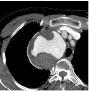
puted tomography (CT) and magnetic resonance imaging (MRI) demonstrated a 7 cm sized proximal right subclavian artery aneurysm (Fig. 2, 3). Thoracic and subclavian aortog- raphy revealed a tortuous right subclavian arterial anatomy with a saccular aneurysm beginning near the subclavian ori- gin (Fig. 4). The preoperative diagnosis was atherosclerotic right subclavian artery aneurysm.
Repair of the aneurysm was performed through a median sternotomy. We dissected at the aneurysmal afferent loop and efferent loop. We made end-to-end anastomosis by Gore Tex (6 mm sized) graft interposition. In the operation field, we could not find the recurrent laryngeal nerve due to severe adhesions. After the debridement, a partially undetached aneurysm was left.
Cultures from the wall of the aneurysm and thrombus were negative. Pathology was consistent with an atherosclerotic aneurysm filled with thrombus (Fig. 5).
After one month follow-up, she still complains of hoarse- ness and laryngoscopic examination revealed right vocal cord palsy.
DISCUSSION
Aneurysms of the subclavian artery represent about 1% of all peripheral arterial aneurysms (4, 5). They fall into two dis-
Hong Gun Bin, Myoung Sook Kim, Seok Chan Kim, Jong Bum Keun*, Jong Ho Lee*, Seung Soo Kim
Departments of Internal Medicine, Thoracic Surgery*, College of Medicine, The Catholic University of Korea, Seoul, Korea
Address for correspondence Seung Soo Kim, M.D.
Department of Internal Medicine, College of Medicine, The Catholic University of Korea, St. Mary’s Hospital, 520-2 Daeheung-dong, Jung-gu, Daejeon 301-723, Korea
Tel : +82.42-220-9811, Fax : +82.42-255-8663 E-mail : [email protected]
674 J Korean Med Sci 2005; 20: 674-6
ISSN 1011-8934
Copyright � The Korean Academy of Medical Sciences
Intrathoracic Aneurysm of the Right Subclavian Artery Presenting with Hoarseness
: A Case Report
Intrathoracic segment of the subclavian artery is an unusual location for peripheral arterial aneurysms. They are normally caused by atherosclerosis, medial degener- ation, trauma, and infection. We report a case of a patient with right subclavian artery aneurysm presenting with hoarseness. Chest radiograph demonstrated a superior mediastinal mass. Laryngoscopy showed a fixed right vocal cord. By chest computed tomography, magnetic resonance imaging, and angiography, preopera- tive diagnosis was established as a saccular aneurysm with afferent loop and effer- ent loop. Patient underwent complete resection of the aneurysm followed by end- to-end anastomosis via median sternotomy. Postoperative pathology was consis- tent with an atherosclerotic aneurysm filled with thrombus. After surgical operation, hoarseness is still continued.
Key Words : Aneurysm; Hoarseness; Subclavian Artery
Received : 28 July 2004 Accepted : 14 September 2004
Intrathoracic Aneurysm of Right Subclavian Artery 675
tinct groups in terms of etiology, presentation, and treatment:
those of the intrathoracic and those of the extrathoracic por- tion of the subclavian artery. Although aneurysms of extratho- racic subclavian artery are related to thoracic outlet syndrome or to previous trauma, intrathoracic segmental involvement is mainly due to atherosclerosis (6).
Intrathoracic aneurysms are most often asymptomatic but can present with symptoms caused by compression or acute
aneurysm expansion such as upper chest or shoulder pain, Horner’s syndrome, venous congestion, and hoarseness. Symp- toms due to distal embolization to the arm are unusual. Extra- thoracic aneurysm most commonly presents with a pulsatile mass in the superior fossa and is often tender, which may be noted by the patient or examiner. Brachial plexopathy is anoth- er complication of the extrathoracic aneurysm and Horner’
syndrome is not infrequent (5-7).
Fig. 1.PA chest roentgenogram shows well-defined, paratracheal mass of about 7 cm in the upper zone of the right hemithorax.
R
Fig. 2.Contrast enhanced computed tomography (CT) demon- strates a 7 cm sized proximal right subclavian artery aneurysm and intraluminal yin-yang appearance due to thrombus.
Fig. 3.Reconstructed three-dimensional magnetic resonance imaging (MRI) demonstrated positional relationships between right subclavian artery aneurysm and surrounding vessel.
Fig. 4.Preoperative digital subtraction angiography of mass reveal- ing an saccular aneurysm of right subclavian artery with afferent and efferent loop and intraluminal filling defects.
676 H.G. Bin, M.S. Kim, S.C. Kim, et al.
Because isolated true aneurysm of the subclavian artery is rare, the natural history of subclavian artery aneurysm is un- clear. It has been reported that an aneurysm of the aberrant right subclavian artery grew at the rate of 0.42 cm/yr mea- sured by CXR (8). In another report, growth rate was report- ed at 1.31 cm/yr in a true left subclavian artery aneurysm measured by CT scan (9).
Elective surgical repair is the treatment of choice for most subclavian aneurysms, because they have an increased risk of rupture, embolization, thrombosis, and other complications.
Intrathoracic subclavian artery aneurysms on the right are best approached by median sternotomy, whereas a high lat- eral thoracotomy is preferred for the left sided aneurysm (6, 10). Resection or aneurysmorrhaphy is preferred to simple ligation because continued growth and rupture of ligated aneurysm have been reported (11). Recently, as less invasive alternative to surgical repair, endovascular stent-graft treat- ment is possible (12-15).
We describe a case of a patient who complained of hoarse- ness and right shoulder pain due to an aneurysm of the intra- thoracic subclavian artery. We treated her with graft inter- position and left the aneurysm tissue unattached because of severe adhesion.
REFERENCES
1. Chung YC, Jeong UG, Cho YK. A case report of subclavian arteri- al aneurysm. J Korean Surg Soc 1978; 20: 83-6.
2. Kim HJ, Kim SS, Huh JD, Chun BH, Joh YD, Cho SR. Intratho- racic aneurysm of the right subclavian artery. J Korean Radiol Soc 1989; 25: 725-7.
3. Kim HK, Kim KH, Park YS, Lee WH, Chug EC, Han WS. Subcla- vian artery aneurysm: Report of a case. Korean J Thorac Cardio- vasc Surg 1993; 26: 557-9.
4. Dougherty MJ, Calligaro KD, Savarese RP, DeLaurentis DA. Athero- sclerotic aneurysm of the intrathoracic subclavian artery: a case report and review of the literature. J Vasc Surg 1995; 21: 521-9.
5. Witz M, Yahel J, Lehmann JM. Subclavian artery aneurysms. A report of 2 cases and a review of the literature. J Cardiovasc Surg 1998; 39: 429-32.
6. Davidovic LB, Markovic DM, Pejkic SD, Kovacevic NS, Colic MM, Doric PM. Subclavian artery aneurysms. Asian J Surg 2003; 26: 7- 11.
7. Utikal P, Bachleda P, Kocher M, Novotny J, Drac P, Drac P. Aneurysm of the subclavian artery. Acta Univ Palacki Olomuc Fac Med 1999;
142: 107-9.
8. Hogg JP, Dominic AJ, Counselman RL, Hurst JL. Expanding aneu- rysm of aberrant right subclavian artery. Case report and imaging evaluation. Clin Imaging 1997; 21: 195-9.
9. Takagi H, Mori Y, Umeda Y, Fukumoto Y, Yoshida K, Shimokawa K, Hirose H. Proximal left subclavian artery aneurysm presenting hemoptysis, hoarseness, and diplopia: repair through partial car- diopulmonary bypass and perfusion of the left common carotid artery.
Ann Vasc Surg 2003; 17: 461-3.
10. Salo JA, Ala-Kulju K, Heikkinen L, Bondestam S, Ketonen P, Luos- to R. Diagnosis and treatment of subclavian artery aneurysms. Eur J Vasc Surg 1990; 4: 271-4.
11. McCann RL. Basic data related to peripheral artery aneurysms. Ann Vasc Surg 1990; 4: 411-4.
12. Kim DK, Yoon YS, Choi SH, Lee DI, Lee DY, Chang BC, Shim WH. A case of transluminal stent-graft implantation at right subcla- vian artery pseudoaneurysm in Behcet’s syndrome. Korean Circ J 1999; 29: 1240-4.
13. Ko KH, Won JW, Won JY, Lee DY, No KS, Lee JT. Endoluminal placement of stent-graft for the treatment of peripheral saccular aneurysm. J Korean Radiol Soc 2002; 46: 213-9.
14. Schoder M, Cejna M, Holzenbein T, Bischof G, Lomoschitz F, Funo- vics M, Nobauer-Huhmann I, Sulzbacher I, Lammer J. Elective and emergent endovascular treatment of subclavian artery aneurysms and injuries. J Endovasc Ther 2003; 10: 58-65.
15. Kasirajan K, Matteson B, Marek JM, Langsfeld M. Covered stents for true subclavian aneurysms in patients with degenerative connec- tive tissue disorders. J Endovasc Ther 2003; 10: 647-52.
Fig. 5.(A) Microscopically, atheromatous plaque contains amor- phous pink material with slit-like “cholesterol clefts” of lipid mate- rial and calcification. There is recent hemorrhage on right side (H&E stain, ×20). (B) At higher magnification, many foam cells and a cholesterol cleft are seen (H&E stain, ×200).
A
B