INTRODUCTION
An increasing number of patients are suffering from verte- bral compression and destructive lesions from osteoporosis and cancer in the aging society. Although the optimum time for the procedure is controversial, percutaneous vertebroplasty (PVP) or kyphoplasty is a the key technique to reduce life- threatening pneumonia, urinary tract infection and other complications from prolonged bed rest and unrelieved back and radiating flank, abdomen and groin pain (1). PVP with or without uni- or bi-lateral facet joint blocks gives the patient immediate pain relief and it also allows fast recovery of ambu- lation.
Excluding 30 cases of operations by bipedicular approaches, we reviewed 500 patients who had undergone PVP at 612 vertebral levels in terms of the criteria of patient selection, operation technique, clinical outcomes and the follow-up course including medication and the medial branch lesion- ing of the posterior ramus.
MATERIALS AND METHODS
This study was approved by the Ethics Committee for Hu- man Study of Pusan National University Hospital, and in- formed consent was obtained from each patient. After exclu- ding 30 cases of operations by bilateral pedicular approach
without the concept of radicular pain due to facet joint load- ing, a total of 500 patients suftering from vertebral compres- sion and destructive fractures from osteoporosis and cancer had PVP with or without facet joint blocks for two years.
We analyzed the period of pain, numeric rating scale (NRS) scores for pain, Oswestry Disability Index (ODI) except the data about sex life, and Karnofsky performance status (KPS) before and after the procedure.
Neurologic examinations were performed to rule out other disorders. Supraspinous tenderness was marked with a paper clip for vertebroplasty, and adjacent facet joint tenderness was pointed with a marking pencil for facet joint block in prone position. CT or MRI and radioactive bone scan were done. For the performance of the unipedicular approach, we measured the angle and distance from the midline on the skin from the CT or MRI.
Basic laboratory tests for preoperative workup and for post- operative follow-up included coagulation tests, liver and renal function tests, and bone mineral density (BMD).
On operation, we performed uni- or bilateral facet joint blocks before PVP according to the complaint of patients, usually neighboring the lesion of the vertebrae.
Lidocaine (1%) was infiltrated from the skin to periosteum near the pedicle under the fluoroscopic guidance. We perfor- med epidural analgesia before or during the procedure around the level of stable vertebral fractures in some patients who could not maintain a prone position. And we injected 30 mg
Tae-Kyun Kim, Kyung-Hoon Kim, Cheul-Hong Kim, Sang-Wook Shin, Jae-Young Kwon, Hae-Kyu Kim, Seong-Wan Baik
Department of Anesthesia and Pain Medicine, College of Medicine, Pusan National University, Busan, Korea
Address for correspondence Kyung-Hoon Kim, M.D.
Department of Anesthesia and Pain Medicine, College of Medicine, Pusan National University, 1-10 Ami-dong, Seo-gu, Busan 602-739, Korea
Tel : +82.51-240-7394, Fax : +82.51-242-7466 E-mail : [email protected]
*This study was supported by a Grant-in Aid from Pusan National University and Hospital in 2003.
1023
Percutaneous Vertebroplasty and Facet Joint Block
It is surprising that about 24% of patients with benign osteoporotic vertebral fracture die within a year from respiratory infection and urinary tract infection because of coughing and voiding difficulties, depending on the sites of compression fractures.
We reviewed 500 patients on whom percutaneous vertebroplasty (PVP) was per- formed, at 612 levels in terms of patient selection, operation technique, medication, and clinical outcomes during the follow-up course for 2 yr study period. To confirm the most painful level among the multiple fracture sites, physical examination after facet joint block under the fluoroscope was the most reliable method. The mean total lumbar spine fracture threshold of bone mineral density was 0.81±0.05 g/cm2. The mean changes of numeric rating scale scores, Oswestry Disability Index except sex life, and Karnofsky performance status were -72.00, -83.50 and +60.62% in the os- teoporosis group and -51.89, -45.02, and 69.03% in the tumor group. Complications related to the procedure were lateral spinal cord damage, transient paresthesia and transient hypotension. PVP with facet joint block is a profitable method for the ver- tebral compression fracture because of low risk and short duration of procedure with a high chance to result in pain relief and early mobilization.
Key Words : Zygapophyseal Joint; Facet Joint Block; Percutaneous Vertebroplasty; Pain Clinics; Spinal Fractures
Received : 24 March 2005 Accepted : 31 May 2005
ketorolac and 50 g of fentanyl intravenously. In some pa- tients who complained of severe anxiety about the procedure, we gave 2-3 mg of midazolam intramuscularly before the procedure or intravenously during the procedure.
Under the fluoroscope, the entry point of local anesthetic needle and 11 gauge out-diameter 10 cm length vertebroplasty needle (J-type Bone Marrow Needle�, Manan Medical Prod- uct, Wheeling, IL, U.S.A.) became the meeting point of tra- jectory line from the pedicle to the body in the lateral view and sagittal line away from the midline in the antero-poste- rior (AP) view. The distance from the midline to sagittal line was determined by CT or MRI.
From the AP view, the target point was the upper lateral part of the pedicle. A small incision was made with No. 15 scalpel. Under the lateral view of fluoroscope, a vertebral nee- dle was advanced carefully through the pedicle by manual turning and pressing of a handle without a hammer. The final reaching point would be anterior 1/4 to 1/3 on the lateral view and midline of the vertebral body on the AP view. Under con- tinuous monitoring using the fluoroscope, the contrast medi- um was injected through the vertebral needle and any evidence of leakage of contrast media to intravenous and epidural space was checked. After testing of the filling into the body and leak- age into the intravenous and epidural space of contrast media, antibiotics-containing polymethyl methacrylate (PMMA) ce- ment was injected by using an injector. The whole injection procedure was monitored with a continuous fluoroscopy to avoid possible leakage. The skin was closed by skin closure (Steri-Strip�, 3M, St. Paul, MN, U.S.A.) and piroxicam patch (Trast Patch�, SK Pharma., Seoul, Korea).
For 2 hr after the procedure, hypotension, dyspnea, and sensory or motor problems were checked in the recovery room, and we asked patients to walk with help, and when possible the patient was discharged.
The patients were followed-up at 1 week, 1, 2, 3, 6, 9, and 12 months after the procedure. We measured the BMD of lumbar spine and femur every 3 months.
After the procedure, the major analgesics for the pain con- trol were changed from codeine-paracetamol-ibuprofen mix- ture (Myprodol�, Adcock Ingram Pharmaceuticals, Johannes- burg, South Africa) to tramadol-acetaminophen mixture (Ul- tracet�, Janssen Korea, Whasung, Korea) for 1 week, and then we usually stopped the analgesic administration. Medication for osteoporosis was 70 mg of alendronate sodium (Fosamax�,
Merck Sharp & Dohne, de Mexido, S. A de C.V, Mexico) week- ly with or without 60 mg of raloxifene HCl (Evista�, Eli Lily
& Co, Hamsphire, U.K.) daily. BMD was measured by dual energy radiography absorptiometry (DEXA, Lunar PRODI- GY, GE System, Madison, WI, U.S.A.) at 3-month intervals.
After facet joint blocks, we evaluated the period of relapse of symptoms and the need for denervation of medial branch of posterior ramus.
Unless indicated otherwise, the data were presented as mean
±SD. The data were analyzed by using Wilcoxon signed rank test of nonparametric test for therapeutic effects includ- ing NRS, ODI, and KPS before and after PVP with facet joint block between the osteoporosis and tumor groups and for the comparison of combination effects of alendronate and raloxi- fene in osteoporosis.
RESULTS
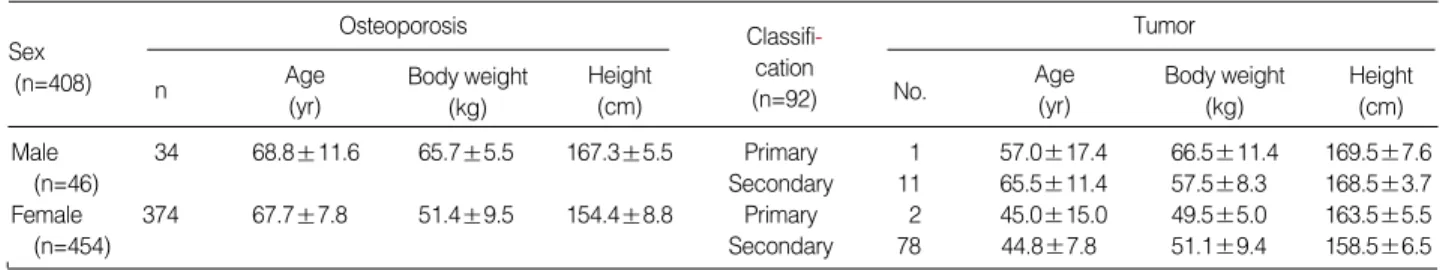
In 500 patients, PVP was performed on 612 vertebral levels with or without facet joint blocks during the 2-yr study peri- od. The demographic data of the patients are shown in Table 1.
Before the procedure, 10 patients suffered from pneumo- nia, 7 patients complained of voiding difficulties, and 4 pa- tients had mild dementia due to posttraumatic organic dis- order after the osteoporotic vertebral compression fractures.
After conservative treatment with corset and medication for 2 weeks, 96.2% (500/520) of patients wanted to receive PVP because of insufficient pain control.
Eighty eight percent of patients (440/500) who complained of radicular pain to the flank, abdomen, or the buttock, recei- ved uni- or bilateral facet joint block around the fractured ver- tebrae before PVP. In 30 cases of patients by bilateral pedic- ular approach without the concept of the radicular pain due to facet joint loading, the mean reduction of ODI except sex life, NRS scores for pain and KPS were 56.1±8.8%, 3.5± 1.2, and 42.2±12.6, compared with 82.2±11.2%, 5.4± 1.3 and 52.3±14.5 in subsequent 500 cases of patients with who had simultaneous facet joint block.
The distribution of vertebral levels of operations and pedicle width, angle and distance from the midline on the skin from the CT or MRI targeting to the anterior 1/3 to 1/4 for PVP with unipedicular approach are shown in Table 2 and Table 3.
It was possible to perform unipedicular approach from T4
Osteoporosis
Male 34 68.8±11.6 65.7±5.5 167.3±5.5 Primary 1 57.0±17.4 66.5±11.4 169.5±7.6
(n=46) Secondary 11 65.5±11.4 57.5±8.3 168.5±3.7
Female 374 67.7±7.8 51.4±9.5 154.4±8.8 Primary 2 45.0±15.0 49.5±5.0 163.5±5.5
(n=454) Secondary 78 44.8±7.8 51.1±9.4 158.5±6.5
Table 1.Demographic data of the study subjects (n=500)
Values represent mean ±SD. n, number of patients.
Sex
(n=408) n Age
(yr)
Body weight No. (kg)
Classifi- cation (n=92) Height
(cm) Body weight
(kg)
Age (yr)
Height (cm) Tumor
to L5 level. During the procedure, 74.3% (455/612) levels showed epidural and/or vascular leakage of contrast media under the fluoroscopy.
The mean changes of NRS scores for pain, ODI except sex life, and KPS were -72.00, -83.50 and +60.62% in the osteo- porosis group and -51.89, -45.02, and +69.03% in the tumor group (Table 4).
As for the diagnostic tools to pinpoint the painful levels among the multiple fractures, plane radiography film, CT or MRI, and bone scan showed discrepant results. To confirm the most painful level among the multiple fracture sites and to get the clinical success to relieve pain, physical examina- tion after facet joint block under the fluoroscope was the most reliable method.
During the follow-up course, 60% (264/440) of the pa- tients who received facet joint block complained of recurring radicular pain in 3.5±1.2 months (range, 1.8 to 11.2 mon- ths). Among them, 50% (132/264) received conventional ra-
diofrequency ablation.
Complications related to the procedure were 1 case of later- al spinal cord syndrome on L1, 3 cases of transient paresthe- sia for 1 week, and 1 case of severe hypotension (70/40 mmHg) with bradycardia related to anterior leakage to the sympathetic nerves, which was immediately recovered by ephedrine.
The mean changes of lumbar spine and femoral neck BMD after 3 and 6 months of treatment with alendronate (ALN) with or without raloxifene (RLX) in the osteoporotic group were shown in Table 5.
DISCUSSION
With regard to conservative treatment which includes bed rest, analgesic medication, and orthotics, conservative thera- py takes a long time and can cause difficult problems. Uncon- trolled continuous pain can cause difficulties in coughing and mobilization, which finally leads to respiratory and urinary tract infection and eventual possible dementia. trauma toward dementia. We should consider spinal fracture as a major trau-
Level of site
T4 0 0 2 124
T5 0 0 1 (20.26%)
T6 21 1 0
T7 10 1 1
T8 21 0 0
T9 42 1 0
T10 22 1 0
Subtotal 116 4 4
(18.95%) (0.65%) (0.65%)
T11 63 0 32 370
T12 84 0 21 (60.46%)
L1 105 0 11
L2 41 0 13
Subtotal 293 0 77
(47.88%) (12.58%)
L3 41 0 5 118
L4 33 0 6 (19.28%)
L5 21 0 12
Subtotal 95 0 23
(15.52%) (3.76%)
Total 504 108 612
(82.35%) (17.65%) (100%)
Table 2.The distribution of 612 vertebral levels in 500 patients who underwent percutaneous vertebroplasty with joint block ac- cording to the patient group
Osteo-
porosis Primary Secondary Tumor
Subtotal
Width of Smallest Largest Shortest Longest Level pedicle angle angle distance distance
(cm) (°) (°) (cm) (cm)
T4 0.29±0.10 19.25±1.10 28.06±2.70 2.45±0.50 3.55±0.50
T5 0.30 19.50 29.00 2.53 3.60
T6 0.33±0.10 17.42±2.14 28.85±1.87 2.51±0.39 4.09±0.48 T7 0.39±0.12 18.72±2.11 28.82±1.87 2.46±0.56 3.56±0.39 T8 0.44±0.11 17.33±3.20 29.20±1.97 2.37±0.55 3.26±0.60 T9 0.55±0.13 16.50±2.60 28.86±1.88 2.27±0.45 3.39±0.51 T10 0.64±0.90 16.25±2.00 29.33±1.72 2.21±0.52 3.41±0.41 T11 0.70±0.15 18.42±3.06 32.05±3.22 2.38±0.36 3.81±0.49 T12 0.74±0.20 20.62±2.62 34.20±2.86 2.61±0.52 4.30±0.81 L1 0.78±0.23 20.37±2.93 32.54±2.28 2.58±0.45 4.17±0.61 L2 0.81±0.22 21.00±2.88 33.96±3.29 2.91±0.39 4.80±0.98 L3 0.84±0.35 21.37±2.96 34.53±3.61 3.40±0.78 5.50±1.28 L4 0.88±0.44 22.18±3.98 36.96±3.86 3.38±0.71 5.87±1.01 L5 1.21±0.48 23.95±4.68 44.45±8.52 3.43±0.58 6.82±1.25 Table 3.Pedicle width, angle and distance from the midline on the skin on CT or MRI targeting to the anterior midline 1/3 to 1/4 for PVP with unipedicular approach
Osteoporosis (n=408) Tumor (n=92) p value NRS (-5.4/7.5) * 100=-72.00% (-4.1/7.9) * 100=-51.89% 0.0015 ODI (-25.3/30.3) * 100=-83.50% (-27.1/60.2) * 100=-45.02% 0.0017 KPS (+29.4/48.5) * 100=+60.62%(+29.2/42.3) * 100=+69.03% 0.0056 Table 4. Mean changes of numeric rating scale (NRS), Oswestry disability index (ODI), and Karnofsky performance status (KPS) after percutaneous vertebroplasty (PVP) with uni- or bi-lateral fa- cet joint blocks
Site Duration ALN ALN + RLX p
(n=100) (n=100) value Lumbar Baseline (g/cm2) 0.70±0.06 0.69±0.10 0.5834
spine 3 month (%) 4.74±0.60 6.81±0.53 0.1714 (L1-4) 6 month (%) 7.11±0.61 8.72±0.44 0.1524 Femoral Baseline (g/cm2) 0.56±0.07 0.57±0.72 0.4537 neck 3 month (%) 4.63±0.52 5.28±0.07 0.2117 6 month (%) 5.62±0.91 8.01±1.21 0.0108 Table 5. Mean changes from the baseline in lumbar spine and femoral neck bone mineral density after 3 and 6 months of treat- ment with alendronate (ALN) with or without raloxifene (RLX) in the osteoporotic group by DEXA
ma in one’s life. Therefore, we tried to reduce hospital stay much as possible to minimize patients’ maladaptation from life-style including space (home to hospital), caring person (family to nursing personnel), and time (including sleeping and feeding).
For multiple osteoporotic or metastatic fractures, it was to decide the order of PVP procedure. Gaughen et al. (2) took the evidence of edema on MRI or increased uptake on bone scan rather than preoperative spinous process tenderness as affecting the clinical success of PVP. In our experience, the level of PVP in multiple spine fractures was not radiography film, active lesions on the bone scan, or MRI. Checking the thoracic or lumbar plain series with paper clip markings on the back had a gap because maximal tenderness point on phy- sical examination in prone position was different from that of the films in erect position. There were usually intractable spinous tenderness below two or three levels from the levels on plain films. Despite multiple levels of active lesions on bone scan and edema on MR, the range of intractable painful sites requiring vertebroplasties were smaller.
Facet joint block before PVP had two major roles, one was to let patients lie down during the procedure under the local anesthesia and the other was to let the operator find out the exact level among the multiple fractures by elimination of ra- diating pain to the flank, abdomen, groin or buttock. It should be reminded that performing PVP over the three levels simul- taneously will increase the risk of asymptomatic leakage of cement, and most of patients cannot endure the prone posi- tion during the procedure.
In 88% (440/500) of patients who complained of radicular pain to the flank, abdomen, or buttock, uni- or bilateral facet joint block around the fractured vertebrae was essential prior to PVP because the injected bone cement obscured the adja- cent facet joints, and radicular pain would be decreased to a tolerable level during PVP. Our choice to select the correct level among the multiple fractures was spinous process ten- derness under the fluoroscope by the aid of facet joint block before PVP for the best clinical outcomes.
Leakage of cement into the intravenous and epidural space would be a threatening condition. To reduce the risk and oper- ating time, first of all, continuous monitoring of filling and leakage of contrast media and cement under the fluoroscopic guidance is most important, even though the risk of radiation hazard is increased. Second, if only leakage of contrast media occurs without the evidence of filling into the body, inserting 2-3 slices of absorbable gelatin sponge (Spongostan�, John- son & Johnson Medical Limited, Gargrave, Skipton, U.K.) through the vertebral needle is helpful to reduce the chance of leakage into the venous and epidural space. Epidural and/or vascular leakages of contrast media occurred on 455/612 levels (74.3%). After filling with gelatin in the vertebroplasty nee- dle, 2-3 mL of normal saline was injected and the appearance of filling and leakage of contrast media was observed again.
At least, confirmation of filling the vertebral body first and
subsequent small leakage into the venous and epidural space would be an acceptable condition to inject cement. Third, it is better to push the needle in rather than pull back when the needle contacted with vessels and filled with blood in aspira- tion despite the insertion of absorbable gelatin sponge. Fourth, abandon to inject more volume of cement for the intensity of the body of the vertebra. The introduction of 20% bone ce- ment by volume results in a significant increase in the com- pressive strength of intact lumbar vertebrae; however, upper thoracic vertebrae do not demonstrate a similar improvement in strength. There was no difference in the stiffness of the ver- tebrae injected with cement regardless of location. Cement leakage was frequently noted with 20% cement injection, es- pecially in the specimens with higher BMD. The location of the cement did not appear to have an effect on the loading behavior of the bone but should be controlled to minimize the chance of cement escaping into the spinal canal (3). From to this article and our experience, in the metastatic compres- sion fractures with higher BMD, the increase of the temper- ature and resultant ablation of the nerve endings of the sino- vertebral and sympathetic nerves around the body could be the important mechanisms to reduce pain than the strength improvement due to a volume increase. After the diagnosis of bone metastasis, the median survivals were 12 months in patients with breast carcinoma, 6 months in patients with prostate carcinoma, and 3 months in patients with lung car- cinoma (4). According to other reports, the median survival rate was 48 months when metastases were confined to the skeletal system in patients with breast carcinoma, but it de- creased to only 9 months if visceral metastases were also pre- sent (5). Finally, barium sulfate mixed with the cement would be helpful for observing the appearance of filling the body despite unknown foreign body reaction and somewhat reduced degree of strength augmentation.
During the follow-up course, 60% (264/440) of the pa- tients who received facet joint block complained of recurring radicular pain in 3.5±1.2 months (range, 1.8 to 11.2 mon- ths). Among them, 50% (132/264) received conventional ra- diofrequency ablation, 90℃for 90 sec with 10 mm active tip and 10 cm long, on the medial branches of the posterior ramus. The patients who received radiofrequency ablation did not experience recurrence of the symptom or neuropathic pain related the neural ablation within 6 months. Further long- term follow-up study is necessary.
For past several years, a patient who could not lie in prone position during the procedure was not considered a good can- didate for PVP. However, we kept a continuous epidural ca- theter and reduced pain with local anesthetics before the pro- cedure after checking MRI and CT films and then removed it after the procedure. Intractable pain and inability to take a suitable position during PVP procedure are not contraindi- cations any more.
The entry point of the needle should be the upper lateral part of the pedicle on the AP view of fluoroscope to reduce
the risk of nerve root damage below the pedicle and cord da- mage medial to the pedicle. If a needle with single bevel sha- ped stylet (6) is used for PVP, it is important to turn the direc- tion of bevel upward or downward, which will determine the final end point of the needle.
Before the injection of cement, a small amount of local anes- thetics, 1 mL of 1% lidocaine, reduced the pain associated with volume expansion without complications. For an esthetic view and wound care, a small transverse incision along the crease of the back for introducing the vertebral needle with
# 15 scalps was better than bruising with a dull 11-gauge needle without incision. And after finishing the procedure, skin closure without suture and piroxicam patch for 2 days’
analgesia were applied on the wound. There was no skin wound infection or osteomyelitis despite no more dressing on the wound and no antibiotic medication. Most of patients could take a shower one week after their first visit to the outpatient department.
Preoperative medications were a codeine-containing mix- ture including acetaminophen and ibuprofen, muscle relax- ants, and alendronate with or without raloxifene according to their economic status and contraindications. After the pro- cedure, the major analgesic medication was changed into tra- madol-acetaminophen mixture. Patients who complained of radicular pain related to facet joint syndrome were prescribed gabapentin and/or amitriptyline or nortrityline before and after the facet joint blockade.
Complications related to the procedure were 1 case of lat- eral spinal cord syndrome on L1 that occurred while trying to aim the vertebral needle to the midline of the body on the unipedicular approach, 3 cases of transient paresthesia for 1 week, and 1 case of severe hypotension (70/40 mmHg) with bradycardia related to anterior leakage to the sympathetic ner- ves, which was immediately recovered by ephedrine. Accord- ing to the Society of Interventional Radiology (SIR) Quality Improvement Guidelines for PVP using SIR Standards of Practice Committee Classification of Complications of by Out- come, there were 1 case in the class D of major complications (Require major therapy, unplanned increase in level of care, prolonged hospitalization over 48 hr) and 3 cases in the class B of minor complications (Normal therapy, no consequence, includes overnight admission for observation only) (7).
As for the mechanism of pain relief in PVP, the sensory role of nerve endings within compact bone has become the focus for two reasons. First, increasing the rigidity of the bone throu- gh the injection and subsequent hardening of PMMA cement is expected to reduce bone deformation during weight bear- ing, and hence to reduce the mechanical forces applied to no- ciceptive endings within the bone. Second, since the cement itself is toxic for the nerve tissue, PVP causes at least partial denervation of the bone matrix (8). Facet joint pain radiating to the dermatome is a neuropathic pain of the medial branch of posterior ramus probably originating from the tightened narrow space between the superior and inferior articular pro-
cesses.
A few patients showed mixed features of osteoporosis and tumor on the vertebrae. We tried to identify the nature of the disease-causing compression fracture by using bone marrow aspiration biopsy during PVP. There was no tumor cell on the aspirate smear and cell block among all the specimens. Non- specific acute and chronic inflammations were found in only three osteoporotic specimens. It was impossible to get tumor cells in the primary or secondary tumorous vertebral compres- sion fracture by aspiration biopsy. Instead of using the aspira- tion biopsy, bone tissue punch biopsy by trephine needle would be helpful to prove the cause of vertebral compres- sion fracture (9).
Some doctors believe that PVP causes new fractures adja- cent to the initial level of PVP because diminishment of the compliance of one vertebra by means of cement injection may place the remainder of the axial skeleton at greater risk for collapse. However Kallmes and Jensen (6) reported that one- half of new fractures were occurring adjacent to the initial PVP level, but 68% of fractures in multiple osteoporotic frac- tures were at contiguous levels before the PVP. We do agree that there is no evidence of increasing risk of new fracture by preceding PVP. In our opinion, it is more important rather to improve the BMD score than to be concerned about the risk of subsequent fracture after PVP.
Similar to the results from a to previous study (10), combi- ned treatment with alendronate and raloxifene resulted in greater increase of BMD than the single-agent group (11). If the combination regimen is not affordable, alendronate is our choice to increase by one standard deviation BMD and to re- duce the risk of new fracture after PVP. Known predictive fracture risk decrease in BMD by one standard deviation at spine and hip were 2.3 and 2.6 respect 1.5 to all other sites (12, 13). Raloxifene was added to alendronate, except in male patients and patients with breast cancer (14).
REFERENCES
1. Haczynski J, Jakimiuk A. Vertebral fractures: a hidden problem of osteoporosis. Med Sci Monit 2001; 7: 1108-17.
2. Gaughen JR Jr, Jensen ME, Schweickert PA, Kaufmann TJ, Marx WF, Kallmes DF. Lack of preoperative spinous process tenderness does not affect clinical success of percutaneous vertebroplasty. J Vasc In- terv Radiol 2002; 13: 1135-8.
3. Higgins KB, Harten RD, Langrana NA, Reiter MF. Biomechanical effects of unipedicular vertebroplasty on intact vertebrae. Spine 2003;
28: 1540-8.
4. Sherry MM, Greco FA, Johnson DH, Hainsworth JD. Breast cancer with skeletal metastases at initial diagnosis. Distinctive clinical char- acteristics and favorable prognosis. Cancer 1986; 58: 178-82.
5. Sherry MM, Greco FA, Johnson DH, Hainsworth JD. Metastatic breast cancer confined to the skeletal system. An indolent disease. Am J Med 1986; 81: 381-6.
′
6. Kallmes DF, Jensen ME. Percutaneous vertebroplasty. Radiology 2003; 229: 27-36.
7. McGraw JK, Cardella J, Barr JD, Mathis JM, Sanchez O, Schwar- tzberg MS, Swan TL, Sacks D; SIR Standards of Practice Commit- tee. Society of Interventional Radiology quality improvement guide- lines for percutaneous vertebroplasty. J Vasc Interv Radiol 2003; 14:
827-31.
8. Niv D, Gofeld M, Devor M. Causes of pain in degenerative bone and joint disease: a lesson from vertebroplasty. Pain 2003; 105: 387-92.
9. Minart D, Vallee JN, Cormier E, Chiras J. Percutaneous coaxial trans- pedicular biopsy of vertebral body lesions during vertebroplasty. Neu- roradiology 2001; 43: 409-12.
10. Johnell O, Scheele WH, Lu Y, Reginster JY, Need AG, Seeman E.
Additive effects of raloxifene and alendronate on bone density and
biochemical markers of bone remodeling in postmenopausal women with osteoporosis. J Clin Endocrinol Metab 2002; 87: 985-92.
11. Kim KH, Lee HJ, Baik SW, Kim HK, Kwon JY, Kim CH. Compar- ison of bone mineral density of lumbar spine in osteoporotic patients treated with percutaneous vertebroplasty. Korean J Anesthesiol 2004;
46: 302-5.
12. Marshall D, Johnell O, Wedel H. Meta-analysis of how well measures of bone mineral density predict occurrence of osteoporotic fractures.
BMJ 1996; 312: 1254-9.
13. Kim KH, Yoon JY. Measurement of bone mineral density of lumbar spine in osteoporotic patients treated with percutaneous vertebroplas- ty. Korean J Anesthesiol 2003; 45: 749-53.
14. Fabian CJ, Kimler BF. Chemoprevention of breast cancer: implica- tions for postmenopausal women. Drugs Aging 2002; 19: 43-78.