Skin Damage Sustained During Head-and-Neck and Shoulder Radiotherapy Due to the Curvature of
Skin and the Use of Immobilization Mask
Sookil Kim*, Tae Sig Jeung†, Sangwook Lim†, Yeong Mouk Park‡, Dahl Park§ Departments of *Biophysics, †Radiation Oncology, Kosin University College of Medicine,
‡Department of Physics, Kyungsung University, §Department of Radiation Oncology, Pusan National University Hospital, Busan, Korea
The purpose of this study was to measure curvature contour skin dose using radiochromic film and TLD for a conventional open field. We also attempted to quantify the degradation of skin sparing associated with use of immobilization devices for high energy photon beams and to calculate the skin dose with a help of Monte Carlo (MC) simulation. To simulate head-and-neck and shoulder treatment, a cylindrical solid water phantom 11 cm in diameter was irradiated with 6 MV x-rays using 40×40 cm2 field at 100 cm source axis distance (SAD) to the center of the phantom. Aquaplastic mesh mask was placed on the surface of the cylindrical phantom that mimicked relevant clinical situations. The skin dose profile was obtained by taking measurements from 0o to 360o around the circumference of the cylindrical phantom. The skin doses obtained from radiochromic film were found to be 47% of the maximum dose of Dmax at the 0o beam entry position and 61% at the 90o oblique beam position without the mask. Using the mask (1.5 mm), the skin dose received was 59% at 0o incidence and 78%
at 80o incidence. Skin dose results were also gathered using thin thermoluminescent dosimeters (TLD). With the mask, the skin dose was 66% at 0o incidence and 80% at 80o incidence. This method with the mask revealed the similar pattern as film measurement. For the treatments of the head-and-neck and shoulder regions in which immobilization mask was used, skin doses at around tangential angle were nearly the same as the prescription dose. When a sloping skin contour is encountered, skin doses may be abated using thinner and more perforated immoblization devices which should still maintain immoblization.
Key Words: Curvature of skin, Skin damage, Immobilization mask, Oblique incidence, Monte Carlo simu- lation
This study was supported by a grant from Kosin University College of Medicine (2008).
Submitted February 16, 2010, Accepted March 15, 2010
Corresponding Author: Sookil Kim, Department of Biophysics, Kosin University College of Medicine, 34, Amnam-dong Seo-gu, Busan 602-703, Korea
Tel: 051)990-6427, Fax: 051)990-3081 E-mail: [email protected]
INTRODUCTION
Recent studies have focused on the skin dose associated with intensity modulated radiation therapy (IMRT). Some of the head-and-neck patients who were treated with extended field IMRT that includes both the primary tumor along with all regional lymph nodes, had severe skin reactions.1) The con- cern due to the use of IMRT is a high total dose distribution
to the patient’s skin from multiple IMRT fields which are of- ten tangential to the patient’s surface.2) Other work has drawn attention to the increased superficial dose resulting from obliq- uity effects when multiple tangential beams are used during head-and- neck treatment, as is the general case in IMRT planning.3)
More severe skin reactions than those that were observed with cobalt 60 have been reported on sloping surfaces (neck and axilla) for patients treated with extended fields on linear accelerators.4)
The increased skin dose in the head-and-neck and axilla re- gions is attributed to several extrinsic factors, namely the obli- que incidence of beams, the use of immobilization devices, the curvature of the skin surface, and the proximity of target vol- umes to the surface.
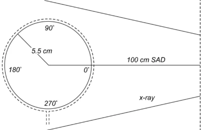
Fig. 1. Schematic diagram of setup of cylindrical phantom with diameter of 11 cm for simulating head-and- neck and shoulder treatment. 6 MV photon beams with 40×40 cm2 open field was irradiated to the cylindrical phantom at SAD 100 cm. Cylindri- cal phantom axis was perpendicular to the 0o incidence. The dotted line represents the perforated thermoplastic mask with 1.5 mm thickness.
Fig. 2. Calibration curve for radiochromic film. Each radiochro- mic film was irradiated by increasing dose. The solid line shows a fitted curve.
As for obliquely incident beams, the surface dose is a result of both scattered electrons from the air, the accelerator, as well as forward scattered electrons produced by interactions of the incident photon beam with the material of the immobiliza- tion mask. The studies with obliquely incident photon beams have shown that skin sparing with oblique beams was reduced from what is observed for a normally incident photon beam, and the depth of maximum dose of oblique incidence (dmax) moves closer to the surface with increasing angle of obli- quity.5)
The purpose of this study was to measure the surface and superficial dose on the angle of incidence and the effect of immobilization devices on cylindrical phantom. By using TLDs or radiochromic films to measure the skin dose deliv- ered by conventional open field, the skin dose was compared between different dosimeters. Skin dose values obtained from the measurements were then compared against the calculated values from the Monte Carlo simulation.
MATERIALS AND METHODS
The angular dependence check of the curvature contour skin dose with immobilization mask was done using 6 MV x-rays from a Varian 21iX (Varian Medical Systems, CA, USA) at
100 cm source axis distance (SAD) to the center of cylindrical phantom. The cylindrical phantom's axis was perpendicular to normal beam incidence.
Skin dose measurements were conducted on a 11-cm-dia.
and 20-cm-long water-equivalent (Solid Water, Gammex RMI, Middleton, WI) cylindrical phantom which was marked from 0o (i.e., beam entry) to 350o in 10o increments around circum- ference of the phantom (Fig. 1). Radiochromic film and TLD rods employed as two radiation detector types were used in the research. Radiation measurements were undertaken for both detectors using a 40×40 cm2 field with a 300 MU. At the cen- tral axis of the field the dose at Dmax (=1.6 cm) was 381 cGy and the prescribed dose at the center of cylindrical phantom was 311 cGy for 300 MU.
1. Radiochomic film
Radiochromic films (GafChromicⓇ EBT, ISP Technologies, USA) with a thickness of 0.23 mm were used in the investi- gation. Radiochromic film is potentially useful tool as an in- tegrative skin dosimeter in radiotherapy. It can measure a sur- face dose profile at a physical effective depth of 0.15 mm hence, superficial dose can be determined.6)
In order to get the calibration curve for radiochromic film, 15 pieces of film (2.0×5.5 cm2) were irradiated separately from 0 cGy to 500 cGy at Dmax in a solid water (Radiation Measurements Inc. RMI, USA) phantom with a 6 MV photon beam. The calibration curve could be fitted to the function as
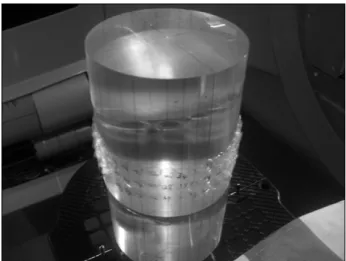
Fig. 3. To obtain the surface dose profile as a function of angle of incidence, a strip of radiochromic film with the dimensions of 12×35 cm, was wrapped tightly around cylindrical phantom.
Fig. 4. The TLD rods were placed around circle on the surface of the cylindrical phantom. The 3 packets were attached to the phantom in each 10o angular position. The TLD rods were arranged into 108 packets, with one TLD rod per packet.
Fig. 5. With the immobilization mask, the surface doses were measured by placing the TLDs on the surface of a cylindrical phantom in each 10o angular position.
followings,
Dose=3E−07(Pixel Value)2−0.0311×794.3
The calibration curve was used to convert the pixel value to the absorbed dose (Fig. 2).
To measure skin dose, a strip of radiochromic film of 12×35 cm, was wrapped tightly around the cylindrical phan- tom to minimize errors due to air gap (Fig. 3). The exposed films were scanned with a flat bed scanner at a resolution of 75 dpi and analysed with Image J software (Scanmaker 9800XL, Microtek International Inc, USA).
The skin dose data was obtained from 0o to 360o around the circumference of the phantom with and without the immobili- zation mask.
2. Thermoluminescent dosimenters
A new batch of 108 lithium fluoride (LiF: Mg, Ti) rods (Harshaw TLD- 100 extruded rod; Solon/Harshaw, USA) were used as thermoluminescent dosimeters. Due to their small physical size, TLD rods are usually treated as point detectors.
However, in locations where strong dose gradients are encoun- tered, the physical dimensions of the TLD rods cannot be neglected. The TLD rods with a length 6 mm and a diameter of 1 mm were calibrated by being placed inside a milled sec- tion of water-equivalent slab phantom and were irradiated at a depth of 5 cm with 20×20 cm2 field of 100 cm SSD and 200
MU. The variability of the reading of 108 rods was on the average±3% (1 s.d.).
The surface doses were measured by placing the TLDs on the surface of the cylindrical phantom (Fig. 4) and underneath the immobilization mask (Fig. 5). The TLD rods were ar- ranged into 108 thin plastic packets, with one TLD rod per packet. The 3 packets were taped to the phantom in each 10o angular position shown in Fig. 4. A total of 108 TLD rods which were wrapped individually in thin plastic packets, were
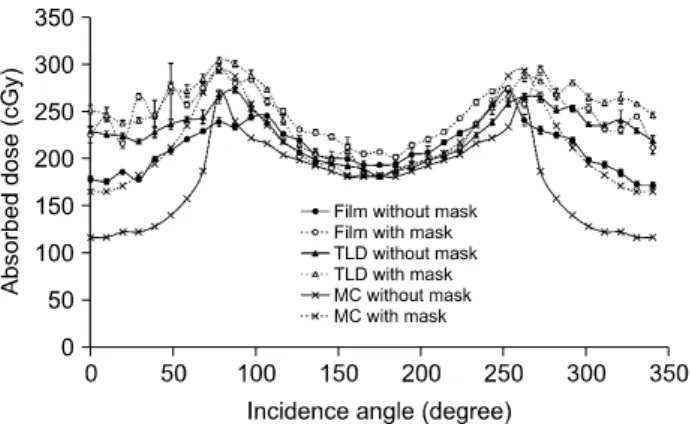
Fig. 6. The surface dose profile as a function of angle of incidence (θ) measured using radiochromic film and TLD rods for 40×40 cm2 open field with and without mask for a 300 MU.
The angular distribution curve of the skin dose calculated with Monte Carlo program. Using the EGSnrc code package the absorbed dose in the outer thin (0.15 mm) layer of the cylindrical phantom was calculated.
attached on the phantom's surface during treatment delivery.
Skin dose data was obtained from 0o to 350o, in 10o incre- ments around the circumference of the phantom with and with- out immobilization mask.
3. Immobilization mask
The immobilization mask used for this study was the Aquaplast (KGF Enterprise Inc., Chesterfield, MI). It has a density of approximately 1.08 g/cm3.7) It was also perforated by the manufacturer in the form of a mesh that is softened and stretched in hot water and then molded over the head of the patient for immobilization during head-and-neck treatments.
The immobilization mesh mask that was used had a thickness of approximately 1.9 mm and 1.5 mm before and after being stretched. The effect on skin dose that may result from the use of an immobilizing mask was investigated.
4. Monte Carlo program
A Monte Carlo study was performed to understand the de- pendence of the skin dose on the incident angle. The program package used for this purpose was the EGS code system which simulated transport of electrons and photons in any me- dium with an arbitrary geometry. The particular version of the EGS adopted in this study was EGSnrc which was suitable for studies of medical physics.
The energy spectrum from Varian Clinac 2100C treatment heads,8) which were assumed to be similar to the model Clinac 21iX, was used in generating photons. The photons were pro- duced radially from a point source at SAD 100 cm and their azimuthal direction were chosen randomly within the angle subtended by the boundary of the cylindrical phantom with the diameter of 11 cm.
Through the various electromagnetic interactions, for exam- ple, Bremsstrahlung, scattering, pair creation and annihilation, the photons incident on the surface of the phantom lost and deposited most of their energies in the medium. The lower bound energies of the electrons and the photons for trans- porting were set to 0.52 MeV and 0.001 MV respectively.
The energies deposited in the outmost layer of the medium, with the thickness of 0.15 mm which is the same as the effec- tive film measurement depth, was summed to give the dose in the film.
RESULTS
1. Skin dose measurements
The dose to the superficial region of the skin relative to Dmax, is commonly referred to as skin dose.9) The skin doses were measured as a function of incident angle of the beam with and without the immobilization mask. In this paper, we refer to circumference angles θ as the (nominal) incident an- gle for convenience. We illustrate the (nominal) incident angle on the circumference of cylindrical phantom in Fig. 1 in order to avoid any confusion with real incident angle φ.
The skin dose was normalized to the dose at Dmax for a 40×40 cm2 field at 0o incidence at a 100 cm SAD for 6 MV.
At the central axis of the field the dose at Dmax (=1.6 cm) was 381 cGy and the prescribed dose at the center of phantom was 311 cGy for 300 MU.
2. Radiochromic film measurement
In Fig. 6 the two curves with scattered circles show the sur- face profile as a function of the angle of incidence (θ) meas- ured using radiochromic film for a 40×40 cm2 open field with and without the 1.5 mm of mesh mask present for a 300 MU.
The variability of film measurements was on the average ±4%
(1 s.d.) and ±3% (1 s.d.) with and without the immobilization
mask respectively. It shows the tung and groove for the skin dose with the mask due to the perforations of the mask.
The skin dose curves for film increase from normal incident position (0o) to around tangential position (90o), because there is a large shift in the shallowed peak buildup depth (dmax) of oblique angles toward the surface as the angle of beam in- cidence becomes more oblique.3)
Skin doses without the mask were found to be 47% (at 0o incidence), 61% (at 90o incidence) and 64% (at 100o in- cidence) of the Dmax for a conventional open field using radio- chromic film. Skin doses with the mask were found to be 59%
(at 0o incidence) and 78% (at 80o incidence). For the skin dose comparison at different locations, the skin dose is greatest at the oblique incidence of around 90o.
When using the mask, the increase in dose on the entrance side was due to the fact that the mask served as buildup material. Adding the mask also caused the surface doses to in- crease at exit side (100o to 180o) due to the fact that it func- tioned as back scattering material.
For film measurement, the skin dose curve of film without mask is much lower than that measured by the TLD at en- trance side. The main reason for this is that the film has shal- lower effective measurement depth than that of the TLD rod.
3. TLD measurement
For each set of readings in this studies, three TLDs were used per measurement. The variability of the reading between different rods of the same set was calculated as standard deviation. The variability was on the average ±5% (1 s.d.) and
±3% (1 s.d.) with and without the immobilization mask re- spectively in Fig. 6. It shows the exceptional error bar for the skin dose with the mask due to the uneven thickness of the perforated mask.
The results of skin dose measurements, taken at 10o incre- ments, using TLD are also included in Fig. 6. The skin dose curves for TLDs increase from normal incident position (0o) to around 90o with and without mask respectively.
Skin doses without mask were found to be 60% (at 0o in- cidence), 72% (at 90o incidence) of the Dmax for a conven- tional open field using TLDs. Skin doses with the mask were found to be 66% (at 0o incidence), 80% (at 90o incidence).
The skin dose curve of TLD with mask revealed the similar
pattern and the dose as that of the film at entrance side.
4. Monte Carlo calculation
The resulting angular distributions of the skin dose with and without the mask of thickness 1.5 mm in the outmost layer of the thickness 0.15 mm are shown in the Fig. 6. The skin dose with the mask is almost systematically (about 50%) higher than the one without the mask at all incident angles.
The dose is at its minimum at normal incidence (0o) and in- creases with incident angle rather sharply and reaches its max- imum at glancing incidence.
In comparison with the data taken with a film shown as the curves in the Fig. 6, it is fair to say that the general tendency is consistent with each other although the MC results are more sensitive to the incident angle than the experimental data. This behavior is more appreciable when the mask is not used.
Especially at normal incidence the dose without the mask ap- peared to be much lower than the one with the mask. One of the important causes leading to such discrepancies is specu- lated to be the absence of electron contamination in the MC study. On the other hand electrons in the real beam deposited most of their energies in shallow layers of the medium. In particular the dose at smaller angles would increase due to the mask placed in front of the detector film and thus be visible on top of the lower contribution from the photons. This would make over all angle dependence in the real data less pro- minent.
DISCUSSION
Uneven air gaps, beam obliquity, or sloping contours create complex dosimetry problems in photon beam treatment planning. Radiation may affect the basal cells to the extent of producing observable desquamation with moderate doses. The superficial layers of interest include the dermal lymphatics which extend to about 1 mm depth and the basal cell layer at about 70μm.10)
In this paper we measured the dose at a shallow depth which is more consistent with the skin’s dermal layer which exposed to a conventional open field. The study has outlined the different responses to surface dose measurement using dif- ferent detectors. Because the differences in measured super-
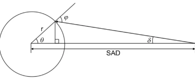
Fig. 7. The real incident angle φ as a function of (nominal) incident angle θ, where r is a radius of the cylindrical phantom and SAD is the source to axis distance.
ficial doses without mask were significant and it was beyond those related to any experimental error, these differences were assumed to be mainly due to the effective depth of measure- ment for each detector.
For MC calculation, when we didn't use mask, the max- imum skin dose was not at the most oblique incidence angle (θ=90o). The maximum skin dose was 71% at 80o incidence compared to 63% at 90o. This can be explained as follows (Fig. 7).
The real incident angle, denoted by φ, can be related with the (nominal) incident angle θ as,
The relationship between the angles, δ and θ is as follows
where r is a radius of the cylindrical phantom and SAD is the source to axis distance. We can write the real incident an- gle φ as a function of θ as follows,
where we substitute r=5.5 cm and SAD=100 cm in the last equation for our experimental setup.
For (nominal) incidence angle θ=87o, the real incidence an- gle φ could be tangential incidence 90o where skin dose could have maximum dose. Consequently in our experimental setup the maximum dose would be at between θ=80o and θ
=90o. MC calculation curve without mask showed the max- imum dose at the angle less than 90o. But the experimental data showed a discrepancy a bit to the theoretical assumption.
CONCLUSION
Excess radiation to which the skin is exposed during ex- ternal beam megavoltage radiation therapy has reportedly caused excessive erythema in patients treated with the linear accelerator on the skin. Curvature contour skin doses at the epidermal level were measured under geometries simulating sloping surfaces for a Varian 21iX.
We also studied the effects of immobilization mesh mask on curvature contour skin. Comparing the surface doses at differ- ent points, the maximum skin dose was found to be 98% of the prescribed dose at the oblique beam angle around 90o. This increase in curvature contour skin dose must be taken into ac- count to avoid undesired skin reactions in the area of the head-and neck or axilla.
As a general rule, skin doses may be abated by using thin- ner and more perforated immobilization devices especially if a sloping skin contour is encountered.
Further work is needed to determine the discrepancies be- tween calculations, theoretical assumption and measurements in the superficial region.
REFERENCES
1. Hsu SH, Roberson PL, Chen Y, Matsh RB, Pierce LJ, Moran JM: Assessment of skin dose for breast chest wall ra- diotherapy as a function of bolus material. Phys Med Biol 53:2593-2606 (2008)
2. Dogan N, Glasgow GP: Surface and build-up region dosim- etry for obliquely incident intensity modulated radiotherapy 6 MV x rays. Med Phys 30:3091-3096 (2003)
3. Higgins PD, Han EY, Yuan JL, Hui S, Lee CK: Evaluation of surface and superficial dose for head and neck treatments us- ing conventional or intensity-modulated techniques. Phys Med Biol 52:1135-1146 (2007)
4. Gagnon WF, Peterson MD: Comparison of skin doses to large fields using tangential beams from Cobalt-60 gamma rays and 4-MV x rays. Radiology 127:785-788 (1978)
5. Gerbi BJ, Meigooni AS, Khan FM: Dose buildup for ob- liquely incident photon beams. Med Phys 14:393-399 (1987) 6. Butson MJ, Cheung T, Yu PKN, Price S, Biley M:
Measurement of radiotherapy superficial X-ray dose under eye shields with radiochromic film. Physica Medica 24:29-33 (2008) 7. Mellenberg DE: Dose behind various immobilization and beam-modifying devices. I J Radiation Oncology Biol Phys 32:1193-1197 (1995)
머리-목 그리고 어깨의 방사선 치료 시 피부곡면과 고정장치로 인한 피부손상연구
고신대학교 의과대학 *생물물리교실, †방사선종양학교실,
‡경성대학교 물리학과, §부산대학교병원 방사선종양학과
김수길*ㆍ정태식†ㆍ임상욱†ㆍ박영목‡ㆍ박 달§
곡면 형태의 피부표면의 방사선량을 방사선크롬 필름과 열형광 선량계를 이용하여 측정하고자 한다. 또한 고정 장치의 사용으로 인한 고에너지 방사선의 피부보존효과의 감쇠를 정량적으로 측정하여 Monte-Carlo 프로그램으로 계산한 값과 비교하고자 한다. 머리-목 그리고 어깨의 곡면 형태를 모의하여 만든 11 cm 직경의 원통 팬텀에 40×40 cm2의 조사야, SAD 100 cm, 6 MV의 방사선을 쪼였다. 또한 관련된 치료 상황과 유사한 조건으로 만들기 위해 그물망 형태의 고정 마 스크를 원통형 팬텀에 씌워서 실험하였다. 원통 팬텀의 원둘레 주위를 따라 0o에서 360o까지의 피부선량곡선을 구하였 다. 방사선크롬 필름을 이용하여 구한, 정면 입사위치(0o)에서의 피부선량은 최대값 깊이(Dmax) 방사선량의 47%, 접선 각 도인 90o에서는 61%로 측정되었다. 1.5 mm의 고정마스크를 씌운 경우 0o 입사지점에서는 59%, 80o에서는 78%였다. TLD 를 통한 결과는 고정마스크를 씌운 경우 0o 입사지점에서는 66%, 80o에서는 80%였고 필름의 경우와 유사한 형태를 보였 다. 고정 마스크를 머리-목 그리고 어깨 부위에 부착시켜서 치료를 하는 경우에 접선 부근 각도에서의 피부선량이 치료 선량과 거의 같은 값을 보였다. 곡면 부위의 피부에는 고정성을 잃지 않는 범위 안에서 보다 더 얇고 더 구멍이 많이 뚫린 고정마스크를 사용해야 과도한 피부선량을 줄일 수 있을 것으로 사료된다.
중심단어: 피부의 곡율, 피부손상, 고정마스크, 사선 입사, 몬테카를로 모의치료 8. Baumgartner A, Steurer A, Maringer FJ: Simulation of
photon energy spectra from Varian 2100C and 2300C/D Liniacs:
Simplified estimates with PENELPOPE Monte Carlo models.
App Radiat Isot 67:2007-2012 (2009)
9. Gagnon WF: Physical factors affecting absorbed dose to the
skin from cobalt-60 gamma rays and 25-MV x rays. Med Phys 6:285-290 (1979)
10. Quach KY, Morales J, Butson MJ, Rosenfeld AB, Metcalfe PE: Measurement of radiotherapy x-ray skin dose on a chest wall phantom. Med Phys 27:1676-1680 (2000)