84 Copyright 2006 by the Korean Society for Clinical Neurophysiology 대한임상신경생리학회지 8(1):84~87, 2006 ISSN 1229-6414
MELAS syndrome is characterized by mitochon- drial encephalopathy, lactic acidosis, and stroke- like episodes.
1-7The stroke-like episodes associ- ated with infarcts may appear on head computed tomography (CT) or magnetic resonance imaging (MRI). But the precise pathogenic mechanism of MELAS is still controversial. There are two hypothesis for pathophysiologic mechanism. One is nonvascular, transient oxidative phosphoryla- tion dysfunction within the brain parenchyme.
The other is ischemic angiopathy. In recent study, the former are more acceptable for mechanism than the later.
2,3We report a patient of MEALS syndrome with
clinical and radiological improvement in follow- up study, and we tried to figure out the relation between clinical symptom, diffusion MRI and pathophysiologic mechanism of stroke-like lesions of MELAS syndrome.
CASE
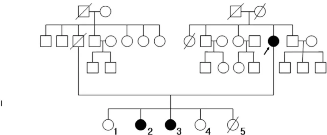
A 16 year-old girl (Ⅲ-3, Fig. 1) was transferred to our hospital for headaches and left sided hemianopsia. She was normal at birth and had normal development.
Since the age of 13, she complained about last- ing unilateral headache. Headache occurred intermittently and was exacerbated by physical activity or fatigue. But, 7th days before first admission, right-sided headache was steady in duration, pulsatile in nature, and accompanied by nausea or vomiting. Two days later, left sided hemianopsia occurred. Her mother has been diag- nosed as having sensorineural hearing loss and migraine headache, and the second elder sister
가역적인 뇌기능장애를 보인 MELAS 증후군
원광대학교 신경과학교실, 원광의과학 연구소
정진성∙이학승∙박현영∙장 혁∙김요식∙조광호
Reversible Brain Dysfunction in MELAS Syndrome
Chung Jin-Seong, M.D., Hak-Seung Lee, M.D., Hyun-Young Park, M.D., Hyuk Chang, M.D., Yo-Sik Kim, M.D., Kwang-Ho Cho, M.D.
Department of Neurology, Wonkwang University School of Medicine, Institue of Wonkwang Medical Science
The MELAS (Mitochondrial Encephalomyopathy with Lactic Acidosis, and Stroke-like episodes) syndrome is one of the inherited mitochondrial disorder. We have experienced a 16-year-old girl with headaches and left hemianopsia.
Diagnosis of MELAS syndrome with multiple brain parenchymal lesions was confirmed by gene study. The stroke-like lesion of MELAS syndrome showed significant improvement in radiological follow up study. Therefore, MRI findings in MELAS could be interpreted as metabolic cellular dysfunction rather than ischemic vasculopathy
Key Words: MELAS syndrome, Stroke-like lesion
Address for correspondence Hyun-young Park, M.D.
Department of Neurology, Wonkwang University Hospital 344-2 Shinyong-dong, Iksan, Jeonbuk 570-180, Korea.
Tel: +82-63-850-1143, Fax: +82-63-842-7379 E-mail : [email protected]
� This study was supported by the Wonkwang University Reserch Grant 2005.
(Ⅲ-2, Fig. 1) also suffered migraine headache.
At the time of admission, blood pressure was 120/80 mmHg, pulse rate was 68 beats per minute with a regular rhythm and respiratory rate was 22 beats per minute. Body temperature was 36.8� C.
The patient had a normal physique without dia- betes or any other history except for headache.
The neurological examination reveled that mental status was slightly drowsy with slurred speech.
Left homonymous hemianopsia was detected in a visual field test. No other neurological deficits and, signs of meningeal irritation were found.
Laboratory results showed a white blood cell count of 11.800/mm
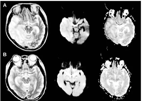
3and an ESR was normal. Test for hepatitis surface antigen, antibodies to human immunodeficiency virus (HIV), anti-nuclear anti- body, rheumatoid factor, anti-DNA antibody and VDRL were all negative. Blood chemical studies were normal except that lactates was increased to 32.6 mg/dl (normal 4.5 to 19.8 mg/dl). Chest radi- ograph and echocardiogram revealed no abnormal findings. The brain MRI showed high signal intensity on T2 weighted image and diffusion weighted image in right temporo-occipital and left temporal cortices, and scattered enhancement on contrast enhanced MR images (Figure 2-A). A transfemoral cerebral angiogram showed no abnormality in the arterial or venous phase of the studies. Electroencephalography showed general- ized intermittent slowing with multiregional epileptiform discharges with out conwmitant clinical seizure. Muscle biopsy from the medial gastrocnemius processed for histochemistry
(Hematoxylin-Eosin, modifided Gomori’s trichrome stains), showed mild increased varia- tion in fiber size, suggesting myopathy, but no ragged red fibers. Mitochondral DNA analysis for the patient, mother and second elder sister were performed, and revealed an A→G point mutation in the transfer RNALeu gene at base pair 3243 of the patient and second elder sister, thereby con- firming the clinical diagnosis of MELAS. But, we could not find abnormal finding in mother’s mitochondrial DNA.
She was treated with multi-vitamin and nicotinamide. The patient was discharged from the hospital without headache, but left homony- mous hemianopsia remained. A follow up brain MRI was performed two months later from the initial study and showed significant improvement in right temoporo-occipital lesion and cerebral edema (Figure 2-B).
DISCUSSION
The MELAS (Mitochondrial Encephalomyopathy with Lactic Acidosis, and Stroke-like episodes) syndrome is one of inherited mitochondrial disor- ders.
1-7The stroke like episodes associated with infarcts may appear on head CT or MRI, and occur in more than 90% of patients with MELAS syndrome.
3The focal neurological deficits of abrupt onset landmarking the evolution of MELAS are clinical- ly indistinguishable from stroke events and the precise mechanism of neurological symptoms is
가역적인 뇌기능장애를 보인 MELAS 증후군
J Korean Society for Clinical Neurophysiology / Volume 8 / June, 2006 85
Figure 1. Pedigrees of family. The black symbols indicate affected individuals; diagonal lines across symbols, deceased individuals;
and arrows, proband.
I
II
III
still controversial. Thus, they are generally named “stroke-like”events.
2,4Brain MRI of patients with MELAS classically shows signal changes involving both gray and white matter predominantly in the occipital and parietal lobes that strongly mimic stroke lesion.
But, why this region is preferentially affected remains unclear, and distribution of these infarct-like lesions on MRI does not usually fol- low vascular territory and pathological studies do not find lesion of the major cerebral blood ves- sel.
2,4In acute ischemic infarction, intracellular diffusion of proton is restricted or reduced, cor- responding to low signal on ADC map (cytotoxic edema) caused by energy failure.
2However, in stroke like lesion of MELAS, the ADC map demonstrates a higher proton mobility, which corresponds to the high signal on ADC map (vasogenic edema) and thus not favour ischemic damage as the main mechanism explaining focal neurological deficit in MELAS.
2,4,6,8So nonvascu- lar, oxidative phosphorylation dysfunction within the brain parenchyma, which cause lactic acidosis
and vasodilatation, is a more susceptible mecha- nism of stroke like episode of MELAS syndrome.
2But, Ohama et al stated the importance of anoth- er mechanism for stroke like episodes in cases of MELAS, and described as mitochondrial angiopa- thy due to abnormal mitochondrial accumulation of endothelial cells and smooth muscle cells of blood vessels.
3,8Also, the coexistence of basal ganglia calcifications and multifocal atrophy may suggest a slowly progressive degenerative process.
6In our case, we found the high signal intensity on T2-weighted image and diffusion weighted image in the right temporo-occipital cortices and left temporal cortices and scattered enhancement on contrast enhanced MR images. However, parenchymal lesion did not follow vascular terri- tory and improved in follow up study. This may suggest that stroke like lesion may reflect a breakdown of the blood-brain barrier and attribute to metabolic dysfunction in cell, not angiopathy, and also related to vasogenic edema.
4,6,8But, we experienced only one patients
정진성∙이학승∙박현영∙장 혁∙김요식∙조광호
86 J Korean Society for Clinical Neurophysiology / Volume 8 / June, 2006
Figure 1. A and B Brain MRI (T2 weighted image, diffusion weighted image, ADC map, in order of figure). (A) Axial T2-weighted and diffusion weighted images show high signal intensity in right temporo-occipital and left temporal cortices and delayed normal signal intensity on ADC map in same lesion. (B) Follow-up images obtained 2 months later show resolution of the lesions.
A
B
가역적인 뇌기능장애를 보인 MELAS 증후군
J Korean Society for Clinical Neurophysiology / Volume 8 / June, 2006 87
and our study is not enough for explain the mechanism of stroke like lesion on MELAS syn- drome. So, more investigations should be done for finding mechanism of stroke like lesion of MELAS syndrome.
REFERENCES
01. Sue CM, Crimmins DS, Soo YS, Pamphlett R, Presgrave CM, Kotsimbos N et al. Neuroradiological features of six kindreds with MELAS tRNALeu A3243G point mutation:
implications for pathogenesis. J Neurol Neurosurg Psychiatry 1998;65: 233-240.
02. Oppenheim C, Galanaud D, Samson Y, Sahel M, Dormont D, Wechsler B et al. Can diffusion weighted magnetic response imaging help differentiate stroke from stroke-like events in MELAS? J Neurol Neurosurg Psychiatry 2000;
69: 248-250.
03. Kiminobu Y, Yasuhiro H, Kazumi K, Kazuo M, Takenori Y. Diffusion-weighted MR imaging in a case of mitochon-
drial myopathy, encephalopathy, lactic acdosis, and strokelike episodes. AJNR 2001; 22: 269-272 .
04. Iizuka T, Sakai F, Suzuki N, Hata T, Tsukahara S, Fukuda M et al. Neuronal hyperexcitability in stroke-like episodes of MELAS syndrome. Neurology 2002;59: 816-824.
05. Park KH, Kim SH, Lee YJ, Kim HT, Kim JH, Kim MH et al. MELAS syndrome : Point mutation at nt3243(A3243G) in tRNAleu(UUR) Gene of mtDNA. J Korean Neurol Ass 1998;16: 585-589.
06. Madhav T, DPhil, Nancy JN, Jonathan DG, Michael RF.
A practical approach to the diagnosis and management of MELAS : case report and review. Neurologist 2002;8:
302-312.
07. Kwon SJ, Park SS, Kim JM, Ahn TB, Kim SH, Kim J et al. Investigation of common mitochondrial point muta- tions in Korea. Ann N Y Acad Sci 2004;1011:339-344 08. Yoneda M, Maeda M, Kimura H, Fujii A, Katayama K,
Kuriyama M. Vasogenic edema on MELAS : A serial study with diffusion-weighted MR imaging. Neurology 1999;53:2182-2184