능이버섯 효소 추출물의 항산화 활성 및 H
2O
2로 유도된 스트레스에 대한 신경보호 효과
이승재·김은경·오현정·권혁주·황진우·문상호*·전병태*·박표잠†·임병우**†
건국대학교 의료생명대학 생명공학전공, *건국대학교 녹용연구센터, **건국대학교 의료생명대학 응용생화학전공
Free Radical Scavenging Activity and Protective Effect against H
2O
2-Induced Stress in Neuronal Cells of Enzymatic Extracts from Sarcodon aspratus
Seung Jae Lee, Eun Kyung Kim, Hyun Jung Oh, Hyuck Ju Kwon, Jin Woo Hwang, Sang Ho Moon
*, Byung Tae Jeon
*, Pyo Jam Park
†and Beong Ou Lim
**†Department of Biotechnology, Konkuk University, Chungju 380-701, Korea.
*
Korea Nokyong Research Center, Konkuk University, Chungju 380-701, Korea.
**
Department of Applied-biochemistry, Konkuk University, Chungju 380-701, Korea.
ABSTRACT : The antioxidative activity of various enzymatic extracts from
Sarcodon aspratus(
S. aspratus) was evaluated by measuring 1,1-diphenyl-2-picrylhydrazyl (DPPH), and alkyl radical scavenging activity using an electron spin resonance (ESR) spectrometer. For this study, the
S. aspratuswere enzymatically hydrolyzed by seven carbohydrases (Viscozyme, Cel- luclast, Dextrozyme, AMG, Promozyme, Maltogenase, and Termamyl) and eight proteases (
α-chymotrypsin, Alcalase, Fla- vourzyme, Neutrase, papain, pepsin, Protamax, and trypsin). The DPPH radical scavenging activities of Viscozyme and pepsin extracts were the highest, and the half maximal inhibitory concentration (IC
50) values were 0.896 and 0.734
㎎/mL, respectively. The Celluclast and trypsin extracts showed the highest scavenging activities on alkyl radical, and their IC
50val- ues were 0.278 and 0.575
㎎/mL, respectively. The Celluclast extracts was decreased cell apoptosis in PC-12 cells against H
2O
2-induced oxidative damage. The findings of the present study suggest that enzymatic extracts of
S. aspratusexhibit anti- oxidative activity against oxidative stress on PC-12 cells.
Key Words
: Antioxidative Activity, Enzymatic Hydrolysates, Free Radical,
S. aspratus, Neuroprotective Effect
INTRODUCTION
Recently, many researches tried to find new physiologically effective materials from medicinal plants for prevention and/or remedy of diseases (Lee et al ., 2004, 2008). The etiology of a range of diseases is associated with the generation of excess reactive oxygen species (ROS). Steady-state maintenance of ROS/antioxidant ratio is, however, essential for cell signaling.
ROS generated in cells include the superoxide anion radical (O2
·−) (Boveris and Cadenas, 1997). ROS are generated by biochemical reactions in the cell. Indeed, leakage of electrons from the mitochondrial electron transport chain is a significant source of mitochondrial ROS (Cashman, 1997). H
2O
2peroxide is produced by mitochondrial monoamine oxidase (Fridovich,
1995) and by the superoxide dismutase (MnSOD and Cu/
ZnSOD)-catalyzed dismutation of O2·
-(Reubsaet et al ., 1988). In addition, peroxisomal acyl-CoA oxidases also generate H
2O
2(Sheu et al ., 2006). ROS generation may also be associated with external stimuli. UV and high-energy radiation, the metabolism of some xenobiotics, air pollutants and the redox cycling of quinones and nitroaromatics are all associated with ROS generation (Boveris and Cadenas, 1997). Currently, mushrooms are drawing attention as beneficial foods for human health, and have been valued as edible and medical resources for some time (Jeoung et al ., 2009).
In an early work, Kaneda and Tokuda reported the hypocholesterolemic action of edible mushrooms (Kaneda and Tokuda, 1966) from cholesterol lowering properties of Lentinus
†
Corresponding author: (Phone) +82-43-840-3588 (E-mail) [email protected] / (Phone) +82-43-840-3570 (E-mail) [email protected]
Received 2010 December 7 / 1st Revised 2011 February 24 / 2nd Revised 2011 February 28 / Accepted 2011 March 4
edodes, Auricularria polytricha, Flammulina velutipes and Agaricus bisporus (Bobek et al ., 1991, 1994; Bobek and Galba, 1999). Besides, some mushrooms was found hypotensive effect when blood pressure is already high (Kabir et al ., 1987, 1988;
Kabir and Kimura, 1989; Miyazawa et al ., 2008). As well as, many species of mushrooms were turns out to include the compounds of antioxidant (Cheung and Cheung, 2003; Wong and Chye, 2009) and anti-inflammatory (Jose et al ., 2004; Kohno et al ., 2008). In addition, Eva et al . (2010) predicted these effects of antioxidant and anti-inflammatory involved with the management of heart and circulation health complications. Yoon et al. (2006) reported the antioxidative activities and antimicrobial effects of water and ethanol extracts from the S.
aspratus . The enzymes high specificity, nontoxicity, water solubility, biodegradability, and mild operational conditions of pH, temperature, and pressure, are major advantages over inorganic catalysts (Taylor, 1991). However, while literature on the chemistry of S. aspratus and clinical trials reporting its effects are vast, research into the antioxidative effect of enzymatic extracts from S. aspratus is scarce, or even non-existent. Thus, the present study aimed to investigate the free radical scavenging activities of enzymatic extracts from S. aspratus by ESR spectroscopy and their possible protective effects on PC-12 cells against oxidative stress.
MATERIALS AND METHODS
1. Materials
PC-12 (ATCC CRL-1721), the standard model for neuronal function studies, was obtained from American Type Culture Collection (Manassas, VA, USA). Dulbecco's Modified Eagle Medium (DMEM), fetal bovine serum (FBS), and penicillin- streptomycin were obtained from Invitrogen Corporation (Carlsbad, CA, USA). The following substances were obtained from Novo Co. (Novozyme Nordisk, Bagsvaerd, Denmark):
Dextrozyme, AMG, Promozyme, Maltogenase, Termamyl, Viscozyme, Celluclast, Flavourzyme, Neutrase, Protamex, and Alcalase. 5,5-dimethyl-1-pyrroline N-oxide (DMPO), 2,2-azobis (2-amidinopropane) hydrochloride (AAPH), 2,2-diphenyl-1- picrylhydrazyl (DPPH), (4-pyridyl-1-oxide)-N-tert-butylnitrone (4-POBN), papain, pepsin, trypsin,
α-chymotrypsin, propidium iodide (PI) were obtained from Sigma Chemical Co. (St. Louis, MO, USA). Tween-20 was supplied by USB (Cleveland, OH, USA). All other reagents were of the highest grade available commercially.
2. Preparation of enzymatic hydrolysates from
S. aspratusThe mushroom was pulverized into powder with the use of a grinder (Hanil, Seoul, Korea), and the enzymatic hydrolysates were obtained according to the methods described by Park et al . (2005) and Cumby et al . (2008). The optimum hydrolysis conditions of particular enzymes are as follows: Dextrozyme, pH 4.5, 60
℃; AMG, pH 4.5, 60
℃; Promozyme, pH 5.0, 60
℃; Maltogenase, pH 5.0, 60
℃; Termamyl, pH 6.0, 60
℃; Viscozyme, pH 4.5, 50
℃; Celluclast, pH 4.5, 60
℃; BAN, pH 7.0, 70
℃; Flavourzyme, pH 7.0, 50
℃; Neutrase, pH 6.0, 50
℃; Protamex, pH 6.0, 40
℃; Alcalase, pH 8.0, 50
℃; trypsin, pH 7.0, 37
℃; papain, pH 7.0, 37
℃; pepsin, pH 7.0, 37
℃; and
α-chymotrypsin, pH 7.0, 37
℃. Briefly, 100 mL of buffer solution were added to 2 g of each powder sample, and then 40
µL (or
㎎) of each enzyme were added after a pre-incubation period of 30 min. The enzymatic hydrolysis reactions were performed for 8 h to achieve an optimum hydrolytic level, and the samples were then immediately heated to 100
℃for 10 min to inactivate the enzyme.
Finally, the supernatant was filtered to obtain the enzymatic extracts, which were then lyophilized and stored at –20
℃until use.
3. DPPH radical scavenging activity
DPPH radical scavenging activity was measured with the use of the method described by Nanjo et al . (1996). Briefly, 60
µL of each enzymatic extracts at various concentrations were added to 60
µL of DPPH (60
µM) in a methanol solution. After the solution was mixed vigorously for 10 sec, it was then transferred into a 100
µL Teflon capillary tube, and the scavenging activity of each enzymatic extracts with regard to DPPH radicals was measured with the use of an ESR spectrometer. A spin adduct was measured on an ESR spectrometer exactly 2 min later.
Experimental conditions were as follows: central field, 3475 G;
modulation frequency, 100
㎑; modulation amplitude, 2 G;
microwave power, 5
㎽; gain, 6.3 × 10
5; and temperature, 298 K.
4. Alkyl radical scavenging activity
Alkyl radicals were generated by AAPH. The PBS (pH 7.4)
reaction mixtures that contained 0.1 mL of 10 mM AAPH, 0.1
mL of 10 mM 4-POBN, and 0.1 mL of the indicated concentra-
tions of the tested samples were incubated at 37
℃in a water bath
for 30 min (Hiramoto et al ., 1993), and they were then
transferred to 100-
µL Teflon capillary tubes. The spin adduct was
recorded on an ESR spectrometer. Measurement conditions were
as follows: central field, 3475 G; modulation frequency, 100
㎑;
modulation amplitude, 2 G; microwave power, 1
㎽; gain, 6.3 ×
10
5; and temperature, 298 K.
5. Cell Culture
PC-12 cells were cultured and maintained in DMEM supplemented with 100 U/mL penicillin, 100
㎍/mL streptomycin and 10% FBS and maintained at 37
℃under a humidified atmosphere with 5% CO
2. All the treatments were performed at 30% confluence.
6. Apoptosis analysis
The PC-12 cells were seeded at 2.1 × 10
5cells/well in 6-well plates in a complete medium, DMEM with 10% FBS. After 24 h incubation in a humidified 5% (v/v) CO
2/air environment at 37
℃, 990
µL of the enzymatic hydrolysate solution in DMEM was transferred to the well to give a final concentration of 1
㎎/mL.
Following 1 h incubation with the extracts, 10
µL of 100 mM H
2O
2was added the medium. After 24 h the cell was harvested, and the harvested cells were suspended in ethanol with 0.5%
Tween-20 and left for 24 h at 4
℃. The cells were then harvested by centrifugation and re-suspended in 1.0 mL of PBS with 0.05
㎎
/mL of propidium iodide and 10
㎍/mL of RNase A and incubated at 37
℃for 30 min. The analysis of apoptotic cell death was performed by measuring the hypodiploid DNA contents using a flow cytometer (FACS-caliber, Becton Dickinson, NJ).
The cells in the sub-G1 population were considered as apoptotic cells, and the percentage of each phase of cell cycle was determined.
7. Statistical analysis
The experimental results were recorded as the mean ± the standard deviation of three parallel measurements
RESULTS AND DISCUSSION
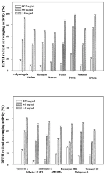
1. DPPH scavenging activity
DPPH is a stable free radical that has been used to evaluate the free-radical scavenging activity of natural antioxidants. The capacity of the enzymatic extracts of S. aspratus to scavenge DPPH was measured by ESR spectrometry, and the results are shown in Fig. 1. It was observed that carbohydratic extracts by Promozyme, Dextrozyme, Maltogenase, AMG, Celluclast, Termamyl, and Viscozyme had scavenging activity 70.17, 72.94, 71.36, 65.30, 69.14, 59.09, and 80.44% at 1.0
㎎/mL on DPPH radical. The proteolytic extracts by Alcalase, Protamex,
α- chymotrypsin, trypsin, papain, Neutrase, Flavourzyme, and
pepsin from the leaves scavenged 69.68, 71.39, 90.37, 95.01, 85.82, 64.47, 67.88, and 95.84% at 1.0
㎎/mL on DPPH radical.
The radical scavenging activity was concentration-dependent manners. In addition, Viscozyme and pepsin extracts exhibited the strongest scavenging activity among the various carbohy- drases and proteases with IC
50values of 0.896 and 0.734
㎎/mL, respectively.
In the earlier reports, S. aspratus have been known for several biological activities (Kalonia et al ., 2009; Halliwell, 1999; Je et al ., 2007). However, to date, there have been no studies on radical scavenging activity by using ESR spectroscopy and protective effects of neuronal cells of enzymatic extracts from S.
aspratus . Therefore, we prepared enzymatic extracts from S.
Fig. 1.
DPPH radical scavenging activity of various enzymatic extracts by proteolytic (Upper) and carbohydratic (Lower) hydrolysis from
S. aspratus. Values are given as the means
± SD of determinations were made in triplicate experi-
ments.
aspratus using various enzymes to take water soluble extracts.
The antioxidative activities of the enzymatic extracts were investigated using radical scavenging activities on DPPH radical.
The earlier results were DPPH radical scavenging activity of another positive control of vitamin C, and the IC
50values was under the 10
㎍/mL. Although DPPH radical scavenging activity of S. aspratus was lower than vitamin C, these results indicate that enzymatic extracts from S. aspratus appear to be good potential candidates for DPPH radical scavenger. Further studies are required for identification of the antioxidative compound from Viscozyme and pepsin extracts.
2. Alkyl radical scavenging activity
The alkyl radical spin adduct of 4-POBN/free radicals was generated from AAPH at 37
℃for 30 min, and the decrease in ESR signals was observed with the dose increment of all enzymatic extracts. All enzymatic extracts of S. aspratus scavenged alkyl radical in a dose-dependent manner. The extracts from S. aspratus exhibited the alkyl radical scavenging activities, and the scavenging activities of Viscozyme, Celluclast, Dextrozyme, AMG, Maltogenase, Promozyme, and Termamyl were 68.21, 78.56, 74.33, 75.87, 71.85, 74.16, and 66.61% at 1.0
㎎
/mL. In addition, same concentration of the extracts hydrolyzed from S. aspratus by the 8 types of proteases such as
α
-chymotrypsin, Flavourzyme, Neutrase, Protamex, pepsin,
Alcalase, trypsin, and papain also scavenged 64.16, 76.63, 66.49, 69.52, 42.55, 63.02, 78.54, and 62.99% at 1.0
㎎/mL. It was observed that Celluclast and trypsin extracts exhibited the strongest scavenging activities among the various carbohydrases and proteases with IC
50values of 0.278 and 0.575
㎎/mL, respectively (Fig. 2).
Lee et al . (2010) reported that the free radical scavenging activity of various enzymatic extracts prepared from Hericium erinaceum was evaluated by using an ESR spectrometer, and all enzymatic extracts of Hericium erinaceum scavenged alkyl radical with dose-dependent manners. It was observed that pepsin and Viscozyme extracts exhibited the strongest scavenging activity among the various proteases and carbohydrases with the IC
50values of 0.419 and 0.236
㎎/mL, respectively. These facts suggest that enzymatic extracts of S. aspratus might be potential source of alkyl radical scavenger with another mushroom.
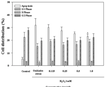
3. Cell cycle and apoptosis analysis by flow cytometer Some previous research has identified a link between neurodegenerative disorders such as, Alzheimer’s, Parkinson’s and Huntington’s disease and oxidative stress (Nakajima et al ., 2007; Fischer and Glass, 2010). Due to strong reactivity with biomolecules,
·OH is probably capable of doing more damage to biological systems than any other ROS (Kalonia et al ., 2009). In the present study, the Celluclast extracts were selected to
Table 1.
Optimum hydrolysis conditions of various enzymes.
Enzyme Optimum conditions Buffer used
aEnzyme composition
pH Temperature
Protamex 7.0 50 0.1 M PB
bHydrolysis of food proteins
Flavourzyme 7.0 50 0.1 M PB Containing both endoprotease and exopeptidase activities Neutrase 7.0 50 0.1 M PB An endoprotease
trypsin 7.0 37 0.1 M PB A serine protease
papain 7.0 37 0.1 M PB A digestive enzyme in the juice of papaya fruits and leaves, used for tenderizing meat pepsin 2.2 37 0.1 N GH
cA digestive enzyme produced by the gastric glands that catalyses the partial
breakdown of dietary protein
α
-chymotrysin 7.0 37 0.1 M PB A serine protease Alcalase 7.0 50 0.1 M PB A endo protease
Promozyme 5.0 60 0.1 M SB
dDebranching enzymes known as pullulanases
Celluclast 4.5 50 0.1 M SB Catalyzing the breakdown of cellulose into glucose, cellobiose and higher glucose polymer Maltogenase 5.0 60 0.1 N SB An alpha-amylase
Viscozyme 4.5 50 0.1 M SB Arabanase, celluiase,
β-glucanase, hemi-cellulase and xyianase Termamyl 6.0 60 0.1 M PB A heat-stable
α-amylase
Dextrozyme 4.5 60 0.1 M SB A glucoamylase and pullulanase AMG 4.5 60 0.1 M SB An exo-1,4-
α-d-glucosidase
a
In Enzymatic hydrolysis,
bPhosphate buffer,
cGlycine-Hcl,
dSodium acetate - acetic acid buffer
investigate neuroprotective effects on H
2O
2-induced damage, as the extracts had the highest alkyl radical scavenging activities among the various carbohydrases and proteases extracts. The neuroprotective effect of the Celluclast extracts was determined by apoptosis analysis using a flow cytometer. The cells were treated with the extracts prior 1.0 mM H
2O
2. In the Celluclast extracts , the percentage of apoptotic cells was observed 32.62%
at 1.0 mM H
2O
2,while the percentages of the Celluclast extracts treated cells were 24.23 and 21.52% at 0.5 and 1.0
㎎/mL, respectively (Fig. 3). Therefore, the Celluclast extracts protect neuronal cells against H
2O
2-induced oxidative damage. In the present study, we focused on natural water-soluble antioxidants from S. aspratus , which prepared by enzymatic hydrolysis using different carbohydrate degrading enzymes and proteases. Their antioxidative effects were evaluated in two different reactive oxygen species assays including DPPH radical and Alkyl radical
scavenging assays by an ESR spectrophotometer.
ACKNOWLEDGMENTS
This research was financially supported by the Ministry of Education, Science Technology (MEST) and Korea Industrial Technology Foundation (KOTEF) through the Human Resource Training Project for Regional Innovation.
LITERATURE CITED
Bobek P, Ginter E, Jur ovi ová M and Kuniak L. (1991).
Cholesterol-lowering effect of the mushroom Pleurotus ostreatus in hereditary hypercholesterolemic rats. Annals of Nutrition and Metabolism. 35:191-195.
Bobek P, Ondreicka R, Klvanova J and Ozdín L. (1994). Oyster mushroom (
Pleurotus ostreatus) decreases serum and liver cholesterol and increases cholesterol 7á-hydroxylase activity and fecal excretion of neutral sterols and bile acids in hypercholesterolemic rats. Nutrition Research. 14:1683-1688.
Bobek P, Ozdín L and Kuniak L. (1994). Mechanism of hypocholesterolemic effect of oyster mushroom (
Pleurotus ostreatus) in rats: reduction of cholesterol absorption and increase of plasma cholesterol removal. Zeitschrift für Ernährungswissenschaft. 33:44-50.
Bobek P and Galbavý S. (1999). Hypocholesterolemic and antiatherogenic effect of oyster mushroom (
Pleurotus ostreatus) in rabbits. Nahrung. 43:339-42.
Boveris A and Cadenas E. (1997). Cellular sources and steady-
cê cê Fig. 2.
Alkyl radical scavenging activity of various enzymatic
extracts by proteolytic (Upper) and carbohydratic (Lower) hydrolysis from
S. aspratus. Values are given as the means ± SD of determinations were made in triplicate experiments.
Fig. 3.