pISSN 2234-778X, eISSN 2234-5248 http://dx.doi.org/10.7602/jmis.2014.17.2.30
This is an open-access article distributed under the terms of the Creative Commons Attribution Non-Commercial License (http://creativecommons.org/licenses/
by-nc/3.0) which permits unrestricted non-commercial use, distribution, and reproduction in any medium, provided the original work is properly cited.
Laparoscopic Inguinal Hernia Repair by Intraperitoneal Onlay Mesh (IPOM) Technique in Specific Cases as an
Alternative Method
Young Bae Jeon, M.D., Myung Jin Kim, M.D., Kyung Yul Hur, M.D., Ph.D.
Department of Surgery, Soonchunhyang University College of Medicine, Seoul, Korea
Purpose: Despite advancem ents in surgery, laparoscopic to- tally extraperitoneal (TEP) repair for inguinal hernia in patients w ith previous low er abdom inal surgeries has been a burden to surgeons. This study was conducted in order to assess the feasibility of laparoscopic intraperitoneal onlay m esh (IPO M ) hernia repair as an alternative m ethod for these cases.
Methods: From M ay 2006 to Novem ber 2010, 48 IPOM re- pairs were perform ed in 43 patients. All m edical records were review ed retrospectively.
Results: M ean age of patients w as 61 years old and m ale to female ratio was 37:6. Five were direct and 43 were indirect hernias. There were 15 recurrent inguinal hernias after either open or laparoscopic hernia repair, and five of 15 w ere re-
current cases m ore than two tim es. M ean operative tim e was 44.5 m inutes, and m ean postoperative hospital stay was 1.41 days. There w ere tw o cases of postoperative swelling at the groin area and two patients com plained of pain that required oral pain-killers during out-patient follow-ups. Recurrence de- veloped in one case.
Conclusion: Even though laparoscopic IPO M repair is not a preferred m ethod for inguinal hernia, it can be applied as an alternative m ethod in specific cases involving difficulties in approaching the usual plane of repair.
Key words: Inguinal hernia, Hernia repair, Laparoscopic in- traperitoneal onlay m esh repair
Received February 17, 2014, Revised 1st, March 22, 2014; 2nd, May 19, 2014, Accepted May 24, 2014
※ Corresponding author:Kyung Yul Hur
Department of Surgery, Soonchunhyang University Hospital, 657, Hannam-dong, Yongsan-gu, Seoul 140-743, Korea
Tel:+82-2-790-9476, Fax:+82-2-749-0449 E-mail:[email protected]
INTRODUCTION
Inguinal hernia repair is one of the most frequently per- formed operations in the field of general surgery. Following the advance in laparoscopic surgical technique and equipment, lap- aroscopic surgery using synthetic mesh has introduced as one of the method for inguinal hernia repair. In the 1990s, common methods for such repairs could be classified under three broad categories: extraperitoneal (TEP) repair, transabdominal preper- itoneal (TAPP) repair and the intraperitoneal onlay mesh (IPOM) repair.1 Nowadays, among them, TAPP and TEP have become the most common procedures to repair inguinal hernias.2 Although IPOM repair is technically the simplest and fastest methods for inguinal hernia repair, but there is a rela- tively high frequency of postoperative complications and early recurrences. The TEP and TAPP repair have great advantages in laparoscopic inguinal hernia repair due to escape from expos- ing synthetic mesh to intraperitoneal contents, but they require considerable surgical skills. In addition, laparoscopic TEP or
TAPP procedure take very heavy burden to surgeons in cases of patients who have undergone previous abdominal proce- dures, especially surgeries approached via extraperitoneal space or lower midline area of abdomen. This study was undertaken to assess the outcomes of laparoscopic intraperitoneal onlay mesh (IPOM) hernia repair in these cases as an alternative method.
MATERIALS AND METHODS
Patients who underwent elective IPOM inguinal hernia repair by single staff surgeon in the tertiary medical institute between May 2006 and November 2010 were reviewed retrospectively.
We tried TEP hernia repair first and then TAPP repair in cases of recurrent hernias after extraperitoneal repair or patients who have history of lower abdominal surgery. We performed IPOM inguinal hernia repair if TEP and TAPP repairs failed.
The inclusion criteria were all patients who have been diag- nosed inguinal hernia, including recurrent hernia with history of previous lower abdomen and pelvic surgery. The exclusion criteria were incarcerated or strangulated hernia. All patients gave their informed consent prior to surgery and understood that this procedure is not a standard method and has the possi- bility of complications such as bowel adhesion and obstruction.
The patients’ demographics, perioperative course and out- patient follow-up records were reviewed. The following data
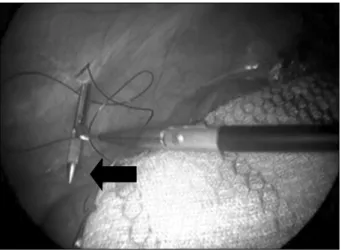
Fig. 2. Additive fixation of mesh by metal tacker after transfascial fixation. Additive fixation of mesh is performed by metal tackers (Protac, Tyco Healthcare, Norwalk, CT; white arrow) along with iliopubic tract (upper border of mesh) after transfascial fixation at infero-lateral corner of the mesh (black arrow).
Fig. 1. Transfascial fixation of mesh. The mesh is inserted into peritoneal cavity after suturing at infero-lateral corner of the mesh with non-absorbable suture material. After threads of suture material are withdrawn by suture passer (black arrow) (suture passerTM, 2.8 mm, KARL STORZ GmbH & Co., KG, Tuttlingen, Germany) through the abdominal wall, they are tied within subcutaneous layer.
collected prospectively was reviewed: age, gender, type of her- nia, duration of operation, intraoperative complications, post- operative complications and recurrence. Variables are presented as mean and standard deviation.
1) Laparoscopic IPOM repair
The procedure was performed with the patient under general anesthesia. Antibiotic prophylaxis was administered with first generation cephalosporin. The patient was laid supine in Trendelenburg position. An 11 mm trocar (Endopath XCELTM, Ethicon Endo-surgery, Inc., Somerville, NJ, USA) was inserted into the peritoneal cavity at supraumbilical area by open Hasson technique, through which 45-degree laparoscope was intro- duced. After the abdominal cavity was explored, we confirmed hernia defects and examined contralateral inguinal area. Two additional 5 mm working trocars were inserted at umbilical lev- el and lower abdomen of contralesional side respectively, under direct vision. The contents of the sac, if present, were carefully reduced into the peritoneal cavity with nontraumatic graspers.
Any redundant floppy umbilical ligament was incised or stapled to the anterior abdominal wall to prevent it from disturbing the view. The sac was left in situ without ligation or incision at the internal ring. A 15×12 cm sized dual facing surgical mesh (PROCEEDTM Mesh, Ethicon Endo-surgery, Inc., Somerville, NJ) with long non-absorbable stitch on the corner of the mesh for transfascial fixation was rolled and passed through the 11 mm trocar into the peritoneal cavity, and then the mesh was
manipulated to lie flat against the posterior inguinal floor. We tried to make as many transfascial fixation as possible, because in case of obese patient with abundant preperitoneal fat, fix- ation with tackers had more chances to fail. If the anatomical structures such as pubic bone, vessels, nerves were concerned, we made only one transfascial suture on the infero-medial cor- ner of the mesh. An instrument (suture passerTM, 2.8 mm, KARL STORZ GmbH & Co., KG, Tuttlingen, Germany) was passed through abdominal wall, grasped each thread of trans- fascial stitches, pulled it out and then tied them at subcutaneous layer (Fig. 1). Several metal tacking devices (Protac, Tyco Healthcare, Norwalk, CT) were used to secure the mesh to the Cooper ligament and to anterior abdominal wall, avoiding tack- ing below iliopubic tract, where lies the so-called triangle of doom and triangle of pain (Fig. 2). Several stitches were done on inferior border of the mesh including infero-medial corner of the mesh with peritoneum to fix the lower edge of the mesh (Fig. 3). Tacks were positioned on the rest of borders of the mesh avoiding deep epigastric vessels.
RESULTS
1) Patient demographics and hernia characteristics
Between May 2006 and November 2010, a total of 48 lapa- roscopic IPOM inguinal hernia repairs in 43 patients were performed. There were 36 patients with unilateral hernias (22 rights and 14 left) and 6 patients with bilateral hernias. There were 37 male patients and 6 female patients with a mean age
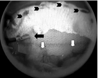
Fig. 3. Intraabdominal view after completion of mesh fixation.
There is a transfascial fixation at infero-lateral corner of the mesh (black arrow). There are two sutures between mesh and peritoneum by absorbable suture material (white arrows). There are multiple metal tackers (Protac, Tyco Healthcare, Norwalk, CT; black arrow heads) at upper border of the mesh along with iliopubic tract.
Table 1. Patient demographics and hernia sites
Variable Number
Number of patients Number of hernias Age (years, mean±SD) Gender
Male Female Site of hernias Right Left Bilateral Type of hernia Indirect hernia Direct hernia Pantaloon Femoral Recurrent hernia† Laparoscopic‡ Open‡ Previous surgery Prostatectomy§ Open bladder surgery Others∥
43*
50 61±16
37 6 22 14 7 43 5 1 1 15 10 12 16 3 5
*3 patients had concomitant operation with hernia repair: removal of undescended testis, removal of foreign bodies in groin region and pelvis, resection of appendiceal mucocele 1 patient misdiag- nosed as spigelian hernia; †5 patients of 15 patients with recurrent inguinal hernia had more than 2 times of recurrence; ‡Indicate previous hernia operation; §9 patients had open radical prostatec- tomy, 5 patients had robotic prostatectomy and 2 patients had TUR-P (transurethral resection of prostate); ∥3 patients had hys- terectomy, 1 patient had uretero-nephrectomy and 1 patient had bowel resection.
Table 2. Summary of perioperative data Operation time (minutes)
Unilateral Bilateral
Intraoperative complications (n) Urinary bladder injury Small bowel injury Conversion
Hospital stay (days, mean±SD) Complications (n)
41±22 56±18 2 1 1 0 1.4±0.9
7 of 61 years. They had various previous operative histories (16
patients of prostatectomy, 3 patients of bladder surgery, and 5 patients of abdominal surgery such as ureteronephrectomy, bowel resection, and hysterectomy) and 15 patients had re- current inguinal hernia. One patient had been misdiagnosed as spigelian hernia preoperatively, so intraperitoneal approach was performed but it came out direct inguinal hernia. Patients’ dem- ographics, site of the hernias, and the types of the previous sur- geries are shown in Table 1. The number of male patients was much higher and more right hernias were observed.
2) Perioperative data
The perioperative data are summarized in Table 2. The mean operative time of unilateral hernia is 41 minutes and bilateral hernia is 56 minutes. Conversion to open repair was not re- quired in any case. There were two intraoperative complica- tions, bladder and small bowel injury. The bladder injury was occurred in patients with recurrent inguinal hernia. We tried TEP repair first, but converted to TAPP repair because of pre- peritoneal space adhesion. During peritoneal dissection, defect on bladder wall was noticed, and repaired immediately without need for conversion. The small bowel injury was occurred in patients with history of previous open bladder resection. During insertion of trocar by open Hasson technique, serosal injury of small bowel was occurred owing to postoperative adhesion. We extended incision and repaired injured small bowel imme-
diately. We performed IPOM repair because there was no soil- ing of bowel contents.
Table 3. Intra- and postoperative complications Intraoperative complications (n)
Urinary bladder injury Small bowel injury Total
Postoperative complications (n) Pain*
Swelling or seroma Hematoma or bleeding Total
Recurrence (n)
1 1 2 2 2 0 4 1
*Pain requiring oral analgesics at out-patient clinics on posto- perative day 7.
3) Morbidity
The morbidity data are summarized in Table 3. There were four early postoperative complications of mild entity: 2 seromas with swelling and 2 transient inguinal pain. As far as post- operative pain is concerned, only 2 patients required analgesics at outpatient clinic after the surgery. One patient had no more pain after a week of medicaiton and the other one recovered completely in a month. In cases of seromas, they were resolved spontaneously without any intervention in 4 to 6 weeks. After a median follow up of 51 months (range 22∼77), we have ob- served only one case of recurrence in the patient operated for re-recurrent hernia. The recurrence was treated by anterior ap- proach using bilayer mesh (Prolene Hernia SystemTM, Ethicon Endo-surgery, Inc., Somerville, NJ, USA).
DISCUSSION
Nowadays laparoscopic surgery has become the gold stand- ard procedure in many surgical fields. Likewise, after decades of experience in laparoscopic hernia surgery, this method has gained worldwide acceptance and has become the first choice for inguinal hernia repair in many centers.2 The advantages of laparoscopic hernia repair include reduced pain and hospital stay, rapid convalescence, quicker return to work, better func- tional and cosmetic results.3,4 In addition, laparoscopic approach allows viewing the entire myopectineal orifice and repairing any unexpected hernias and more specifically in case of re- current hernia. It also allows reducing risk of spermatic cord damage, chronic pain and especially further recurrence (observed in up to 35% of the cases).5
In the early periods of laparoscopic hernia repairs, ex- traperitoneal (TEP) repair, transabdominal preperitoneal (TAPP)
repair and intraperitoneal onlay mesh (IPOM) repair had been performed generally. But recently, TEP and TAPP are being performed widely because IPOM has shown relatively high re- currence rate and complications such as small bowel ob- struction and formation of fistula.6
Among laparoscopic techniques for the inguinal hernia re- pair, our preferential approach has been TEP procedure. We have repaired inguinal hernias after previous surgery of lower abdomen also via laparoscopic TEP approach. We believe this approach is safe and feasible because we have no particular complications. However, despite of a thousand and hundreds of experience, there are some difficult cases of being repaired via TEP or TAPP approach such as after prostatectomy and bladder surgery or recurrent hernia after hernia repair with bilayer mesh. So we employed the IPOM technique in cases of hernia repairs failed via TEP or TAPP repair.
The mean operative time was longer than our data of TEP inguinal hernia repair.7 It is probably because we performed IPOM repair if TEP or TAPP failed.
We could not find any fatal intraoperative complications. We have observed only 4 cases of minor postoperative morbidity (2 seromas, 2 inguinal pains) and a case of recurrence. TAPP and TEP, due to the wide dissection of preperitoneal space, are rather more complicated procedure requiring a long learning curve with a higher risk of morbidity.8-10 In addition, the IPOM has same advantage to other laparoscopic techniques in terms of postoperative pain and faster recovery. Furthermore, IPOM repairs also allow exploration of the contralateral side that may show a non-symptomatic hernia in 11.2∼32.3% of the cases.11 The second hernia may be repaired simultaneously without af- fecting postoperative pain and course.11
The most important point in the evaluation of the efficacy of hernia repair is certainly recurrence.12 After a median follow up of 51 months, we have experienced one recurrence out of 50 hernias. The recurrence occurred in an obese patient with large direct re-recurrent hernia after TEP repair in 3 month.
After another 2 month after IPOM repair, the recurrence oc- curred again, and then we performed bilayer mesh repair via open anterior approach. The reason of recurrence was migration of mesh. The first migration was due to relatively smaller sized mesh than hernia defect. We used commercialized mesh but it was too small to cover the patient’s hernia defect and entire myopectineal orifice. The second migration of the mesh was due to weak fixation of the mesh. We think tacks on upper border of the mesh might have failed to fix the mesh because the patient was so obese that tackers couldn’t reach to muscular structure but preperitoneal adiopose tissue layer.
The features of mesh are another fundamental point. There have been reports about several properties of theoretically ideal mesh: It should be strong enough to withstand physiologic stresses over a long period of time, conform to the abdominal wall, promote strong host tissue ingrowth, which mimics nor- mal tissue healing, resist the formation of bowel adhesions and erosions into visceral structures, not induce allergic or adverse foreign body reactions, resist infection, and be non-car- cinogenic.13-15 No single product possesses all the properties of the ideal mesh. And the characteristics of the mesh are more important in intraperitoneal hernia repair. Sometimes mesh-re- lated complications such as bowel obstruction and even fistula formation have been reported.16 We used dual facing surgical mesh. This mesh has different characteristics on both sides: the parietal surface is rough with micro-pores in order to stimulate a strong fibroblastic tissue reaction and the visceral surface is coated with anti-adhesive material in order to minimize tissue reaction, thus avoiding intestinal adhesions. For period of our all follow up, there has been neither bowel obstruction nor fis- tula formation due to bowel adhesion to mesh.
The size of the mesh is another crucial point. It should not be less than 10×15 cm so that entire myopectineal orifice (pos- sible inguino-crural hernia sites) can be largely covered.12 This is utmost important in order to prevent recurrence.17
The appropriate fixation of the mesh is also important point.
The staples should ensure tight fixation to the underlying naïve abdominal wall musculature and this is better achieved by using tacks. But in cases of obese patients, the thickness of fat tissue can interfere with appropriate fixation of the staples to the un- derlying fascia. The recurrence rate for transfascial suture plus tacks was lower than the use of tacks alone.18
The IPOM repair is also acceptable in terms of operative times, easiness.1,12 However, compared to open surgery, laparo- scopic repair is expensive and needs general anesthesia,12 and so does the IPOM repair. Even more, IPOM repair has big con- cerns about relatively high recurrence rate and complication rate other than other laparoscopic hernia repair.19,20
It is very important not to depend on fascial structures and to avoid use of scarred tissue to repair recurrent inguinal hernia.21 It is believed that to avoid approaching previously damaged plane is crucial for better results. If recurrent inguinal hernia has occurred after hernia repair with bilayer mesh, or after anterior approaching procedure in a patient who has had previous lower abdominal surgery, another alternative method could be helpful.
There are limitations of this study. Since our study have been performed retrospectively to relatively small number of pa-
tients, large-numbered prospective study must be required for valuable results.
CONCLUSION
Though laparoscopic IPOM repair for inguinal hernia is not a standard method, it can be performed for specific cases of recurrent hernias which have difficulties in approaching usual plane of repair as an alternative option with proficient skills and care.
REFERENCES
1) Hatzitheofilou C, Lakhoo M, Sofianos C, Levy RD, Velmahos G, Saadia R. Laparoscopic inguinal hernia repair by an intra- peritoneal onlay mesh technique using expanded PTFE: a prospective study. Surgical Laparoscopy & Endoscopy 1997;7:
451-455.
2) Dulucq JL, Wintringer P, Mahajna A. Totally extraperitoneal (TEP) hernia repair after radical prostatectomy or previous lower abdominal surgery: is it safe? A prospective study.
Surgical Endoscopy 2006;20:473-476.
3) Toy FK, Smoot RT, Jr. Toy-Smooth laparoscopic hernio- plasty. Surgical Laparoscopy & Endoscopy 1991;1:151-155.
4) Spaw AT, Ennis BW, Spaw LP. Laparoscopic hernia repair:
the anatomic basis. Journal of Laparoendoscopic Surgery 1991;1:269-277.
5) Lichtenstein IL, Shulman AG, Amid PK. The cause, preven- tion, and treatment of recurrent groin hernia. The Surgical clinics of North America 1993;73:529-544.
6) Prasad P, Tantia O, Patle NM, Khanna S, Sen B. Laparoscopic ventral hernia repair: a comparative study of transabdominal preperitoneal versus intraperitoneal onlay mesh repair. Journal of Laparoendoscopic & Advanced Surgical Techniques Part A 2011;21:477-483.
7) Choi YY, Hur KY. Simultaneous laparoscopic totally extrape- ritoneal repair of bilateral inguinal hernia: review of 1 surgeon experiences. Surgical Laparoscopy, Endoscopy & Percuta- neous Techniques 2011;21:264-266.
8) Fitzgibbons RJ, Jr., Camps J, Cornet DA, et al. Laparoscopic inguinal herniorrhaphy. Results of a multicenter trial. Annals of Surgery 1995;221:3-13.
9) Schultz C, Baca I, Gotzen V. Laparoscopic inguinal hernia repair. Surgical Endoscopy 2001;15:582-584.
10) Ramshaw B, Shuler FW, Jones HB, et al. Laparoscopic inguinal hernia repair: lessons learned after 1224 consecutive cases. Surgical Endoscopy 2001;15:50-54.
11) Sayad P, Abdo Z, Cacchione R, Ferzli G. Incidence of incipient contralateral hernia during laparoscopic hernia repair.
Surgical Endoscopy 2000;14:543-545.
12) Catani M, De Milito R, Pietroletti R, et al. Is there a place
for intraperitoneal onlay mesh repair (IPOM) of inguinal hernia among laparoscopic techniques? Hepatostroenterology 2004;51:1387-1392.
13) Robinson TN, Clarke JH, Schoen J, Walsh MD. Major mesh-related complications following hernia repair: events reported to the Food and Drug Administration. Surgical Endoscopy 2005;19:1556-1560.
14) Matthews BD, Pratt BL, Pollinger HS, et al. Assessment of adhesion formation to intra-abdominal polypropylene mesh and polytetrafluoroethylene mesh. The Journal of Surgical Research 2003;114:126-132.
15) Schumpelick V, Klinge U. Prosthetic implants for hernia repair. The British Journal of Surgery 2003;90:1457-1458.
16) Haltmeier T, Groebli Y. Small bowel lesion due to spiral tacks after laparoscopic intraperitoneal onlay mesh repair for incisional hernia. International Journal of Surgery Case Reports 2013;4:283-285.
17) Phillips EH, Rosenthal R, Fallas M, et al. Reasons for early recurrence following laparoscopic hernioplasty. Surgical Endoscopy 1995;9:140-144; discussion 144-145.
18) LeBlanc KA. Laparoscopic incisional hernia repair: are transfascial sutures necessary? A review of the literature.
Surgical Endoscopy 2007;21:508-513.
19) Sarli L, Pietra N, Choua O, Costi R, Cattaneo G. Laparoscopic hernia repair: a prospective comparison of TAPP and IPOM techniques. Surgical Laparoscopy & Endoscopy 1997;7:472- 476.
20) Kingsley D, Vogt DM, Nelson MT, Curet MJ, Pitcher DE.
Laparoscopic intraperitoneal onlay inguinal herniorrhaphy.
American Journal of Surgery 1998;176:548-553.
21) Itani KM, Fitzgibbons R, Jr., Awad SS, Duh QY, Ferzli GS.
Management of recurrent inguinal hernias. Journal of the American College of Surgeons 2009;209:653-658.