https://doi.org/10.12750/JET.2017.32.4.287
Endoplasmic Stress Inhibition during Oocyte Maturation Improves Preimplantation Development of Cloned Pig Embryos
Fazle Elahi
1, Hyeji Shin
1, Joohyeong Lee
2and Eunsong Lee
1,2†1
College of Veterinary Medicine, Kangwon National University, Chuncheon, Gangwon 24341, Korea
2
Institute of Veterinary Science, Kangwon National University, Chuncheon, Gangwon 24341, Korea
ABSTRACT
Mitochondrial dysfunction is found in oocytes and transmitted to offspring due to maternal obesity. Treatment of obese mothers with endoplasmic reticulum (ER) stress inhibitors such as salubrinal (SAL) can reverse the mitochondrial dysfunction and result in normal embryonic development. Pig oocytes have also shown ER stress mostly in metaphase II stage. ER stress in oocytes may hinder the in vitro production of pig embryos. This study investigated the effect of ER stress inhibition by SAL treatment during in vitro maturation (IVM) of porcine oocytes at 1, 10, 50 and 100 nM concentrations. Firstly, we tested various concentrations of SAL. SAL at 10 nM showed higher (P
< 0.05) developmental competence to the blastocyst stage (55.6%) after parthenogenesis (PA) than control (44.2%) while not different from other concentrations (49.2, 51.6, and 50.8% for 1, 50, and 100 nM, respectively). Secondly, we performed time-dependent treatment at 10 nM of SAL for IVM of oocytes. It revealed that treatment with SAL during 22 to 44 h of IVM significantly improved PA embryonic development to the blastocyst stage compared to control (40.5, 46.3, 51.7 and 60.2% for control, 0 to 22 h, 22 to 44 h and 0 to 44 h of IVM, respectively, P < 0.05).
Glutathione (GSH) content is an indicator of cytoplasmic maturation of oocytes. Reactive oxygen species (ROS) have a harmful effect on developmental competence of oocytes. For this, we determined the intraoocyte levels of GSH and ROS after 44 h of IVM. It was found that SAL increased intraoocyte GSH level and also decreased ROS level (P
< 0.05). Finally, we performed somatic cell nuclear transfer (SCNT) after treating oocytes with 10 nM SAL during IVM. SAL treatment significantly improved blastocyst formation of SCNT embryos compared to control (39.6% vs.
24.7%, P < 0.05). Our results indicate that treatment of pig oocytes with ER stress inhibitor SAL during IVM improves preimplantation development PA and cloned pig embryos by influencing cytoplasmic maturation in terms of increased GSH content and decreased ROS level in IVM pig oocytes.
(Keywords: Oocyte maturation, Endoplasmic reticulum stress, Embryonic development, Pig)
†Correspondence: Eunsong Lee (ORCID: 0000-0001-9654-7788) Phone: +82-33-250-8670, Fax: +82-33-259-5625
E-mail: [email protected].
INTRODUCTION
Unfolded or misfolded proteins accumulate in the endoplasmic reticulum (ER), triggering activation of the unfolded protein response (UPR) to allow cells to respond to stress conditions (Kaufman, 1999; Ron and Walter, 2007). The ER is a major site of synthesis of transmembrane proteins and lipids and is involved in maintenance of intracellular calcium homeostasis (Larner et al., 2006; Raffaello et al., 2016). When ER homeostasis is perturbed or the control mechanism overloaded, prolonged stress causes apoptosis. Although the exact mechanisms remain poorly described in mammals, three major sensors are thought to activate the UPR during ER stress specifically, EIF2AK3
(PERK), ERN1 (IRE1α), and ATF6 (Tang and Yang, 2015).
It has been reported that controlling ER stress influences embryonic development. In the mice, ER stress signaling was detected at the 1-cell stage and was very high at the blastocyst stage (Kim et al., 1990). HSPA5 (GRP78/BiP), a stress- induced ER chaperone, was required to ensure cell proliferation and to protect the inner cell mass (ICM) from apoptosis during early mouse embryonic development (Luo et al., 2006).
XBP1s regulates transcription of a group of core genes that are
involved in constitutive maintenance of ER function in all cell
types (Acosta-Alvear et al., 2007). The xbp1 gene product is
essential for embryonic development in Drosophila (Souid et
al., 2007). A loss-of-function Xbp1 mutant caused mouse
embryonic lethality, with liver hypoplasia as the principal symptom (Reimold et al., 2000). In addition, the UPR contributed to preimplantation mouse embryonic death that was associated with inability to resolve the ER stress (Hao et al., 2009).
PA embryos were normally used as model system for studying in vitro culture (IVC) condition or environmental stresses due to its physiological alike with fertilized embryos in early development and experimentally simple production of PA embryos with less ethical problems (Tseng et al., 2006;
Paffoni et al., 2008). Pig oocytes and embryos are hypersensitive to various stress during in vitro maturation (IVM) and IVC.
Interestingly, lipid content of oocytes differs in different species.
Triglyceride in pig oocytes shows about three times more than both cow and sheep oocytes (McEvoy et al., 2000). Palmitic, stearic and oleic acids are the most abundant in oocytes of bovine, porcine and sheep, but pig oocytes has higher palmitic acid than oleic acid whereas cow and sheep oocytes has a relatively greater oleic acid (Genicot et al., 2000). Palmitic acid at high levels are known to induce ER stress, impair embryonic development in mice (Wu et al., 2012). In addition, a study confirmed the ER stress in MII oocytes in pig (Zhang et al., 2012). Therefore, it was hypothesized that inhibition of the ER stress during IVM would stimulate embryonic development after PA and somatic cell nuclear transfer (SCNT).
Salubrinal is a well-known ER stress inhibitor, a selective eIF2α dephosphorylation inhibitor, protect cells from lipotoxicity induced by ER stress. It maintains the high in phospho-eIF2α, which the restoration of ER function, help in protein folding and maintain cellular homeostasis (Tian et al., 2011; Kuo et al., 2012). This study investigated the effect of salubrinal during IVM on oocyte maturation, intra-oocytes glutathione (GSH) and reactive oxygen species (ROS) content, and embryonic development after PA and SCNT in pigs.
MATERIALS and METHODS
1. Culture media
All chemicals and reagents were purchased from Sigma- Aldrich (St. Louis, MO, USA) unless otherwise specified. The base IVM medium for oocytes was medium-199 (M199;
Invitrogen, Grand Island, NY, USA). M199 was added with 0.91 mM pyruvate, 0.6 mM cysteine, 10 ng/ml epidermal growth factor, 1 μg/ml insulin and 75 μg/ml kanamycin and
10% (v/v) porcine follicular fluid (PFF). The IVC medium was porcine zygote medium-3 (PZM) (Yoshioka et al, 2002) for embryonic development after PA and SCNT, which consisted of 0.34 mM trisodium citrate, 2.77 mM myo-inositol, and 10 μM β-mercaptoethanol (You et al, 2012).
2. Oocyte collection and IVM
Pig ovaries were obtained from a local abattoir and then transported to the laboratory in warm physiological saline. The cumulus oocytes complex (COCs) were subsequently aspirated from follicles (3–8 mm in diameter) by using an 18-gauge needle connected to a 10-ml syringe. COCs with multiple layers of compact cumulus cells and uniform ooplasm were considered for after washing three times in HEPES-buffered Tyrode's medium containing 0.05% (w/v) polyvinyl alcohol (PVA). The COCs were then cultured in of IVM (500 μl) medium in the presence of 10 IU/ml hCG (Intervet International BV, Boxmeer, Holland) and 80 μg/ml FSH (Antrin R-10; Kyoritsu Seiyaku, Tokyo, Japan). COCs were matured at 39°C with 5% CO
2at maximum humidity for 22 h. For an additional 22 h or 20 h oocytes were cultured in hormone-free IVM medium after washing in fresh hormone- free IVM medium for PA and SCNT, respectively.
3. Somatic cell nuclear transfer and parthenogenesis (PA)
As nuclei donors, porcine fetal fibroblasts were prepared as
described previously (Lee et al., 2013). After IVM for 41 h,
the cumulus cells of COCs were dispersed by gentle pipetting
in the presence of 0.1% (w/v) hyaluronidase. Oocytes having
first polar bodies and uniform ooplasm were selected and
stained with 5 µg/ml Hoechst 33342 for 15 min. Oocytes were
then washed twice in fresh manipulation medium, transferred
into a drop of this media containing 5 μg/ml cytochalasin B
(CB), and overlaid with warm mineral oil. Enucleation was
subsequently performed by a 17-µm beveled glass pipette
(Humagen, Charlottesville, VA, USA) after aspirating the first
polar body (PB) and a small amount of surrounding
cytoplasm. The expelled cytoplasm was then surveyed by
epifluorescence microscopy (TE300; Nikon, Tokyo, Japan) to
verify that the nuclear material had been removed. A single
disaggregated donor cell was injected into the perivitelline
space of the enucleated oocytes, after which oocyte–cell
couplets were placed on a 1 mm fusion chamber overlaid with
1 ml of 280 mM mannitol solution containing 0.001 mM
CaCl
2and 0.05 mM MgCl
2, as previously described (Walker et al, 2002). Cell fusion was performed by using an alternating current field of 2 V cycling at 1 MHz for 2 seconds, followed by two pulses of 170 V/mm direct current (DC) for 30 μsec using a cell fusion generator (LF101; NepaGene, Chiba, Japan). The oocytes were then incubated for 1 h in TLH-BSA, after which they were assessed for confirmation of fusion under a stereomicroscope. The nuclear transferred oocytes were activated with two pulses of 120 V/mm DC for 60 μsec in a 280 mM mannitol solution containing 0.05 mM MgCl
2and 0.1 mM CaCl
2. For PA, MII oocytes were activated as described in SCNT procedures.
4. Post-activation and embryo culture
After electrical activation, the PA were cultured with 5 μ g/ml CB and SCNT embryos were treated with 0.4 μg/ml demecolcine combined with 1.9 mM 6-dimethylaminopurine in IVC medium for 4 h. Afterward, the embryos were washed three times in fresh IVC medium, cultured into 30 μl IVC medium droplets under mineral oil, at 39°C in a humidified atmosphere of 5% O
2, 5% CO
2, and 90% N
2for 7 days.
Cleavage and blastocyst formation were evaluated on Days 2 and 7, respectively. The day of SCNT or PA was designated as Day 0. The total cell count in blastocysts was performed using Hoechst 33342 staining and visualized under an epifluorescence microscope.
5. Measurement of oocyte diameter
After 44 h of IVM, images of denuded oocytes in each group were captured at 200X magnification using a digital camera (DS-L3; Nikon) connected to an inverted microscope (TE 300; Nikon). The size of matured oocytes was determined by the ImageJ software (version 1.46r; National Institutes of Health, Bethesda, MD, USA).
6. Measurement of GSH and ROS contents
After 44 h of IVM, oocytes were examined for GSH and ROS levels. The GSH and ROS contents were measured as previously described (Sakatani et al., 2007). Briefly, (4- chloromethyl-6.8-difluoro-7-hydroxycoumarin, Invitrogen) Cell- Tracker Blue and (2’,7’-dichlorodihydrofluorescein diacetate;
Invitrogen) H2DCFDA were used to detect intraoocyte GSH and ROS with blue fluorescence and green fluorescence for GSH and ROS, respectively. A group of 7–10 oocytes from each
treatment group were cultured for 30 min in TLH-PVA supplemented with 10 μM Cell-Tracker and 10 μM H2DCFDA and in the dark. Embryos treated with Cell-Tracker were then incubated for 30 min with PZM-3 supplemented with 0.3% (w/v) BSA at 39°C in the dark. Following incubation, the embryos were washed with Dulbecco’s phosphate-buffered saline (D-PBS; Invitrogen) containing 0.01% (w/v) PVA, placed into 2-μl droplets. Fluorescence was observed under an epifluorescence microscope (TE300; Nikon) with ultraviolet ray filters at 370 and 460 nm for GSH and ROS, respectively. The fluorescence intensities of oocytes were normalized against the untreated control.
7. Determination of mitochondrial oxidative activity
Denuded oocytes were incubated in M199 containing 200 nM Mitotracker Orange CM-H
2-TMRos (Molecular Probes, Eugene, OR, USA) for 40 min at 39°C in the dark. After washing three times in the fresh M199 medium, oocytes were examined under an inverted epifluorescence microscope (TE300; Nikon). Fluorescence signals were captured with a digital camera (DS-L3; Nikon), and normalized against the untreated control oocytes.
8. Differential count of inner cell mass and trophectoderm cells Differential staining of blastocysts to determine cell number of inner cell mass and trophectoderm was performed as described previously (Thouas et al., 2001). Briefly, blastocysts were stained with 5 μg/ml Hoechst 33342 for 1 h, treated with 0.04% (v/v) Triton X-100 for 3 min and then with 0.005%
(w/v) propidium iodide for 10 min. Stained blastocysts were observed for fluorescence. The propidium iodide and Hoechst 33342-labeled trophectoderm nuclei appeared pink or red and bisbenzimide-labeled ICM nuclei appeared blue.
9. Experimental design
In the first experiment, oocytes were exposed to salubrinal
for 44 h during IVM at 0 (control) 1, 10, 50, and 100 nM
concentrations to determine the optimal concentration for pig
IVM system. Based on the result from the first experiment,
oocytes were treated with salubrinal at 10 nM for 0 to 22 h,
22 to 44 h, or 0 to 44 h of IVM (designated as 022h, 2244h,
and 044h, respectively) to determine the time-dependent effect
in the second and further experiments. The intra-oocytes GSH
and ROS contents were evaluated in the third experiment.
Salubrinal treatment
*% of oocytes that reached metaphase II
No of PA oocytes cultured
% of embryos developed to No of cells in blastocyst
≥ 2-cells Blastocyst
Control 96.8 ± 1.6 128 84.4 ± 3.1
a40.5 ± 4.2
a32.9 ± 1.4
0-22 h 97.3 ± 2.7 131 90.8 ± 2.1
ab46.3 ± 4.9
ab34.8 ± 1.6
22-44 h 96.8 ± 0.6 121 91.5 ± 3.1
ab51.7 ± 4.4
ab37.4 ± 1.5
0-44 h 96.1 ± 1.3 118 95.9 ± 1.5
b60.2 ± 4.5
b37.0 ± 1.8
Four replicates.
*
Oocytes were untreated (control) or treated with 10 nM salubrinal during 0-22, 22-44 and 0-44 h of IVM.
ab
Values with different superscripts denote difference within the same column (P < 0.05).
Table 2. Effect of salubrinal treatment during various stages of in vitro maturation (IVM) on oocyte maturation and parthenogenesis (PA) embryonic development
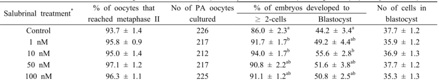
Table 1. Effect of salubrinal treatment during in vitro maturation (IVM) on oocyte maturation and parthenogenesis (PA) embryonic development Salubrinal treatment
*% of oocytes that
reached metaphase II
No of PA oocytes cultured
% of embryos developed to No of cells in blastocyst
≥ 2-cells Blastocyst
Control 93.7 ± 1.4 226 86.0 ± 2.3
a44.2 ± 3.4
a37.7 ± 1.2
1 nM 95.8 ± 0.9 217 91.7 ± 1.7
b49.2 ± 4.4
ab35.9 ± 1.2
10 nM 95.0 ± 1.4 212 94.0 ± 1.7
b55.6 ± 2.8
b36.9 ± 1.3
50 nM 97.1 ± 1.2 217 90.8 ± 2.2
ab51.6 ± 3.8
ab37.7 ± 1.2
100 nM 96.3 ± 1.1 225 91.1 ± 1.2
ab50.8 ± 2.5
ab35.3 ± 1.3
Six replicates.
*
Oocytes were untreated (control) or treated with various concentrations of salubrinal during 0-44 h of IVM.
ab
Values with different superscripts denote difference within the same column (P < 0.05).
Mitochondrial oxidative activity and diameter of IVM oocytes were measured the fourth experiment and the effect on SCNT embryonic development was determined in the fifth experiment.
10. Statistical analyses
Statistical analyses were executed by using the Statistical Analysis System (version 9.3; SAS Institute, Cary, NC, USA).
The general linear model procedure followed by the least significant difference mean separation procedure was used for data analysis. When treatments differed at p < 0.05. The percentage data were considered to arcsine transformation before analysis to maintain homogeneity of variance. The results are present as the mean ± standard error of the mean (SEM).
RESULTS
1. Dose-dependent effects of salubrinal during IVM on embryonic development after PA
To determine the optimal concentration of salubrinal for pig oocyte maturation, oocytes were treated for 0-44 h of IVM with salubrinal at various concentrations. Salubrinal did not
influence nuclear maturation of oocytes after IVM. Among the concentrations tested, 10 nM salubrinal showed significant effect (P < 0.05) on embryo cleavage (94.0 ± 1.7% vs. 86.0
± 2.3%) and blastocyst development (55.6 ± 2.8% vs. 44.2 ± 3.4%) compared to control. Mean cell number in blastocyst was not affected by salubrinal treatment (Table 1).
2. Effects of salubrinal during various stages of IVM on PA embryonic development
Oocytes were untreated or exposed to 10 nM salubrinal for 0 to 22 h, 22 to 44 h and 0 to 44 h of IVM. Cleavage (95.9
± 1.5% vs. 84.4 ± 3.1%) and blastocyst (60.2 ± 4.5% vs. 40.5
± 4.2%) development were significantly (P < 0.05) increased in 044h than the control. Oocyte maturation and average cell number per blastocyst were not influenced by the salubrinal treatment (Table 2).
3. Effect of salubrinal treatment during IVM on the quality of PA blastocysts
Effect of salubrinal treatment during IVM on blastocyst
quality in terms of ICM and trophectoderm cell numbers was
evaluated. Salubrinal treatment in 2244h (9.7 ± 1.0 cells) and
Salubrinal treatment
*No of PA blastocysts examined
Cell number
ICM/total (%)
ICM TE Total
Control 17 6.4 ± 0.5
a31.8 ± 3.4 38.1 ± 4.1 17.8 ± 1.3
22-44 h 20 9.7 ± 1.0
b27.1 ± 2.2 36.1 ± 2.7 25.3 ± 2.0
0-44 h 17 9.8 ± 1.3
b35.6 ± 4.0 45.4 ± 4.2 23.3 ± 3.1
Three replicates.
*
Oocytes were untreated (control) or treated with 10 nM salubrinal during 22-44 and 0-44 h of IVM.
ab
Values with different superscripts denote difference within the same column (P < 0.05).
Table 3. Effect of salubrinal treatment during in vitro maturation on inner cell mass (ICM) and trophectoderm (TE) cell numbers of parthenogenesis (PA) blastocysts
Salubrinal treatment
*No of oocytes GSH content (pixels/oocyte) No of oocytes ROS content (pixels/oocyte)
Control 60 1.00 ± 0.12
a40 1.00 ± 0.18
a22-44 h 60 1.12 ± 0.13
a40 0.59 ± 0.08
b0-44 h 60 1.37 ± 0.12
b40 0.58 ± 0.08
bThree replicates.
*
Oocytes were untreated (control) or treated with 10 nM salubrinal during 22-44 and 0-44 h of IVM.
ab
Values with different superscripts denote difference within the same column (P < 0.05).
Table 4. Effect of salubrinal treatment during in vitro maturation (IVM) on intra-oocyte glutathione (GSH) and reactive oxygen species (ROS) contents
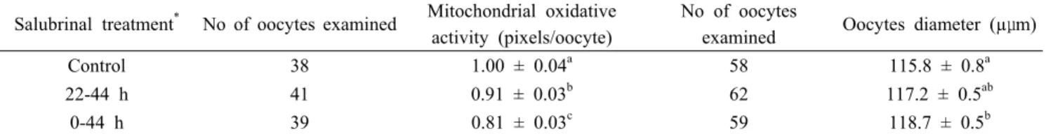
Salubrinal treatment
*No of oocytes examined Mitochondrial oxidative activity (pixels/oocyte)
No of oocytes
examined Oocytes diameter (µμm)
Control 38 1.00 ± 0.04
a58 115.8 ± 0.8
a22-44 h 41 0.91 ± 0.03
b62 117.2 ± 0.5
ab0-44 h 39 0.81 ± 0.03
c59 118.7 ± 0.5
bThree replicates.
*
Oocytes were untreated (control) or treated with 10 nM salubrinal during 22-44 and 0-44 h of IVM.
a-c
Values with different superscripts denote difference within the same column (P < 0.05).
Table 5. Effect of salubrinal on mitochondrial oxidative activity and oocyte diameter after in vitro maturation 044h (9.8 ± 1.3 cells) significantly increased (P < 0.05) the
number of ICM cells compared to control (6.4 ± 0.5 cells).
The number of trophectoderm cells, total cell number, and the ratio of ICM to total cell number was not altered by the treatment (Table 3).
4. Effects of salubrinal on intra-oocyte GSH and ROS contents The intra-oocyte GSH and ROS contents were evaluated after 44 h of IVM. The result revealed that salubrinal increased (P < 0.05) the GSH content of oocytes in 044h compared to those in control and 2244h. In contrast, salubrinal decreased (P < 0.05) the ROS level of oocytes in 044h and 2244h compared to that of oocytes in control (Table 4).
5. Effects of salubrinal on mitochondrial oxidative activity and oocytes diameter
The mitochondrial oxidative activity was determined in the
MII oocytes by Mitotracker Orange CMTMRos, which only stains
the respiring mitochondria based on the oxidative activity of
oocytes. The results indicated that the respiring mitochondria
was significantly higher in control oocytes than in salubrinal-
treated oocytes (1.00, 0.91, and 0.81 pixels/oocyte for control,
044h, and 2244h, respectively). Salubrinal treatment significantly
increased oocyte diameter in 044h (118.7 µm) compared to control
(115.8 µm) while there was no difference between 044h and
2244h (117.2 µm) (Table 5).
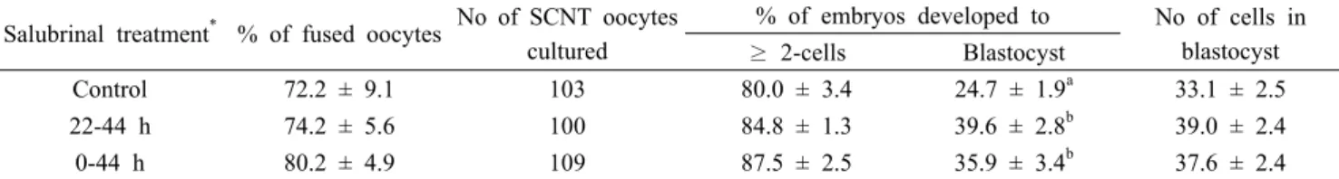
Salubrinal treatment
*% of fused oocytes No of SCNT oocytes cultured
% of embryos developed to No of cells in blastocyst
≥ 2-cells Blastocyst
Control 72.2 ± 9.1 103 80.0 ± 3.4 24.7 ± 1.9
a33.1 ± 2.5
22-44 h 74.2 ± 5.6 100 84.8 ± 1.3 39.6 ± 2.8
b39.0 ± 2.4
0-44 h 80.2 ± 4.9 109 87.5 ± 2.5 35.9 ± 3.4
b37.6 ± 2.4
Four replicates.
*
Oocytes were untreated (control) or treated with 10 nM salubrinal during 22-44 and 0-44 h of IVM.
ab